Abstract
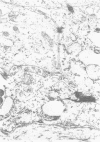
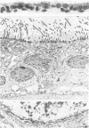
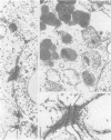
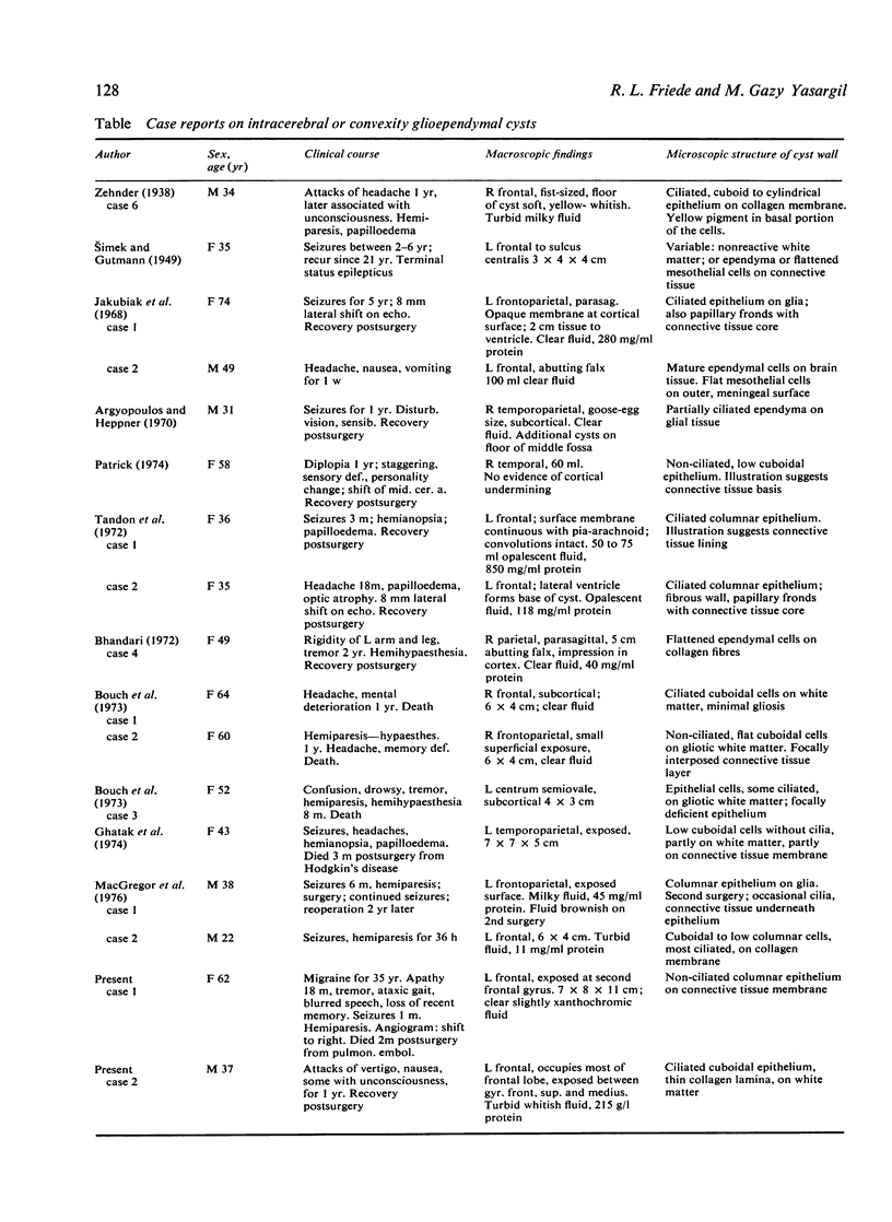
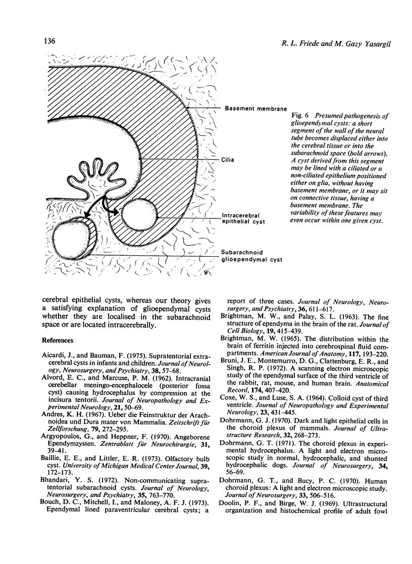
The paper concerns the rare supratentorial, intracerebral or convexity cysts in adults having a wall lined with an epithelium resembling ependyma. The clincopathological aspects of such cysts are reviewed from 15 published cases and two specimens of the authors which could be examined with the electronmicroscope. These cysts manifest at a median age of 46 years as progressive, space occupying lesions with a fairly rapid clinical course of about one to two years. Twelve of 17 cysts were located in the frontal lobes, most were unequivocally intracerebral and none communicated with the lateral ventricle. Microscopic examination of the cyst wall disclosed some variance in structure, the most common feature being a monolayer of ciliated cells sitting on a very thin collagen membrane. One of the present cases was unique in that the compression by the cyst had caused a shell of infarction in the encompassing tissue. The fine structure of the cysts is described and compared with that of potential host tissues from which such cysts may originate. It is concluded that the cysts arise from displaced segments of the wall of the neural tube which correspond to the sites from which the tela chorioidea forms.
Full text
PDF
Images in this article
Selected References
These references are in PubMed. This may not be the complete list of references from this article.
- ALVORD E. C., Jr, MARCUSE P. M. Intracranial cerebellar meningo-encephalocele (posterior fossa cyst) causing hydrocephalus by compression at the incisura tentorii. J Neuropathol Exp Neurol. 1962 Jan;21:50–69. doi: 10.1097/00005072-196201000-00004. [DOI] [PubMed] [Google Scholar]
- Aicardi J., Bauman F. Supratentorial extracerebral cysts in infants and children. J Neurol Neurosurg Psychiatry. 1975 Jan;38(1):57–68. doi: 10.1136/jnnp.38.1.57. [DOI] [PMC free article] [PubMed] [Google Scholar]
- Andres K. H. Uber die Feinstruktur der Arachnoidea und Dura mater von Mammalia. Z Zellforsch Mikrosk Anat. 1967;79(2):272–295. [PubMed] [Google Scholar]
- Argyopoulos G., Heppner F. Angeborene Ependymzysten. Zentralbl Neurochir. 1970;31(1):39–41. [PubMed] [Google Scholar]
- BRIGHTMAN M. W., PALAY S. L. THE FINE STRUCTURE OF EPENDYMA IN THE BRAIN OF THE RAT. J Cell Biol. 1963 Nov;19:415–439. doi: 10.1083/jcb.19.2.415. [DOI] [PMC free article] [PubMed] [Google Scholar]
- Bhandari Y. S. Non-communicating supratentorial subarachnoid cysts. J Neurol Neurosurg Psychiatry. 1972 Dec;35(6):763–770. doi: 10.1136/jnnp.35.6.763. [DOI] [PMC free article] [PubMed] [Google Scholar]
- Bouch D. C., Mitchell I., Maloney A. F. Ependymal lined paraventricular cerebral cysts; a report of three cases. J Neurol Neurosurg Psychiatry. 1973 Aug;36(4):611–617. doi: 10.1136/jnnp.36.4.611. [DOI] [PMC free article] [PubMed] [Google Scholar]
- Brightman M. W. The distribution within the brain of ferritin injected into cerebrospinal fluid compartments. II. Parenchymal distribution. Am J Anat. 1965 Sep;117(2):193–219. doi: 10.1002/aja.1001170204. [DOI] [PubMed] [Google Scholar]
- Bruni J. E., Montemurro D. G., Clattenburg R. E., Singh R. P. A scanning electron microscopic study of the ependymal surface of the third ventricle of the rabbit, rat, mouse and human brain. Anat Rec. 1972 Dec;174(4):407–420. doi: 10.1002/ar.1091740402. [DOI] [PubMed] [Google Scholar]
- COXE W. S., LUSE S. A. COLLOID CYST OF THIRD VENTRICLE. AN ELECTRON MICROSCOPIC STUDY. J Neuropathol Exp Neurol. 1964 Jul;23:431–445. doi: 10.1097/00005072-196407000-00003. [DOI] [PubMed] [Google Scholar]
- Dohrmann G. J., Bucy P. C. Human choroid plexus: a light and electron microscopic study. J Neurosurg. 1970 Nov;33(5):506–516. doi: 10.3171/jns.1970.33.5.0506. [DOI] [PubMed] [Google Scholar]
- Dohrmann G. J. Dark and light epithelial cells in the choroid plexus of mammals. J Ultrastruct Res. 1970 Aug;32(3):268–273. doi: 10.1016/s0022-5320(70)80007-7. [DOI] [PubMed] [Google Scholar]
- Dohrmann G. J. The choroid plexus in experimental hydrocephalus. A light and electron microscopic study in normal, hydrocephalic, and shunted hydrocephalic dogs. J Neurosurg. 1971 Jan;34(1):56–69. doi: 10.3171/jns.1971.34.1.0056. [DOI] [PubMed] [Google Scholar]
- Doolin P. F., Birge W. J. Ultrastructural organization and histochemical profile of adult fowl choroid plexus epithelium. Anat Rec. 1969 Dec;165(4):515–529. doi: 10.1002/ar.1091650407. [DOI] [PubMed] [Google Scholar]
- Friede R. L., Hu K. H. A new approach for determining the volume of cerebral cellular fluid and demonstration of its communication with C.S.F. J Physiol. 1971 Oct;218(2):477–493. doi: 10.1113/jphysiol.1971.sp009629. [DOI] [PMC free article] [PubMed] [Google Scholar]
- Ghatak N. R., Hirano A., Kasoff S. S., Zimmerman H. M. Fine structure of an intracerebral epithelial cyst. J Neurosurg. 1974 Jul;41(1):75–82. doi: 10.3171/jns.1974.41.1.0075. [DOI] [PubMed] [Google Scholar]
- Gilles F. H., Rockett F. X. Infantile hydrocephalus: retrocerebellar "arachnoidal" cyst. J Pediatr. 1971 Sep;79(3):436–443. doi: 10.1016/s0022-3476(71)80153-1. [DOI] [PubMed] [Google Scholar]
- Hirano A., Ghatak N. R., Wisoff H. S., Zimmerman H. M. An epithelial cyst of the spinal cord. An electron microscopic study. Acta Neuropathol. 1971;18(3):214–223. doi: 10.1007/BF00685067. [DOI] [PubMed] [Google Scholar]
- Jakubiak P., Dunsmore R. H., Beckett R. S. Supratentorial brain cysts. J Neurosurg. 1968 Feb;28(2):129–136. doi: 10.3171/jns.1968.28.2.0129. [DOI] [PubMed] [Google Scholar]
- LIST C. F., WILLIAMS J. R. Subdural epithelial cyst in the interhemispheral fissure. Report of a case, with some remaks concerning the classification of intracranial and thelial cysts. J Neurosurg. 1961 Sep;18:690–693. doi: 10.3171/jns.1961.18.5.0690. [DOI] [PubMed] [Google Scholar]
- Landolt-Weber U. M. Ultrastruktur einer Kolloidcyste des dritten Ventrikels. Acta Neuropathol. 1973;26(1):59–70. doi: 10.1007/BF00685523. [DOI] [PubMed] [Google Scholar]
- Loeser J. D., Alvord E. C., Jr Clinicopathological correlations in agenesis of the corpus callosum. Neurology. 1968 Aug;18(8):745–756. doi: 10.1212/wnl.18.8.745. [DOI] [PubMed] [Google Scholar]
- Lopes C. A., Mair W. G. Ultrastructure of the arachnoid membrane in man. Acta Neuropathol. 1974;28(2):167–173. doi: 10.1007/BF00710326. [DOI] [PubMed] [Google Scholar]
- MacGregor B. J., Gawler J., South J. R. Intracranial epithelial cysts. Report of two cases. J Neurosurg. 1976 Jan;44(1):109–115. doi: 10.3171/jns.1976.44.1.0109. [DOI] [PubMed] [Google Scholar]
- Nabeshima S., Reese T. S., Landis D. M., Brightman M. W. Junctions in the meninges and marginal glia. J Comp Neurol. 1975 Nov 15;164(2):127–169. doi: 10.1002/cne.901640202. [DOI] [PubMed] [Google Scholar]
- Oksche A., Vaupel-von Harnack M. Elektronenmikroskopische Studien über Altersveränderungen (Filamente) der Plexus chorioidei des Menschen (Biopsiematerial) Z Zellforsch Mikrosk Anat. 1969;93(1):1–29. [PubMed] [Google Scholar]
- Patrick B. S. Ependymal cyst of the Sylvian fissure. Case report. J Neurosurg. 1971 Dec;35(6):751–754. doi: 10.3171/jns.1971.35.6.0751. [DOI] [PubMed] [Google Scholar]
- Rawlinson D. G., Herman M. M., Rubinstein L. J. The fine structure of a myxopapillary ependymoma of the filum terminale. Acta Neuropathol. 1973 Jun 26;25(1):1–13. doi: 10.1007/BF00686853. [DOI] [PubMed] [Google Scholar]
- SHUANGSHOTI S., ROBERTS M. P., NETSKY M. G. NEUROEPITHELIAL (COLLOID) CYSTS: PATHOGENESIS AND RELATION TO CHOROID PLEXUS AND EPENDYMA. Arch Pathol. 1965 Sep;80:214–224. [PubMed] [Google Scholar]
- STARKMAN S. P., BROWN T. C., LINELL E. A. Cerebral arachnoid cysts. J Neuropathol Exp Neurol. 1958 Jul;17(3):484–500. doi: 10.1097/00005072-195807000-00009. [DOI] [PubMed] [Google Scholar]
- Santolaya R. C., Echandia E. L. The surface of the choroid plexus cell under normal and experimental conditions. Z Zellforsch Mikrosk Anat. 1968;92(1):43–51. doi: 10.1007/BF00339401. [DOI] [PubMed] [Google Scholar]
- Shuangshoti S., Netsky M. G. Histogenesis of choroid plexus in man. Am J Anat. 1966 Jan;118(1):283–316. doi: 10.1002/aja.1001180114. [DOI] [PubMed] [Google Scholar]
- TENNYSON V. M., PAPPAS G. D. An electron microscope study of ependymal cells of the fetal, early postnatal and adult rabbit. Z Zellforsch Mikrosk Anat. 1962;56:595–618. doi: 10.1007/BF00540584. [DOI] [PubMed] [Google Scholar]
- Tandon P. N., Roy S., Elvidge A. Subarachnoid ependymal cyst. Report of two cases. J Neurosurg. 1972 Dec;37(6):741–745. doi: 10.3171/jns.1972.37.6.0741. [DOI] [PubMed] [Google Scholar]
- Waggener J. D., Beggs J. The membranous coverings of neural tissues: an electron microscopy study. J Neuropathol Exp Neurol. 1967 Jul;26(3):412–426. doi: 10.1097/00005072-196707000-00005. [DOI] [PubMed] [Google Scholar]
- Weindl A., Joynt R. J. Ultrastructure of the ventricular walls. Three-dimensional study of regional specialization. Arch Neurol. 1972 May;26(5):420–427. doi: 10.1001/archneur.1972.00490110054005. [DOI] [PubMed] [Google Scholar]


