

Abstract
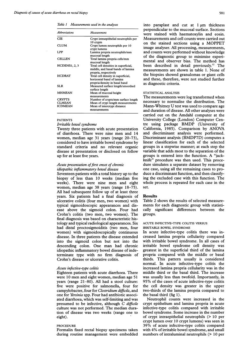
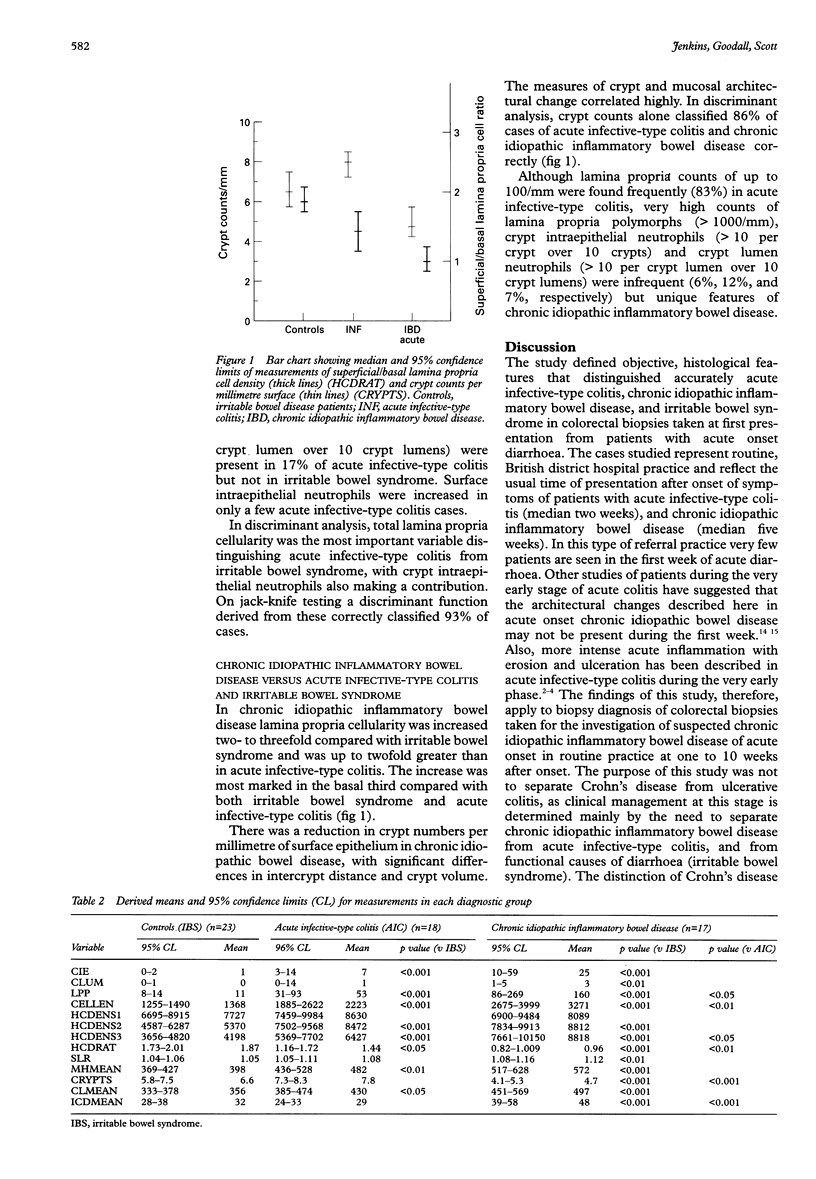
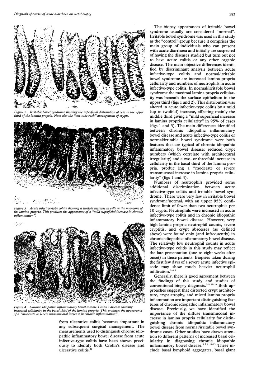
AIM: To identify simple, objective, accurate histological criteria for distinguishing acute infective-type colitis, chronic idiopathic inflammatory bowel disease, and irritable bowel syndrome on rectal biopsy in patients with acute onset diarrhoea at first presentation, one to 10 weeks after onset. METHODS: Cell counts and measurements of mucosal architecture were made on initial rectal biopsies from 18 patients with acute infective-type colitis, 17 patients with first acute presentation of chronic idiopathic inflammatory bowel disease, and 23 patients with irritable bowel syndrome. The data were analysed by ANOVA and discriminant analysis. RESULTS: Lamina propria cells were mainly in the upper third in irritable bowel syndrome patients. Increased lamina propria cellularity, mainly in the middle third, and numbers of crypt intraepithelial neutrophils distinguished acute infective-type colitis from irritable bowel syndrome in 93% of cases. Chronic idiopathic inflammatory bowel disease differed from irritable bowel syndrome and acute infective-type colitis in a decreased number of crypts and altered crypt architecture. Chronic idiopathic inflammatory bowel disease showed higher lamina propria cellularity, especially in the basal third, with an increased number of lamina propria neutrophils. On discriminant analysis, crypt numbers distinguished 86% of the cases of chronic idiopathic inflammatory bowel disease from the other groups. CONCLUSION: At one week or more from onset, acute infective-type colitis is characterised by a superficial increase in lamina propria cellularity, with only a slight increase in the number of polymorphs. At this stage, chronic idiopathic inflammatory bowel disease is characterised by a transmucosal increase in cellularity together with crypt loss and architectural abnormality. Thus, measurement of mucosal architecture establishes simple, accurate, objective criteria for routine biopsy diagnosis of chronic idiopathic inflammatory bowel disease from acute infective-type colitis and irritable bowel syndrome at initial presentation, one to 10 weeks after onset.
Full text
PDF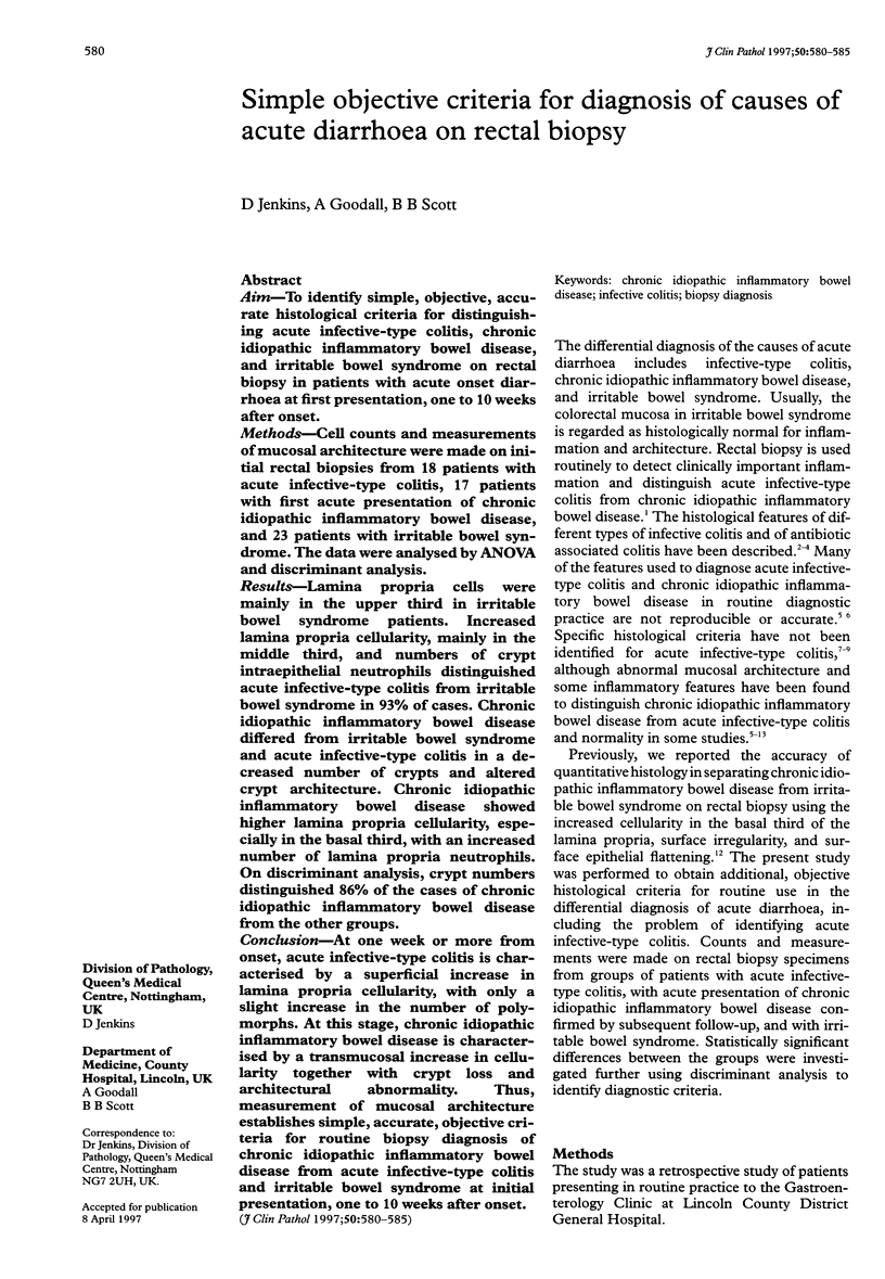
Images in this article
Selected References
These references are in PubMed. This may not be the complete list of references from this article.
- Allison M. C., Hamilton-Dutoit S. J., Dhillon A. P., Pounder R. E. The value of rectal biopsy in distinguishing self-limited colitis from early inflammatory bowel disease. Q J Med. 1987 Dec;65(248):985–995. [PubMed] [Google Scholar]
- Anand B. S., Malhotra V., Bhattacharya S. K., Datta P., Datta D., Sen D., Bhattacharya M. K., Mukherjee P. P., Pal S. C. Rectal histology in acute bacillary dysentery. Gastroenterology. 1986 Mar;90(3):654–660. doi: 10.1016/0016-5085(86)91120-0. [DOI] [PubMed] [Google Scholar]
- Cook M. G., Dixon M. F. An analysis of the reliability of detection and diagnostic value of various pathological features in Crohn's disease and ulcerative colitis. Gut. 1973 Apr;14(4):255–262. doi: 10.1136/gut.14.4.255. [DOI] [PMC free article] [PubMed] [Google Scholar]
- Kumar N. B., Nostrant T. T., Appelman H. D. The histopathologic spectrum of acute self-limited colitis (acute infectious-type colitis). Am J Surg Pathol. 1982 Sep;6(6):523–529. doi: 10.1097/00000478-198209000-00004. [DOI] [PubMed] [Google Scholar]
- Mathan M. M., Mathan V. I. Morphology of rectal mucosa of patients with shigellosis. Rev Infect Dis. 1991 Mar-Apr;13 (Suppl 4):S314–S318. doi: 10.1093/clinids/13.supplement_4.s314. [DOI] [PubMed] [Google Scholar]
- Nostrant T. T., Kumar N. B., Appelman H. D. Histopathology differentiates acute self-limited colitis from ulcerative colitis. Gastroenterology. 1987 Feb;92(2):318–328. doi: 10.1016/0016-5085(87)90124-7. [DOI] [PubMed] [Google Scholar]
- Price A. B., Jewkes J., Sanderson P. J. Acute diarrhoea: Campylobacter colitis and the role of rectal biopsy. J Clin Pathol. 1979 Oct;32(10):990–997. doi: 10.1136/jcp.32.10.990. [DOI] [PMC free article] [PubMed] [Google Scholar]
- Schumacher G. First attack of inflammatory bowel disease and infectious colitis. A clinical, histological and microbiological study with special reference to early diagnosis. Scand J Gastroenterol Suppl. 1993;198:1–24. [PubMed] [Google Scholar]
- Seldenrijk C. A., Morson B. C., Meuwissen S. G., Schipper N. W., Lindeman J., Meijer C. J. Histopathological evaluation of colonic mucosal biopsy specimens in chronic inflammatory bowel disease: diagnostic implications. Gut. 1991 Dec;32(12):1514–1520. doi: 10.1136/gut.32.12.1514. [DOI] [PMC free article] [PubMed] [Google Scholar]
- Surawicz C. M., Belic L. Rectal biopsy helps to distinguish acute self-limited colitis from idiopathic inflammatory bowel disease. Gastroenterology. 1984 Jan;86(1):104–113. [PubMed] [Google Scholar]
- Surawicz C. M. Diagnosing colitis. Biopsy is best. Gastroenterology. 1987 Feb;92(2):538–540. doi: 10.1016/0016-5085(87)90156-9. [DOI] [PubMed] [Google Scholar]
- Surawicz C. M., Haggitt R. C., Husseman M., McFarland L. V. Mucosal biopsy diagnosis of colitis: acute self-limited colitis and idiopathic inflammatory bowel disease. Gastroenterology. 1994 Sep;107(3):755–763. doi: 10.1016/0016-5085(94)90124-4. [DOI] [PubMed] [Google Scholar]
- Theodossi A., Spiegelhalter D. J., Jass J., Firth J., Dixon M., Leader M., Levison D. A., Lindley R., Filipe I., Price A. Observer variation and discriminatory value of biopsy features in inflammatory bowel disease. Gut. 1994 Jul;35(7):961–968. doi: 10.1136/gut.35.7.961. [DOI] [PMC free article] [PubMed] [Google Scholar]
- Therkildsen M. H., Jensen B. N., Teglbjaerg P. S., Rasmussen S. N. The final outcome of patients presenting with their first episode of acute diarrhoea and an inflamed rectal mucosa with preserved crypt architecture. A clinicopathologic study. Scand J Gastroenterol. 1989 Mar;24(2):158–164. doi: 10.3109/00365528909093031. [DOI] [PubMed] [Google Scholar]
- Thompson E. M., Price A. B., Altman D. G., Sowter C., Slavin G. Quantitation in inflammatory bowel disease using computerised interactive image analysis. J Clin Pathol. 1985 Jun;38(6):631–638. doi: 10.1136/jcp.38.6.631. [DOI] [PMC free article] [PubMed] [Google Scholar]