Abstract
MECP2 (methyl CpG binding protein 2) duplication causes syndromic intellectual disability. Patients often suffer from life-threatening infections, suggesting an additional immunodeficiency. We describe for the first time the detailed infectious and immunological phenotype of MECP2 duplication syndrome. 17/27 analyzed patients suffered from pneumonia, 5/27 from at least one episode of sepsis. Encapsulated bacteria (S.pneumoniae, H.influenzae) were frequently isolated. T-cell immunity showed no gross abnormalities in 14/14 patients and IFNy-secretion upon ConA-stimulation was not decreased in 6/7 patients. In 6/21 patients IgG2-deficiency was detected – in 4/21 patients accompanied by IgA-deficiency, 10/21 patients showed low antibody titers against pneumococci. Supra-normal IgG1-levels were detected in 11/21 patients and supra-normal IgG3-levels were seen in 8/21 patients – in 6 of the patients as combined elevation of IgG1 and IgG3. Three of the four patients with IgA/IgG2-deficiency developed multiple severe infections. Upon infections pronounced acute-phase responses were common: 7/10 patients showed CRP values above 200 mg/l. Our data for the first time show systematically that increased susceptibility to infections in MECP2 duplication syndrome is associated with IgA/IgG2-deficiency, low antibody titers against pneumococci and elevated acute-phase responses. So patients with MECP2 duplication syndrome and low IgA/IgG2 may benefit from prophylactic substitution of sIgA and IgG.
Electronic supplementary material
The online version of this article (doi:10.1007/s10875-015-0129-5) contains supplementary material, which is available to authorized users.
Keywords: Xq28-duplication syndrome, methyl CpG binding protein 2 (MECP2), MECP2 duplication syndrome, primary immunodeficiency, intellectual disability, humoral immunodeficiency
Introduction
MECP2 duplication on chromosome Xq28 (#300260) causes a severe form of X-linked intellectual disability (XLID). It was first clinically described as Lubs syndrome, the underlying genetic condition was identified in 2005 [1–3]. Up to now more than 200 patients have been described [1–50]. The exact prevalence of MECP2 duplication syndrome is still unknown, but in synopsis of different screening attempts it is estimated that MECP2 duplication syndrome may explain ~1 % of cases of severe XLID [51]. The core phenotype of MECP2 duplication syndrome includes infantile hypotonia, mild dysmorphic features, developmental delay/severe to profound intellectual disabilty and absent to minimal speech [51]. Spasticity, ataxia and autism are facultative clinical signs that occur in different combinations and to different degrees. Since the description of the first individuals it became evident that besides the striking neurological phenotype patients with MECP2 duplication syndrome are at increased risk for severe infections. About 70–75 % of the affected individuals are reported to develop recurrent infections, especially of the respiratory tract. Respiratory infections are the main cause of early death reported among the population [51, 52]. However a detailed description of the infectious phenotype and an investigation of the immunological phenotype based on a large cohort has been missing to date.
Suspecting an immune dysfunction in patients with MECP2 duplication syndrome we assessed the infectious and immunological phenotype of a cohort of 30 patients. We here provide for the first time a detailed description of the type of infections, the pathogens isolated and the core cellular and humoral immunological phenotype in patients with MECP2 duplication syndrome.
Materials and Methods
Patients
The current study was conducted in accordance with the Helsinki Declaration, with informed consent obtained from each patient or the patient’s family. Samples from the patients were used in this study with approval from the local ethics committee of the Charité (approval # EA2/063/12). All patients enrolled in this study show a genetically confirmed duplication of at least MECP2 and IRAK1. The diagnostics were performed prior to this study and the results were submitted to us by the performing laboratories or geneticists. In patients P1, P2, P8, P9, P10, P11, P12, P13, P14, P17, P19, P20, P21, P25, P27, P28, P29 and P30 the duplication had been detected by use of array-CGH thus revealing the actual size of the duplication on Xq28. In Patient P15 quantitative PCR had been performed and in patients P3, P4, P5, P6, P7, P18, P23, P24 and P26 Multiplex-ligation probe amplification was used for the diagnosis of MECP2 duplication and single neighbouring genes, e.g., IRAK1. So in patients P15, P3, P4, P5, P6, P7 and P18, P23, P24 and P26 the actual size of the duplication is unknown. The following patients have been described previously: P1, P2 in Bartsch et al. [21], P3, P4, P5, P6, P7 in Echenne et al. [18], P8 in Budisteanu et al. [31], P9 in Jezela-Stanek et al. [28], P10 in Mayo et al. [32], P12 in Grasshoff et al. [30], P13 in Bauters et al. [9], P15 in Meins et al. [2], P17 in Madrigal et al. [8], P20, P21 in Xu et al. [43], and P22, P27 in Vignoli et al. [40]. For detailed molecular analyses of these patients see the corresponding publications. Patients P11, P14, P16, P18, P19, P23, P24, P25, P26, P28, P29 and P30 have not been published before. For molecular characterization of these patients see supplementary table 1.
Blood Samples of the Patients
Venous punctures were performed in parallel to routine blood tests. The blood samples were sent to our laboratory to ensure that all patients’ samples were analyzed with the same methods and with the same laboratory equipment.
Assessment of the Infectious Phenotype
A detailed questionnaire on the clinical phenotype was completed by the physicians caring for the patients with MECP2 duplication syndrome and sent to two of the authors (MB and HVB) for thorough review.
Assessment of Immunoglobulin Levels and Antibody Titers Against Tetanus Toxoid and Pneumococci
Total immunoglobulin levels and immunoglobulin subclasses of all patients’ samples in our cohort were measured by ECLIA on a COBAS 6000 (Roche, Switzerland) in our laboratory. Antigen-specific IgG antibodies against Tetanus-Toxoid and PCP as well as anti-PCP-IgG2 of all patients’ samples in our cohort were measured by ELISA according to manufacturer’s protocols (MK013, MK012 and MK010, The Binding Site) in our laboratory.
Assessment of Lymphocyte Subsets
Lymphocyte subsets of patients’ EDTA-blood were stained with fluorescence-labelled monoclonal antibodies against CD14 (RMO52), CD56 (N901), CD16 (3G8), CD4 (SFCI12T4D11), CD19 (J3-119), CD8 (SFCI21Thy2D3), CD3 (UCH-T1), CD45 (J33), CD45R0 (UCHL1), TCRαβ (IP26A), TCRγδ (IMMU510) and CD45RA (2H4LDH11LDB9) (all Beckman Coulter, USA) for 15 min at room temperature and were measured and analyzed after erythrocyte lysis (Versa-Lyse, Beckman Coulter) on a NAVIOS-FACS (Beckman Coulter, USA) in our laboratory.
Lymphocyte Proliferation Assay
Lymphocyte proliferative responses were tested according to standard protocols in heparinised whole blood and were stimulated with phytohemagglutinin (PHA, Sigma-Aldrich, L1668), plate bound anti-CD3 (3, 10, and 30 μg/ml; clone HIT3a, Becton Dicinson), Pokeweed-Mitogen (PWM, 1, 5, and 15 μg/ml, Sigma-Aldrich, L-9379), IL-2 (20 ng/ml, #200-02, PeproTech, Rocky Hill/NJ, USA) and Staphylococcus aureus Cowan-1 strain (SAC, Pansorbin, 1:1000, 1:10,000, Merck, Darmstadt, Germany) for 72 h and with Tetanus-Toxoid RT50 (40, 20, and 10 Lf/ml, Staten Serum Inst. Copenhagen), Candida albicans antigen (0.025, 0.25, and 2.5 μg/ml; CAN001-P; Biologo, Kronshagen, Germany) and Diphtheria-Toxoid (20 LF/ml, Chiron-Behring) for 120 h. After pulsing with 1 μCi [3H]-thymidine per well during the last 12 h incorporated [3H]-thymidine was measured on a β-scintillation counter (MicroBeta 1450 TriLux, PerkinElmer).
Assessment of IFNγ After Stimulation with ConcanavalinA (ConA)
Heparinised blood was stimulated 1:5 diluted in RPMI with 50 μg/ ml ConA (Sigma-Aldrich, #7246) and IFNy was assessed by Th1/Th2-cytokine detection kit (Becton Dicinson Biosciences, #550749) according to the manufacturers protocol. Measurement was performed on a FACS-Navios (Beckman Coulter). For analysis FCAP-Array-Software (Soft Flow Hungary) was used. The normal range of IFNy-secretion upon activation with ConA after 4 h of activation was assessed based on the activation of 50 healthy donors (25 male and 25 female). The normal range of IFNy-secretion upon activation with ConA after 24 h of activation was assessed based on the activation of 10 healthy male donors.
Results
Cohort, Genotypes and Infectious Phenotype
30 patients with MECP2 duplication syndrome from Germany, France, Italy, Poland, Romania, Spain, The Netherlands, Belgium, USA and China were enrolled in the study (28 male patients and two female patients). All patients showed a duplication of MECP2 and neighbouring IRAK1 (Fig. 1). Patients’ age ranged from 3 to 48 years. We had access to the history of 27 of the 30 patients with 343 cumulative patient-years. The occurrence of infections varied in our cohort of patients: In 9/27 patients (including the two girls) no severe infections were reported at all. 18/27 patients developed at least one severe infection requiring intravenous antibiotic treatment. Pneumonia occurred in 17/27 patients. Regarding the type of pneumonia, complete assessment was possible in patient P14 and in this patient all pneumonias were X-ray-proven bronchopneumoniae. 5/27 patients developed at least one episode of sepsis. Urinary tract infections were reported in 6/27 patients. Recurrent fever of unknown origin occurred in three patients. No meningitis was diagnosed in our cohort. The frequency of severe infections also varied with a maximum of 27 episodes requiring intravenous antibiotic treatment since birth in a 16-year old boy (P1). Two patients died of pneumonia during the course of the study (Table 1).
Fig. 1.
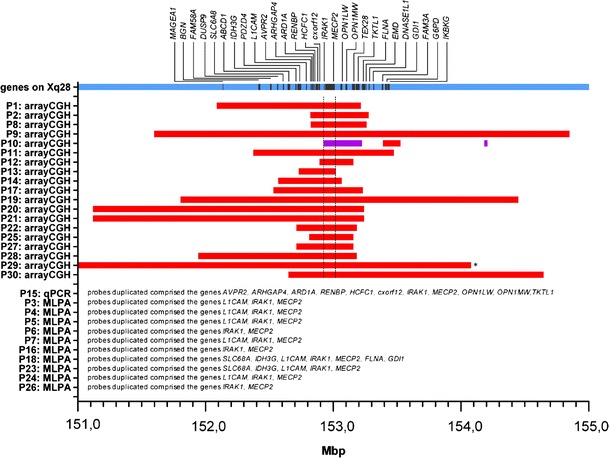
Size of duplications in MbP on chromosome Xq28. The x-axis depicts positions on chromosome Xq28 between 151 and 155 Mbp. Red bars indicate duplicated segments, purple bars indicate triplicated segments in the individual patients. Positions on the X chromosome are based on the UCSC genome browser build hg18. (* Duplication of patient P29 extends the depicted area on Xq28 to the left side until position 147.384.720 bp)
Table 1.
Infectious phenotype of patients with MECP2 duplication syndrome
| P1 | P14 | P9 | P29 | P15 | P23 | P17 | P16 | P2 | P24 | P27 | P18 | P13 | P22 | P28 | P26 | P19 | P11 | P12 | P10 | P3 | P4 | P5 | P6 | P7 | P20 | P21 | P8 | P25 | P30 | |
|---|---|---|---|---|---|---|---|---|---|---|---|---|---|---|---|---|---|---|---|---|---|---|---|---|---|---|---|---|---|---|
| Age | 16years | 15years | 15years | 7years | 15years | 8years | 9years | 3years | 6years | 15years | 12years | 9years | 8years | 18years | 4years | 10years | 22years | 20years | 9years | 11years | 9years | 28years | 23years | 5years | 17years | 19years | 10years | 10years | 48years | 18years |
| Sex | m | m | m | m | m | m | m | m | m | m | m | m | m | m | m | m | m | m | f | f | m | m | m | m | m | m | m | m | m | m |
| Outcome status | alive | † | alive | alive | † | alive | alive | alive | alive | alive | alive | alive | alive | alive | alive | alive | alive | alive | alive | alive | alive | alive | alive | alive | alive | alive | alive | alive | alive | alive |
| Number of infections requiring intravenous antibiotic treatment | 27 | 18 | 13 | 10 | 8 (between 8y-15y) | 7 | 3 | 3 | 3 | 3 | 3 | 2 | 2 | 2 | 2 | 2 | 1 | 1 | 0 | 0 | 0 | 0 | 0 | 0 | 0 | 0 | 0 | n.a. | n.a. | n.a. |
| Invasive infections | ||||||||||||||||||||||||||||||
| Sepsis | 2 | 0 | 3 | 1 | 1 | 0 | 1 | 0 | 0 | 0 | 0 | 0 | 0 | 0 | 0 | 0 | 0 | 0 | 0 | 0 | 0 | 0 | 0 | 0 | 0 | 0 | 0 | n.a. | n.a. | n.a. |
| Meningitis | 0 | 0 | 0 | 0 | 0 | 0 | 0 | 0 | 0 | 0 | 0 | 0 | 0 | 0 | 0 | 0 | 0 | 0 | 0 | 0 | 0 | 0 | 0 | 0 | 0 | 0 | 0 | n.a. | n.a. | n.a. |
| Others | – | – | – | – | – | – | – | – | – | – | – | – | – | 1x peritonitis | – | – | – | – | – | – | – | – | – | – | – | – | – | – | – | – |
| Noninvasive infections | ||||||||||||||||||||||||||||||
| Pneumonia | 48 | 14 | 43 | 8 | 50 | 7 | 2 | 3 | 2 | 3 | 3 | 3 | 2 | 1 | 2 | 2 | 30 | 0 | 0 | 0 | 0 | 0 | 0 | 0 | 0 | 0 | 0 | n.a. | n.a. | n.a. |
| Purulent otitis | 3 | 2 | n.a. | 4 | 3 | 1 | 0 | n.a. | 1 | n.a. | 1 | >8 | >4 | n.a. | n.a. | n.a. | 5 | 0 | n.a. | 0 | 0 | 0 | 0 | 0 | 0 | 0 | 0 | n.a. | n.a. | n.a. |
| Others |
>1 urinary tract infection 1 x ARDS 1 x streptococcal toxic shock syndrome |
5x urinary tract infection 2x pyelonephritis |
1x lung tuberculosis 3x cystitis recurrent idiopathic fever episodes (~1 day lasting) |
3 x pyelonephritis | – | 1 x Tonsillitis |
1x cystitis 1x salmonella gastroenteritis |
– |
1 x pyelonephritis >2 urinary tract infection |
– | – |
recurrent episodes of bronchitis 1 x severe gastroenteritis |
– | – | – | – | 3 x urinary tract infection |
1x adenophlegmon laterocervical 2 x pharyngotonsilitis with high fever |
>4x tonsillitis | – | – | – | – | – | – | – | – | – | – | – |
| Others | ||||||||||||||||||||||||||||||
| Severe fever of unknown origin | 0 | 0 | >4 | 0 | 3 | 0 | 0 | 3 | 0 | 0 | 0 | 0 | 0 | 0 | 0 | 0 | 0 | 0 | 0 | 0 | 0 | 0 | 0 | 0 | 0 | 0 | 0 | n.a. | n.a. | n.a. |
Age, sex, number of infections requiring intravenous antibiotic treatment, occurence of invasive versus non-invasive infections and outcome. Order of patients runs from the most often infected to the least often infected
Of the 25 different pathogens detected in the patients, the majority were bacteria with 84 %. Only 3 viruses (12 %) and 1 fungus (4 %) were among the 25 pathogens. Of the 21 different bacteria, 7 bacteria were Gram-positive (33 %) and 14 bacteria were Gram-negative (66 %). Thirteen of the 21 isolated bacteria (61.9 %) are capable of building a capsule (A.hydrophila [53], B.fragilis [54–56], E.coli [57], H.influenzae [57], K.oxytoca [58], K.pneumonia [59], P.mirabilis [60], P.aeruginosa [59, 61], S.aureus [62–65], S.epidermidis [66, 67], S.agalactiae [57], Streptococcus group A [68–70], S.pneumoniae [57, 64]).
Pathogens were isolated in pharyngeal swab, sputum, bronchoalveolar lavage, bronchial secretion/aspirate, tracheal secretion, ear swab, urine, stool, peritoneal liquid and blood. When considering the isolation of pathogens in these biological materials altogether, the pathogens isolated in most patients were S.pneumoniae (5/27) and H.influenzae (5/27) - both are typical encapsulated bacteria [64]. In 4/27 patients E. coli, in 4/27 C.albicans, in 3/27 patients S.aureus, in 3/27 P.aeruginosa, in 2/27 P.mirabilis and in 2/27 Streptococcus group A were isolated. S.agalactiae, Streptococcus group C, Streptococcus group F, S.epidermidis, K.pneumonia, K.oxytoca, M. tuberculosis, O.anthropi, A.hydrophila, C.freundii, B.fragilis, S.enteritidis, S.marcescens and S.plymuthica were each detected in one patient. Influenza A, Respiratory-Syncytial-Virus and Rhinovirus were also each isolated in only one patient (Fig. 2a).
Fig. 2.
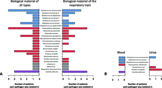
Pathogens isolated in patients with MECP2 duplication syndrome and number of patients each pathogen was detected in. a. All biological material and material of the respiratory tract b. Blood and Urine
When considering only the isolates in biological material of the respiratory tract, the pathogens isolated in most patients were H. influenzae (4/27) and C.albicans (4/27) followed by S.pneumoniae (3/27) (Fig. 2a). The spectrum of pathogens isolated in blood included S.pneumoniae, H.influenzae, S.aureus, S.epidermidis and C.albicans, each only isolated in one patient (Fig. 2b). Pathogens isolated in urine were P.mirabilis (in 2/27 patients), E.coli (in 1/27 patients) and S.pneumoniae (in 1/27 patients) (Fig. 2b). Neither infections by Pneumocystis jirovecii, nor invasive infections by mycobacteria or S.enteritidis were observed. In summary patients with MECP2 duplication syndrome show increased susceptibility for pneumonia and sepsis caused by bacteria that are mainly capable of building a capsule (Fig. 2 and supplementary table 2).
Cellular and Humoral Immunological Phenotype
Peripheral blood smears of 21 patients showed values of total white blood cells that were elevated in two patients (P9, P30). Monocytes were slightly elevated in patient P9 and P22. Neutrophilic granulocytes were increased in patient P9 and P15, and reduced in patient P14. Eosinophilic and basophilic granulocytes were within normal ranges (supplementary figure 1). More detailed immunophenotyping of lymphocytes from 14 patients revealed numbers of total T (CD3+) lymphocytes that were elevated in 1/14 patient. CD4+ T-cells were above age-dependent reference value in 1/14 (different) patient. Absolute numbers of CD8+ T-cells were elevated in 1/14 further patient. In 5 of 14 patients tested naïve CD4+ T-cells (CD4+CD45RA+) were elevated, while memory CD4+ T-cells (CD4+CD45RO+) were reduced. B-cells (CD19+) were reduced in 3 of the 14 patients tested and NK-cells (CD16+CD56+CD3-) were below age-dependent reference value in 1 of the 14 patients tested (supplementary table 3A and supplementary table 3B).
Lymphocyte proliferation upon stimulation with mitogens (PHA, PWM) was normal in 13 patients tested. Stimulation with IL-2 showed attenuated responses in 2/13 patients and stimulation with anti-CD3 revealed a lymphocyte response below reference value in 1/13 patients. When stimulating cells of the 13 patients with SAC, 3 patients showed reduced lymphocyte response. Attenuated lymphocyte proliferation was also observed upon stimulation with candida-antigen (3 of 13 patients tested), diphtheria-antigen (5 of 13 patients tested) and tetanus toxoid antigen (2 of 13 patients tested) (supplementary table 4).
We tested IFNy-secretion upon stimulation with ConA for 24 h in whole blood of 7 patients. We observed a normal production of IFNy in blood that was stimulated soon after the blood withdrawal (n = 1). In blood that had travelled, 4 patients showed an IFNy-level after stimulation that was within the reference values of healthy controls. The IFNy-level of one patient was above the reference value and the IFNy-level of one patient was below reference values. So in total in 6/7 patients tested IFNy-production upon stimulation with ConA was not impaired (Fig. 3). In summary lymphocyte subsets and lymphocyte proliferation assays show no gross abnormalities and IFNy-production upon stimulation with ConA seems to be normal.
Fig. 3.
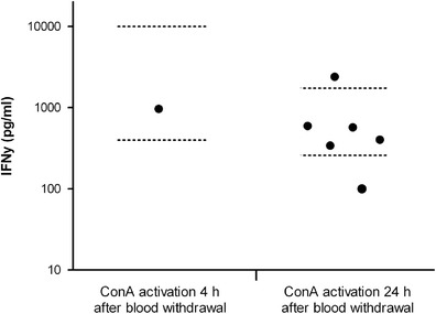
Interferon-y secretion in whole blood upon stimulation with ConA. (Normal ranges are depicted as dotted lines)
Of 30 patients enrolled we could assess immunoglobulin levels (IgG, IgA and IgM), and levels of IgG subclasses (IgG1 – IgG4) and specific antibodies against tetanus and pneumococci in 21 patients. This revealed six patients with IgG2-deficiency – four of them with an additional IgA-deficiency. One of the four patients with IgA/IgG2-deficiency had globalized low levels also for IgG, IgG1, IgG3, IgG4 and IgM. Additionally IgG2 was in low normal range in 3 further patients. IgG4 was below reference values in 4 patients, all of which also had IgG2-deficiency. IgG1 was above age-dependent reference values in 11/21 and IgG3 was above age-dependent reference values in 8/21 patients; 6/21 patients showed supra-normal values for IgG1 and IgG3 (Fig. 4a–g), IgG2-antibodies against S.pneumoniae were below normal values in 4 patients, in whom also IgG2 was low. Additionally S.pneumoniae IgG2-antibodies were in the lower range of normal in 6 further patients. So in total 10/21 patients showed low antibody titers against pneumococci (Fig. 5). Of the 21 patients in whom we assessed the levels of S.pneumoniae IgG2-antibodies, 8 patients had received vaccination against pneumococci, 8 patients had not received vaccination and in five patients information on vaccination status was not available. Interestingly S.pneumoniae IgG2-antibodies were low even in the patients with vaccination against pneumococci (Fig. 5). Both pneumococcal polysaccharide vaccines and pneumococcal conjugate vaccines have been used in the 8 patients with pneumococcal vaccination and 7 of the 8 patients have received several pneumococcal vaccinations. The period between last vaccination and assessment of aPCP-IgG2 ranged from 1 month up to 7 years and 6 month. It is noteworthy that even in the patient with the short interval of 1 month between last vaccination and ascertainment of antibody titers no appropriate antibody production against S. pneumoniae was determined after three vaccinations (Table 2). In contrast to this rather common impairement of IgG2 formation against pneumococci IgG-antibodies against Tetanus-Toxoid were below age-dependent reference values in only 2/21 patients and low in normal range in 2 further patients, so normal in 19/21 patients (supplementary figure 2). In summary 6/21 patients show IgG2-deficiency – four of them with an additional IgA-deficiency - and half of the patients (10/21) show antibodies against pneumococci either below normal range or in the low range of normal.
Fig. 4.
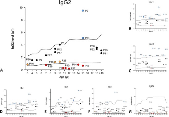
Humoral immunological phenotype of patients with MECP2 duplication syndrome. a. IgG2 levels. b. IgG1 levels. c. IgG3 levels. d. IgG levels. e. IgA levels. f. IgM levels. g. IgG4 levels. (Normal ranges are depicted as dotted lines)
Fig. 5.
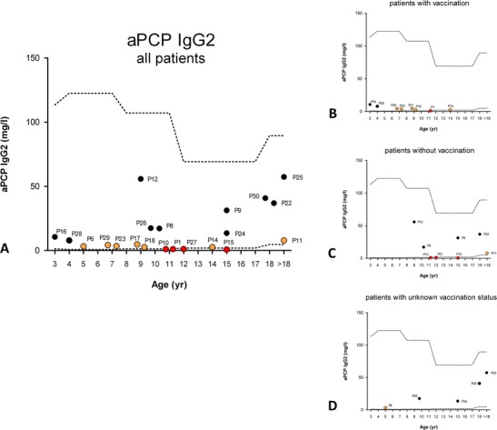
Humoral immunological phenotype of patients with MECP2 duplication syndrome. a. IgG2 levels against S. pneumoniae in all patients tested. b. IgG2 levels against S. pneumoniae of patients with vaccination. c. IgG2 levels against S. pneumoniae of patients without vaccination d. IgG2 levels against S. pneumoniae of patients with unknown vaccination status (Normal ranges are depicted as dotted lines)
Table 2.
Pneumococcal vaccination in patients with MECP2 duplication syndrome
| Patient | Type of vaccine | Number of vaccinations | Time between last vaccination and assessment of aPCP IgG2 | Age at last vaccination |
|---|---|---|---|---|
| P16 | PCV | 5 | 3 months | 3 years, 4 months |
| P17 | PCV | 2 | 7 years, 7 months | 1 year, 8 months |
| P23 | PCV | 3 | 6 years, 10 months | 10 months |
| P28 | PCV | 4 | 3 years, 4 months | 1 year, 1 month |
| P29 | PCV | 4 | 5 years, 9 months | 2 years |
| P1 | PPV23 | 3 | 1 month | 11 years, 1 month |
| P14 | PPV23 | 2 | 1 year, 7 months | 12 years, 11 months |
| P18 | PPV23 | 1 | 7 years, 6 months | 2 years, 3 months |
Type of vaccine, number of vaccinations, time between last vaccination and assessment of aPCP IgG2, age at last vaccination
PCV polysaccharide conjugated vaccine, PPV23 23-valent pneumococcal polysaccharide vaccine
Acute-Phase Responses
As infections in patients with MECP2 duplication syndrome have not only been reported for their high frequency but also for the severity of the single infectious episode we systematically assessed acute-phase responses in these patients. Data on acute-phase responses was available for 10 patients. 7/10 patients experienced non-invasive infections with maximal CRP values above 200 mg/l. In 4/10 patients CRP values above 300 mg/l were reported during non invasive infections. Only one patient (P16) showed no elevated CRP values upon non-invasive infections. The mean value of all reported maximal CRP values during non-invasive infections of all patients was 157,58 mg/l (Fig. 6a). The maximal temperature measured during non-invasive infections ranged from 38.5 to 41.6 °C and the mean value measured during non-invasive infections of all patients was 39.75 °C (Fig. 6b). 2/10 patients experienced non-invasive infections with leucocyte counts above 30/nl and 8/10 patients showed leucocyte counts above 20/nl during single infectious episodes. Mean of all reported maximal leucocyte values was 15.69/nl (Fig. 6c). 5/10 patients showed a neutrophil count above 15/nl during a non-invasive infection. The mean value of the maximal neutrophil count for all patients was 10.51/nl (Fig. 6d). Taken together these data indicate a strong acute-phase response in patients with MECP2 duplication syndrome.
Fig. 6.
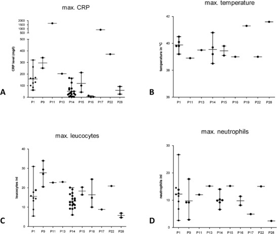
Acute phase responses in patients with MECP2 duplication syndrome. a. Maximal CRP-level during non-invasve infections. Upper reference value is 5 mg/l b. Maximal temperature during non-invasive infections c. Maximal leucocyte count during non-invasive infections. d. Maximal neutrophil count during non-invasive infections
Discussion
The duplication of MECP2 on chromosome Xq28 leads to a rather common syndromic form of XLID (estimated ~1 % of x-linked patients). Besides the neurological phenotype it was soon noted that many of the patients suffer from recurrent infections [51, 52]. We here describe for the first time, systematically the infectious and immunological phenotype of MECP2 duplication syndrome based on data derived from a standardized study-questionnaire filled out by the physicians together with the parents of 30 patients. Our findings show that patients with MECP2 duplication syndrome are at an increased risk for infections by bacteria that are able to build capsules (61.9 % of isolated pathogens); in particular for infections by S.pneumoniae and H.influenzae. In our cohort we have not observed increased susceptibility to mycobacteria, pneumocystis spp, fungi and viruses. We could not identify a correlation between the size of the duplication (genotype) and the infectious or immunological phenotype. The predominance of respiratory and severe invasive infections caused by bacteria that are able to build capsules, suggested an impairment of humoral immunology.
IgG2-deficiency was detected in 6/21 analyzed patients - in 4/6 patients accompanied by IgA-deficiency, while 15 out of 21 analyzed patients showed no IgG2 or IgA deficiency. IgG2-deficiency in the 6/21 patients and low antibody titers against pneumococci in 10/21 patients suggest that among patients with MECP2 duplication humoral impairement is more common than in the normal population. 5/6 patients with IgG2-deficiency also developed severe infections. As 3/4 individuals with combined IgA/IgG2-deficiency additionally suffered from a very high frequency of infections and developed the most severe infectious phenotype in our cohort there seems at least an association of IgA-/IgG2-deficiency with severe infections. In line with this hypothesis only 2/15 patients without IgA or IgG2 deficiency developed recurrent severe infections comparable to the phenotype observed in the patients with IgA/IgG2-deficiency. One of them showed low IgG2-antibodies against pneumococci even after vaccination indicating an inability to mount an appropriate antibody production although global levels of immunoglobulins are normal in this patient. The other patient (P9) with a severe infectious phenotype despite having an IgA-/IgG2-deficiency showed massively elevated IgG (32 g/l). Given the fact that this patient has a persistently raised leucocyte count, it seems plausible that he additionally suffers from an unknown chronic inflammatory process. 7 of the 15 patients without IgA or IgG2 deficiency developed few infections and 2 of the 15 patients developed no infections. In 4 of the 15 patients without IgA or IgG2 deficiency the severity of the infectious phenotype could not be assessed as the patient was either still at young age or because the history could not be completed appropriately. The single patient who has to date not developed severe infections despite IgA/IgG2-deficiency is female. So in MECP2 duplication syndrome not all patients develop humoral immunodeficiency but severe infections with potentially encapsulated bacteria are, at least in male patients, strongly associated with a lack of IgA and IgG2.
We further investigated whether patients with MECP2 duplication syndrome showed stronger acute phase responses, which we could confirm in 7/10 patients in terms of elevated CRP values above 200 mg/l during non-invasive infections, mainly pneumoniae. This observation raises the question if the genetic alterations in patients with MECP2 duplication syndrome lead to a hyper-inflammatory immune response. Because all patients show a duplication on Xq28 that includes MECP2 and neighbouring IRAK1 and given the fact that these two genes were assumed to be the minimal critical region leading to the phenotype of MECP2 duplication syndrome until recently [15, 17, 51], it seems plausible to hypothesize a role of IRAK1 in the pathogenesis of strong acute-phase responses. IRAK1 encodes for a protein in the TLR/IL1R-signalling pathway and it could be assumed that its overexpression leads to stronger or constantly triggered NFκB-mediated pro-inflammatory signalling and to a hyper-inflammatory immune response. In our opinion recently published results suggesting normal TLR-mediated signalling in PBMCs of patients with MECP2/IRAK1 duplication do not sufficiently falsify this hypothesis as these experiments were performed in PBMCs (without granulocytes) and as it remains unclear whether LPS-stimulation was performed with or without serum [44]. So it seems at least possible that the previously described similar production of cytokines upon activation with TLR-agonists in patients and controls in these experiments is due to the insufficient stimulation of cells or a lack of stimulation of granulocytes or both. So to date it remains an open question whether the duplication of IRAK1 renders patients with MECP2 duplication prone to strong inflammatory responses.
It has been published that MECP2 duplication causes a lack of TH1-differentiation, elevated levels of CD4+CD45RA+ naïve/ lowered levels of CD4+CD45R0+ memory T-cells and impaired IFNγ-secretion by T-cells in mice and in patients with MECP2 duplication. It was concluded that this lack of TH1 differentiation/ IFNγ-secretion is the major cause for the increased susceptibility for infections in patients with MECP2 duplication [44]. These observations are partially in line with ours. Also in our cohort 5/14 patients showed elevated levels of CD4+CD45RA+ naïve/ lowered levels of CD4+CD45R0+ memory T-cells. The observed ratio of CD4+CD45RA naïve versus CD4+CD45R0+ memory T-cells in our cohort is >1 in 11/14 patients and < 1 in 3/14 patients. However our data do not fully confirm the hypothesis of an impaired IFNγ-secretion as sufficient explanation for the increased susceptibility to infections: 1. Upon ConA-stimulation we observed a normal IFNγ-production in 5/7 patients, an even stronger IFNγ-production in one patient and only a slightly impaired IFNγ-production in one patient compared to healthy controls (Fig. 3); 2. If the observed reduction of IFNγ-secretion in patients with MECP2 duplication was causative for severe infections one would expect disseminated and/or invasive infections with atypical mycobacteria and S.enteritidis as described for molecular precisely–defined partial and complete defects in at least 9 genes leading to the impairment of IFNγ-receptor signalling or IFNγ-secretion [71–74]; 3. Conversely during a total of 343 patient years in our study only one patient (P9) developed a single, non-complicated infection by M.tuberculosis, which remained restricted to the lung and was successfully treated by standard therapy. In a patient with a strongly relevant impairement of IFNγ-secretion an infection with M.tuberculosis would have taken a severe course [71–74]. Interestingly however 13/21 patients developed either supra-normal values of IgG1 of IgG3 or of both, IgG1 and IgG3. This observation suggests a direct or indirect effect of MECP2 duplication on the expression of IgG3 and IgG1 at the γ3/γ1 locus and of IgA, IgG2 and IgG4 at the α1/γ2/γ4 locus. It has been shown that IFNy stimulates the expression of the IgG2a-isotype and inhibits the production of IgG3, IgG1, IgG2b and IgE in mice [75]. So a deficiency in IFNy-secretion may lead to alterations of immunoglobulin levels in humans with MECP2 duplication although the role of IFNy in the regulation of IgG3 is controversial [76, 77]. We did not observe a generally impaired IFNy secretion in our cohort. However previously published results on irregularities in IFNy secretion should be taken into account as being responsible for altered immunoglobulin levels in patients with MECP2 duplication syndrome [44].
Besides the classical encapsulated bacteria S. pneumoniae and H.influenzae several other bacteriae were also isolated in the patients of our cohort, that are indeed able to build capsules but are not clearly linked to capsule-related virulence. So it remains unclear if humoral immunodeficiency is causative also for infections by these facultative-encapsulated pathogens or if additional cellular immune deficits also account for the broad spectrum of observed pathogens in patients with MECP2 duplication syndrome.
In summary we here show for the first time systematically that patients with MECP2 duplication syndrome are at increased risk for in particular non-invasive but also for invasive infections with potentially encapsulated bacteria, that this increased susceptibility to infections may be associated with IgG2-subclass deficiency/ low titers against pneumococci and elevated acute-phase responses, while the precise role of T-cell immunity and in particular the extent of impaired IFNγ-secretion and its role for the observed infectious phenotype is still to be defined. Based on our observation we hypothesize a multifactorial pathogenesis of infections in patients with MECP2 duplication syndrome: Pulmonary infections may be triggered by frequent aspirations due to the severe neurological phenotype and favoured due to humoral immunodeficiency. The observed alterations in the IgG subclass composition (high IgG1/IgG3 and low IgA/IgG2/IgG4) may be caused directly by interaction of MECP2 with the respective loci or indirectly by spatially impaired IFNγ-secretion. An increased susceptibility to hyper-inflammation either caused by IRAK1 duplication or other direct or indirect effects of MECP2 duplication may aggravate infections. Based on the compiled immunological and infectious findings we recommend to vaccinate patients with MECP2 duplication against pneumococci and to evaluate post-vaccination titers. If post-vaccination titers are not sufficient, several boosts might be required. In patients with MECP2 duplication syndrome, suffering from recurrent infections and with an IgG2-subclass deficiency and/or low post-vaccination titers against pneumococci prophylactic substitution of IgG seems a plausible strategy to prevent infections – eventually in combination with the use of prophylactic antibiotics.
Electronic supplementary material
Below is the link to the electronic supplementary material.
Cellular immunological phenotype of patients with MECP2 duplication syndrome. A. Leucocytes. B. Monocytes. C. Neutrophilic leucocytes. D. Eosinophilic leucocytes. E. Basophilic leucocytes. (PPTX 207 kb)
Humoral immunological phenotype of patients with MECP2 duplication syndrome. IgG levels against Tetanus toxoid. (PPTX 129 kb)
Data on duplications of previously non-published patients P11, P14, P16, P18 P19, P23, P24, P25, P26, P28, P29 and P30. (PPTX 64 kb)
Infectious phenotype of patients with MECP2 duplication syndrome. Pathogens isolated and location of isolation. (PPTX 55 kb)
Cellular immunological phenotype of patients with MECP2 duplication syndrome. Age group < 6 years and age group 6–10 years. Lymphocyte subsets are depicted in absolute numbers and percentages. Reference values for the respective age group are given. (PPTX 81 kb)
Cellular immunological phenotype of patients with MECP2 duplication syndrome. Age group 10–18 years and age group >18 years. Lymphocyte subsets are depicted in absolute numbers and percentages. Reference values for the respective age group are given. (PPTX 80 kb)
Cellular immunological phenotype of patients with MECP2 duplication syndrome. Lymphocyte proliferation assay. Proliferation is depicted as count per minute (cpm). (PPTX 66 kb)
Acknowledgments
We thank the patients and their families for participating in this study and Margret Oberreit, Petra Ellensohn (deceased 01/2011), Christine Seib, Kristin Neuhaus, Anne-Hélène Lebrun and Karoline Strehl for discussions, critical reading of the manuscript and technical assistance. This work was supported by the German Research Foundation (DFG BE 3895/3-1), the Federal Ministry of Education and Research, Germany (BMBF PID-NET 01GM1111D/TP-A5) and the Sonnenfeld Stiftung, Berlin.
Abbreviations
- MECP2
Methyl CpG binding protein 2
- XLID
X linked intellectual disability
Contributor Information
Michael Bauer, Email: Michael.Bauer@charite.de.
Horst von Bernuth, Email: horst.von-bernuth@charite.de, Email: horst.von-bernuth@laborberlin.com.
References
- 1.Lubs H, Abidi F, Bier JA, Abuelo D, Ouzts L, Voeller K, et al. XLMR syndrome characterized by multiple respiratory infections, hypertelorism, severe CNS deterioration and early death localizes to distal Xq28. Am J Med Genet. 1999;85(3):243–8. doi: 10.1002/(SICI)1096-8628(19990730)85:3<243::AID-AJMG11>3.0.CO;2-E. [DOI] [PubMed] [Google Scholar]
- 2.Meins M, Lehmann J, Gerresheim F, Herchenbach J, Hagedorn M, Hameister K, et al. Submicroscopic duplication in Xq28 causes increased expression of the MECP2 gene in a boy with severe mental retardation and features of Rett syndrome. J Med Genet. 2005;42(2):e12. doi: 10.1136/jmg.2004.023804. [DOI] [PMC free article] [PubMed] [Google Scholar]
- 3.Van Esch H, Bauters M, Ignatius J, Jansen M, Raynaud M, Hollanders K, et al. Duplication of the MECP2 region is a frequent cause of severe mental retardation and progressive neurological symptoms in males. Am J Hum Genet. 2005;77(3):442–53. doi: 10.1086/444549. [DOI] [PMC free article] [PubMed] [Google Scholar]
- 4.Ariani F, Mari F, Pescucci C, Longo I, Bruttini M, Meloni I, et al. Real-time quantitative PCR as a routine method for screening large rearrangements in Rett syndrome: report of one case of MECP2 deletion and one case of MECP2 duplication. Hum Mutat. 2004;24(2):172–7. doi: 10.1002/humu.20065. [DOI] [PubMed] [Google Scholar]
- 5.Lugtenberg D, de Brouwer AP, Kleefstra T, Oudakker AR, Frints SG, Schrander-Stumpel CT, et al. Chromosomal copy number changes in patients with non-syndromic X linked mental retardation detected by array CGH. J Med Genet. 2006;43(4):362–70. doi: 10.1136/jmg.2005.036178. [DOI] [PMC free article] [PubMed] [Google Scholar]
- 6.Friez MJ, Jones JR, Clarkson K, Lubs H, Abuelo D, Bier JA, et al. Recurrent infections, hypotonia, and mental retardation caused by duplication of MECP2 and adjacent region in Xq28. Pediatrics. 2006;118(6):e1687–95. doi: 10.1542/peds.2006-0395. [DOI] [PubMed] [Google Scholar]
- 7.del Gaudio D, Fang P, Scaglia F, Ward PA, Craigen WJ, Glaze DG, et al. Increased MECP2 gene copy number as the result of genomic duplication in neurodevelopmentally delayed males. Genet Med. 2006;8(12):784–92. doi: 10.1097/01.gim.0000250502.28516.3c. [DOI] [PubMed] [Google Scholar]
- 8.Madrigal I, Rodríguez-Revenga L, Armengol L, González E, Rodriguez B, Badenas C, et al. X-chromosome tiling path array detection of copy number variants in patients with chromosome X-linked mental retardation. BMC Genomics. 2007;8:443. doi: 10.1186/1471-2164-8-443. [DOI] [PMC free article] [PubMed] [Google Scholar]
- 9.Bauters M, Van Esch H, Friez MJ, Boespflug-Tanguy O, Zenker M, Vianna-Morgante AM, et al. Nonrecurrent MECP2 duplications mediated by genomic architecture-driven DNA breaks and break-induced replication repair. Genome Res. 2008;18(6):847–58. doi: 10.1101/gr.075903.107. [DOI] [PMC free article] [PubMed] [Google Scholar]
- 10.Smyk M, Obersztyn E, Nowakowska B, Nawara M, Cheung SW, Mazurczak T, et al. Different-sized duplications of Xq28, including MECP2, in three males with mental retardation, absent or delayed speech, and recurrent infections. Am J Med Genet B Neuropsychiatr Genet. 2008;147B(6):799–806. doi: 10.1002/ajmg.b.30683. [DOI] [PubMed] [Google Scholar]
- 11.Velinov M, Novelli A, Gu H, Fenko M, Dolzhanskaya N, Bernardini L, et al. De-novo 2.15 Mb terminal Xq duplication involving MECP2 but not L1CAM gene in a male patient with mental retardation. Clin Dysmorphol. 2009;18(1):9–12. doi: 10.1097/MCD.0b013e3283157cad. [DOI] [PubMed] [Google Scholar]
- 12.Sanlaville D, Schluth-Bolard C, Turleau C. Distal Xq duplication and functional Xq disomy. Orphanet J Rare Dis. 2009;4:4. doi: 10.1186/1750-1172-4-4. [DOI] [PMC free article] [PubMed] [Google Scholar]
- 13.Kirk EP, Malaty-Brevaud V, Martini N, Lacoste C, Levy N, Maclean K, et al. The clinical variability of the MECP2 duplication syndrome: description of two families with duplications excluding L1CAM and FLNA. Clin Genet. 2009;75(3):301–3. doi: 10.1111/j.1399-0004.2008.01102.x. [DOI] [PubMed] [Google Scholar]
- 14.Clayton-Smith J, Walters S, Hobson E, Burkitt-Wright E, Smith R, Toutain A, et al. Xq28 duplication presenting with intestinal and bladder dysfunction and a distinctive facial appearance. Eur J Hum Genet. 2009;17(4):434–43. doi: 10.1038/ejhg.2008.192. [DOI] [PMC free article] [PubMed] [Google Scholar]
- 15.Lugtenberg D, Kleefstra T, Oudakker AR, Nillesen WM, Yntema HG, Tzschach A, et al. Structural variation in Xq28: MECP2 duplications in 1% of patients with unexplained XLMR and in 2% of male patients with severe encephalopathy. Eur J Hum Genet. 2009;17(4):444–53. doi: 10.1038/ejhg.2008.208. [DOI] [PMC free article] [PubMed] [Google Scholar]
- 16.Prescott TE, Rødningen OK, Bjørnstad A, Stray-Pedersen A. Two brothers with a microduplication including the MECP2 gene: rapid head growth in infancy and resolution of susceptibility to infection. Clin Dysmorphol. 2009;18(2):78–82. doi: 10.1097/MCD.0b013e32831e19cd. [DOI] [PubMed] [Google Scholar]
- 17.Carvalho CM, Zhang F, Liu P, Patel A, Sahoo T, Bacino CA, et al. Complex rearrangements in patients with duplications of MECP2 can occur by fork stalling and template switching. Hum Mol Genet. 2009;18(12):2188–203. doi: 10.1093/hmg/ddp151. [DOI] [PMC free article] [PubMed] [Google Scholar]
- 18.Echenne B, Roubertie A, Lugtenberg D, Kleefstra T, Hamel BC, Van Bokhoven H, et al. Neurologic aspects of MECP2 gene duplication in male patients. Pediatr Neurol. 2009;41(3):187–91. doi: 10.1016/j.pediatrneurol.2009.03.012. [DOI] [PubMed] [Google Scholar]
- 19.Vandewalle J, Van Esch H, Govaerts K, Verbeeck J, Zweier C, Madrigal I, et al. Dosage-dependent severity of the phenotype in patients with mental retardation due to a recurrent copy-number gain at Xq28 mediated by an unusual recombination. Am J Hum Genet. 2009;85(6):809–22. doi: 10.1016/j.ajhg.2009.10.019. [DOI] [PMC free article] [PubMed] [Google Scholar]
- 20.Ramocki MB, Peters SU, Tavyev YJ, Zhang F, Carvalho CM, Schaaf CP, et al. Autism and other neuropsychiatric symptoms are prevalent in individuals with MeCP2 duplication syndrome. Ann Neurol. 2009;66(6):771–82. doi: 10.1002/ana.21715. [DOI] [PMC free article] [PubMed] [Google Scholar]
- 21.Bartsch O, Gebauer K, Lechno S, van Esch H, Froyen G, Bonin M, et al. Four unrelated patients with Lubs X-linked mental retardation syndrome and different Xq28 duplications. Am J Med Genet A. 2010;152A(2):305–12. doi: 10.1002/ajmg.a.33198. [DOI] [PubMed] [Google Scholar]
- 22.Campos M, Jr, Churchman SM, Santos-Rebouças CB, Ponchel F, Pimentel MM. High frequency of nonrecurrent MECP2 duplications among Brazilian males with mental retardation. J Mol Neurosci. 2010;41(1):105–9. doi: 10.1007/s12031-009-9296-2. [DOI] [PubMed] [Google Scholar]
- 23.Belligni EF, Palmer RW, Hennekam RC. MECP2 duplication in a patient with congenital central hypoventilation. Am J Med Genet A. 2010;152A(6):1591–3. doi: 10.1002/ajmg.a.33311. [DOI] [PubMed] [Google Scholar]
- 24.Reardon W, Donoghue V, Murphy AM, King MD, Mayne PD, Horn N, et al. Progressive cerebellar degenerative changes in the severe mental retardation syndrome caused by duplication of MECP2 and adjacent loci on Xq28. Eur J Pediatr. 2010;169(8):941–9. doi: 10.1007/s00431-010-1144-4. [DOI] [PubMed] [Google Scholar]
- 25.Makrythanasis P, Moix I, Gimelli S, Fluss J, Aliferis K, Antonarakis SE, et al. De novo duplication of MECP2 in a girl with mental retardation and no obvious dysmorphic features. Clin Genet. 2010;78(2):175–80. doi: 10.1111/j.1399-0004.2010.01371.x. [DOI] [PubMed] [Google Scholar]
- 26.Honda S, Hayashi S, Imoto I, Toyama J, Okazawa H, Nakagawa E, et al. Copy-number variations on the X chromosome in Japanese patients with mental retardation detected by array-based comparative genomic hybridization analysis. J Hum Genet. 2010;55(9):590–9. doi: 10.1038/jhg.2010.74. [DOI] [PubMed] [Google Scholar]
- 27.Fernández RM, Núñez-Torres R, González-Meneses A, Antiñolo G, Borrego S. Novel association of severe neonatal encephalopathy and Hirschsprung disease in a male with a duplication at the Xq28 region. BMC Med Genet. 2010;11:137. doi: 10.1186/1471-2350-11-137. [DOI] [PMC free article] [PubMed] [Google Scholar]
- 28.Jezela-Stanek A, Ciara E, Juszczak M, Pelc M, Materna-Kiryluk A, Krajewska-Walasek M. Cryptic x; autosome translocation in a boy–delineation of the phenotype. Pediatr Neurol. 2011;44(3):221–4. doi: 10.1016/j.pediatrneurol.2010.10.007. [DOI] [PubMed] [Google Scholar]
- 29.Breman AM, Ramocki MB, Kang SH, Williams M, Freedenberg D, Patel A, et al. MECP2 duplications in six patients with complex sex chromosome rearrangements. Eur J Hum Genet. 2011;19(4):409–15. doi: 10.1038/ejhg.2010.195. [DOI] [PMC free article] [PubMed] [Google Scholar]
- 30.Grasshoff U, Bonin M, Goehring I, Ekici A, Dufke A, Cremer K, et al. De novo MECP2 duplication in two females with random X-inactivation and moderate mental retardation. Eur J Hum Genet. 2011;19(5):507–12. doi: 10.1038/ejhg.2010.226. [DOI] [PMC free article] [PubMed] [Google Scholar]
- 31.Budisteanu M, Papuc SM, Tutulan-Cunita A, Budisteanu B, Arghir A. Novel clinical finding in MECP2 duplication syndrome. Eur Child Adolesc Psychiatry. 2011;20(7):373–5. doi: 10.1007/s00787-011-0184-2. [DOI] [PubMed] [Google Scholar]
- 32.Mayo S, Monfort S, Roselló M, Orellana C, Oltra S, Armstrong J, et al. De novo interstitial triplication of MECP2 in a girl with neurodevelopmental disorder and random X chromosome inactivation. Cytogenet Genome Res. 2011;135(2):93–101. doi: 10.1159/000330917. [DOI] [PubMed] [Google Scholar]
- 33.Carvalho CM, Ramocki MB, Pehlivan D, Franco LM, Gonzaga-Jauregui C, Fang P, et al. Inverted genomic segments and complex triplication rearrangements are mediated by inverted repeats in the human genome. Nat Genet. 2011;43(11):1074–81. doi: 10.1038/ng.944. [DOI] [PMC free article] [PubMed] [Google Scholar]
- 34.Tang SS, Fernandez D, Lazarou LP, Singh R, Fallon P. MECP2 triplication in 3 brothers - a rarely described cause of familial neurological regression in boys. Eur J Paediatr Neurol. 2012;16(2):209–12. doi: 10.1016/j.ejpn.2011.07.011. [DOI] [PubMed] [Google Scholar]
- 35.Honda S, Satomura S, Hayashi S, Imoto I, Nakagawa E, Goto Y, et al. Japanese mental retardation consortium. Concomitant microduplications of MECP2 and ATRX in male patients with severe mental retardation. J Hum Genet. 2012;57(1):73–7. doi: 10.1038/jhg.2011.131. [DOI] [PubMed] [Google Scholar]
- 36.Utine GE, Kiper PO, Alanay Y, Haliloğlu G, Aktaş D, Boduroğlu K, et al. Searching for copy number changes in nonsyndromic X-linked intellectual disability. Mol Syndromol. 2012;2(2):64–71. doi: 10.1159/000334289. [DOI] [PMC free article] [PubMed] [Google Scholar]
- 37.Bijlsma EK, Collins A, Papa FT, Tejada MI, Wheeler P, Peeters EA, et al. Xq28 duplications including MECP2 in five females: expanding the phenotype to severe mental retardation. Eur J Med Genet. 2012;55(6–7):404–13. doi: 10.1016/j.ejmg.2012.02.009. [DOI] [PMC free article] [PubMed] [Google Scholar]
- 38.Honda S, Hayashi S, Nakane T, Imoto I, Kurosawa K, Mizuno S, et al. The incidence of hypoplasia of the corpus callosum in patients with dup (X)(q28) involving MECP2 is associated with the location of distal breakpoints. Am J Med Genet A. 2012;158A(6):1292–303. doi: 10.1002/ajmg.a.35321. [DOI] [PubMed] [Google Scholar]
- 39.Sanmann JN, Bishay DL, Starr LJ, Bell CA, Pickering DL, Stevens JM, et al. Characterization of six novel patients with MECP2 duplications due to unbalanced rearrangements of the X chromosome. Am J Med Genet A. 2012;158A(6):1285–91. doi: 10.1002/ajmg.a.35347. [DOI] [PubMed] [Google Scholar]
- 40.Vignoli A, Borgatti R, Peron A, Zucca C, Ballarati L, Bonaglia C, et al. Electroclinical pattern in MECP2 duplication syndrome: eight new reported cases and review of literature. Epilepsia. 2012;53(7):1146–55. doi: 10.1111/j.1528-1167.2012.03501.x. [DOI] [PubMed] [Google Scholar]
- 41.Shimada S, Okamoto N, Ito M, Arai Y, Momosaki K, Togawa M, Maegaki Y, Sugawara M, Shimojima K, Osawa M, Yamamoto T. MECP2 duplication syndrome in both genders. Brain Dev. 2012. [DOI] [PubMed]
- 42.Hanchard NA, Carvalho CM, Bader P, Thome A, Omo-Griffith L, Del Gaudio D, et al. A partial MECP2 duplication in a mildly affected adult male: a putative role for the 3′ untranslated region in the MECP2 duplication phenotype. BMC Med Genet. 2012;13:71. doi: 10.1186/1471-2350-13-71. [DOI] [PMC free article] [PubMed] [Google Scholar]
- 43.Xu X, Xu Q, Zhang Y, Zhang X, Cheng T, Wu B, et al. A case report of Chinese brothers with inherited MECP2-containing duplication: autism and intellectual disability, but not seizures or respiratory infections. BMC Med Genet. 2012;13:75. doi: 10.1186/1471-2350-13-75. [DOI] [PMC free article] [PubMed] [Google Scholar]
- 44.Yang T, Ramocki MB, Neul JL, Lu W, Roberts L, Knight J, et al. Overexpression of methyl-CpG binding protein 2 impairs T(H)1 responses. Sci Transl Med. 2012;5:4(163). doi: 10.1126/scitranslmed.3004430. [DOI] [PMC free article] [PubMed] [Google Scholar]
- 45.Shimada S, Okamoto N, Hirasawa K, Yoshii K, Tani Y, Sugawara M, et al. Clinical manifestations of Xq28 functional disomy involving MECP2 in one female and two male patients. Am J Med Genet A. 2013;161A(7):1779–85. doi: 10.1002/ajmg.a.35975. [DOI] [PubMed] [Google Scholar]
- 46.Wax JR, Pinette MG, Smith R, Chard R, Cartin A. Second-trimester prenasal and prefrontal skin thickening - association with MECP2 triplication syndrome. J Clin Ultrasound. 2013;41(7):434–7. doi: 10.1002/jcu.22065. [DOI] [PubMed] [Google Scholar]
- 47.Peters SU, Hundley RJ, Wilson AK, Carvalho CM, Lupski JR, Ramocki MB. Brief report: regression timing and associated features in MECP2 duplication syndrome. J Autism Dev Disord. 2013;43(10):2484–90. doi: 10.1007/s10803-013-1796-9. [DOI] [PubMed] [Google Scholar]
- 48.Novara F, Simonati A, Sicca F, Battini R, Fiori S, Contaldo A, et al. MECP2 duplication phenotype in symptomatic females: report of three further cases. Mol Cytogenet. 2014;7(1):10. doi: 10.1186/1755-8166-7-10. [DOI] [PMC free article] [PubMed] [Google Scholar]
- 49.Scott Schwoerer J, Laffin J, Haun J, Raca G, Friez MJ, Giampietro PF. MECP2 duplication: possible cause of severe phenotype in females. Am J Med Genet A. 2014;164A(4):1029–34. doi: 10.1002/ajmg.a.36380. [DOI] [PubMed] [Google Scholar]
- 50.Fukushi D, Yamada K, Nomura N, Naiki M, Kimura R, Yamada Y, et al. Clinical characterization and identification of duplication breakpoints in a Japanese family with Xq28 duplication syndrome including MECP2. Am J Med Genet A. 2014;164A(4):924–33. doi: 10.1002/ajmg.a.36373. [DOI] [PubMed] [Google Scholar]
- 51.Ramocki MB, Tavyev YJ, Peters SU. The MECP2 duplication syndrome. Am J Med Genet A. 2010;152A(5):1079–88. doi: 10.1002/ajmg.a.33184. [DOI] [PMC free article] [PubMed] [Google Scholar]
- 52.Van Esch H. MECP2 duplication syndrome. Mol Syndromol. 2012;2(3–5):128–36. doi: 10.1159/000329580. [DOI] [PMC free article] [PubMed] [Google Scholar]
- 53.Tomás JM. The main Aeromonas pathogenic factors. ISRN Microbiol. 2012;2012:256261. doi: 10.5402/2012/256261. [DOI] [PMC free article] [PubMed] [Google Scholar]
- 54.Gibson FC, 3rd, Tzianabos AO, Onderdonk AB. The capsular polysaccharide complex of Bacteroides fragilis induces cytokine production from human and murine phagocytic cells. Infect Immun. 1996;64(3):1065–9. doi: 10.1128/iai.64.3.1065-1069.1996. [DOI] [PMC free article] [PubMed] [Google Scholar]
- 55.Kalka-Moll WM, Wang Y, Comstock LE, Gonzalez SE, Tzianabos AO, Kasper DL. Immunochemical and biological characterization of three capsular polysaccharides from a single Bacteroides fragilis strain. Infect Immun. 2001;69(4):2339–44. doi: 10.1128/IAI.69.4.2339-2344.2001. [DOI] [PMC free article] [PubMed] [Google Scholar]
- 56.Coyne MJ, Tzianabos AO, Mallory BC, Carey VJ, Kasper DL, Comstock LE. Polysaccharide biosynthesis locus required for virulence of Bacteroides fragilis. Infect Immun. 2001;69(7):4342–50. doi: 10.1128/IAI.69.7.4342-4350.2001. [DOI] [PMC free article] [PubMed] [Google Scholar]
- 57.Buttery J, Moxon ER. Capsulate bacteria and the lung. Br Med Bull. 2002;61:63–80. doi: 10.1093/bmb/61.1.63. [DOI] [PubMed] [Google Scholar]
- 58.Bao G, Zhang Y, Du C, Chen Z, Li Y, Cao Z, Ma Y. Genome sequence of Klebsiella oxytoca M5al, a promising strain for nitrogen fixation and chemical production. Genome Announc. 2013; 1(1). [DOI] [PMC free article] [PubMed]
- 59.Llobet E, Tomás JM, Bengoechea JA. Capsule polysaccharide is a bacterial decoy for antimicrobial peptides. Microbiology. 2008;154(Pt 12):3877–86. doi: 10.1099/mic.0.2008/022301-0. [DOI] [PubMed] [Google Scholar]
- 60.Dumanski AJ, Hedelin H, Edin-Liljegren A, Beauchemin D, McLean RJ. Unique ability of the Proteus mirabilis capsule to enhance mineral growth in infectious urinary calculi. Infect Immun. 1994;62(7):2998–3003. doi: 10.1128/iai.62.7.2998-3003.1994. [DOI] [PMC free article] [PubMed] [Google Scholar]
- 61.Deretic V, Dikshit R, Konyecsni WM, Chakrabarty AM, Misra TK. The algR gene, which regulates mucoidy in Pseudomonas aeruginosa, belongs to a class of environmentally responsive genes. J Bacteriol. 1989;171(3):1278–83. doi: 10.1128/jb.171.3.1278-1283.1989. [DOI] [PMC free article] [PubMed] [Google Scholar]
- 62.O’Riordan K, Lee JC. Staphylococcus aureus capsular polysaccharides. Clin Microbiol Rev. 2004;17(1):218–34. doi: 10.1128/CMR.17.1.218-234.2004. [DOI] [PMC free article] [PubMed] [Google Scholar]
- 63.Coldren FM, Palavecino EL, Levi-Polyachenko NH, Wagner WD, Smith TL, Smith BP, et al. Encapsulated Staphylococcus aureus strains vary in adhesiveness assessed by atomic force microscopy. J Biomed Mater Res A. 2009;89(2):402–10. doi: 10.1002/jbm.a.31973. [DOI] [PubMed] [Google Scholar]
- 64.Spellberg B, Daum R. Development of a vaccine against Staphylococcus aureus. Semin Immunopathol. 2012;34(2):335–48. doi: 10.1007/s00281-011-0293-5. [DOI] [PMC free article] [PubMed] [Google Scholar]
- 65.Nanra JS, Buitrago SM, Crawford S, Ng J, Fink PS, Hawkins J, et al. Capsular polysaccharides are an important immune evasion mechanism for Staphylococcus aureus. Hum Vaccin Immunother. 2012;18:9(3). doi: 10.4161/hv.23223. [DOI] [PMC free article] [PubMed] [Google Scholar]
- 66.Foster TJ. Immune evasion by staphylococci. Nat Rev Microbiol. 2005;3(12):948–58. doi: 10.1038/nrmicro1289. [DOI] [PubMed] [Google Scholar]
- 67.McKenney D, Hübner J, Muller E, Wang Y, Goldmann DA, Pier GB. The ica locus of Staphylococcus epidermidis encodes production of the capsular polysaccharide/adhesin. Infect Immun. 1998;66(10):4711–20. doi: 10.1128/iai.66.10.4711-4720.1998. [DOI] [PMC free article] [PubMed] [Google Scholar]
- 68.Dinkla K, Sastalla I, Godehardt AW, Janze N, Chhatwal GS, Rohde M, et al. Upregulation of capsule enables Streptococcus pyogenes to evade immune recognition by antigen-specific antibodies directed to the G-related alpha2-macroglobulin-binding protein GRAB located on the bacterial surface. Microbes Infect. 2007;9(8):922–31. doi: 10.1016/j.micinf.2007.03.011. [DOI] [PubMed] [Google Scholar]
- 69.Stollerman GH, Dale JB. The importance of the group a streptococcus capsule in the pathogenesis of human infections: a historical perspective. Clin Infect Dis. 2008;46(7):1038–45. doi: 10.1086/529194. [DOI] [PubMed] [Google Scholar]
- 70.Kang SO, Wright JO, Tesorero RA, Lee H, Beall B, Cho KH. Thermoregulation of capsule production by Streptococcus pyogenes. PLoS One. 2012;7(5):e37367. doi: 10.1371/journal.pone.0037367. [DOI] [PMC free article] [PubMed] [Google Scholar]
- 71.Zhang SY, Boisson-Dupuis S, Chapgier A, Yang K, Bustamante J, Puel A, et al. Inborn errors of interferon (IFN)-mediated immunity in humans: insights into the respective roles of IFN-alpha/beta, IFN-gamma, and IFN-lambda in host defense. Immunol Rev. 2008;226:29–40. doi: 10.1111/j.1600-065X.2008.00698.x. [DOI] [PubMed] [Google Scholar]
- 72.de Beaucoudrey L, Samarina A, Bustamante J, Cobat A, Boisson-Dupuis S, Feinberg J, et al. Revisiting human IL-12Rβ1 deficiency: a survey of 141 patients from 30 countries. Medicine (Baltimore) 2010;89(6):381–402. doi: 10.1097/MD.0b013e3181fdd832. [DOI] [PMC free article] [PubMed] [Google Scholar]
- 73.Bogunovic D, Byun M, Durfee LA, Abhyankar A, Sanal O, Mansouri D, et al. Mycobacterial disease and impaired IFN-γ immunity in humans with inherited ISG15 deficiency. Science. 2012;337(6102):1684–8. doi: 10.1126/science.1224026. [DOI] [PMC free article] [PubMed] [Google Scholar]
- 74.Parvaneh N, Casanova JL, Notarangelo LD, Conley ME. Primary immunodeficiencies: a rapidly evolving story. J Allergy Clin Immunol. 2013;131(2):314–23. doi: 10.1016/j.jaci.2012.11.051. [DOI] [PubMed] [Google Scholar]
- 75.Snapper CM, Paul WE. Interferon-gamma and B cell stimulatory factor-1 reciprocally regulate Ig isotype production. Science. 1987;236(4804):944–7. doi: 10.1126/science.3107127. [DOI] [PubMed] [Google Scholar]
- 76.Snapper CM, McIntyre TM, Mandler R, Pecanha LM, Finkelman FD, Lees A, et al. Induction of IgG3 secretion by interferon gamma: a model for T cell-independent class switching in response to T cell-independent type 2 antigens. J Exp Med. 1992;175(5):1367–71. doi: 10.1084/jem.175.5.1367. [DOI] [PMC free article] [PubMed] [Google Scholar]
- 77.Gerth AJ, Lin L, Peng SL. T-bet regulates T-independent IgG2a class switching. Int Immunol. 2003;15(8):937–44. doi: 10.1093/intimm/dxg093. [DOI] [PubMed] [Google Scholar]
Associated Data
This section collects any data citations, data availability statements, or supplementary materials included in this article.
Supplementary Materials
Cellular immunological phenotype of patients with MECP2 duplication syndrome. A. Leucocytes. B. Monocytes. C. Neutrophilic leucocytes. D. Eosinophilic leucocytes. E. Basophilic leucocytes. (PPTX 207 kb)
Humoral immunological phenotype of patients with MECP2 duplication syndrome. IgG levels against Tetanus toxoid. (PPTX 129 kb)
Data on duplications of previously non-published patients P11, P14, P16, P18 P19, P23, P24, P25, P26, P28, P29 and P30. (PPTX 64 kb)
Infectious phenotype of patients with MECP2 duplication syndrome. Pathogens isolated and location of isolation. (PPTX 55 kb)
Cellular immunological phenotype of patients with MECP2 duplication syndrome. Age group < 6 years and age group 6–10 years. Lymphocyte subsets are depicted in absolute numbers and percentages. Reference values for the respective age group are given. (PPTX 81 kb)
Cellular immunological phenotype of patients with MECP2 duplication syndrome. Age group 10–18 years and age group >18 years. Lymphocyte subsets are depicted in absolute numbers and percentages. Reference values for the respective age group are given. (PPTX 80 kb)
Cellular immunological phenotype of patients with MECP2 duplication syndrome. Lymphocyte proliferation assay. Proliferation is depicted as count per minute (cpm). (PPTX 66 kb)


