Abstract
Successful human colonizers such as Candida pathogens have evolved distinct strategies to survive and proliferate within the human host. These include sophisticated mechanisms to evade immune surveillance and adapt to constantly changing host microenvironments where nutrient limitation, pH fluctuations, oxygen deprivation, changes in temperature, or exposure to oxidative, nitrosative, and cationic stresses may occur. Here, we review the current knowledge and recent findings highlighting the remarkable ability of medically important Candida species to overcome a broad range of host-imposed constraints and how this directly affects their physiology and pathogenicity. We also consider the impact of these adaptation mechanisms on immune recognition, biofilm formation, and antifungal drug resistance, as these pathogens often exploit specific host constraints to establish a successful infection. Recent studies of adaptive responses to physiological niches have improved our understanding of the mechanisms established by fungal pathogens to evade the immune system and colonize the host, which may facilitate the design of innovative diagnostic tests and therapeutic approaches for Candida infections.
Introduction
The human body is home to a large number of microbes that play essential roles in maintaining human health. However, under particular host-compromising conditions, they can shift from harmless commensals to opportunistic pathogens to cause inflammation and disease. Fungal communities, which can include Candida species, constitute an integral part of the human microbiota that, under normal conditions, asymptomatically colonize several niches, including the skin, oral cavity, gastrointestinal, and urogenital tracts [1–3]. The remarkable ability to alternate between local current microenvironments within internal host niches such as blood or tissues is often linked with their pathogenic potential. Therefore, environmental changes promoted either by alterations in host microbiota or the host immune system may allow these microorganisms to overgrow, cross the epithelial barriers, and cause severe, life-threatening infections [4].
Among the Candida species that trigger human disease, Candida albicans, C. glabrata, C. parapsilosis, C. tropicallis, and C. krusei are the most common [4–6]. Yet, other emerging species, including C. auris, C. guilliermondii, C. lusitaniae, and C. metapsilosis, are of particular concern because they are rapidly spreading worldwide, with several reported outbreaks [5,7,8]. Moreover, Candida infections are difficult to diagnose, commonly resulting in delayed antifungal treatments that have been associated with hospital mortality [9]. The antifungal drugs available to eradicate these fungal pathogens are also limited and often ineffective, mainly because of the intrinsic multidrug resistance of certain Candida species and their ability to form biofilms on implanted medical devices [10–12]. Considering that each species presents its own distinctive features in relation to invasive potential, morphogenesis, antifungal susceptibility, and biofilm formation, studies focusing on the adaptation to different hosts and environmental factors have the potential to reveal novel molecular players of virulence pathways.
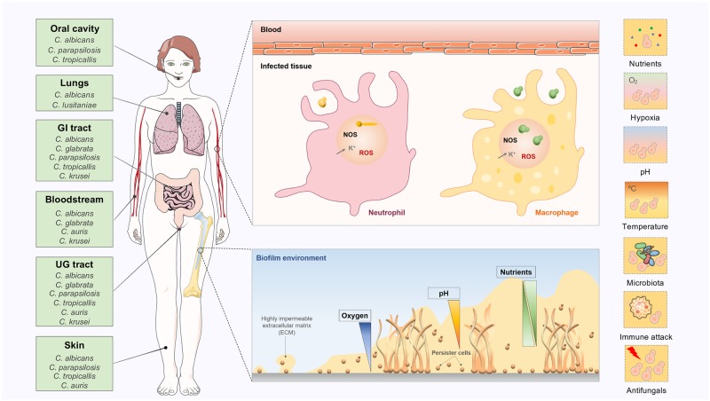
Here, we provide an overview of established and emerging strategies used by Candida to adapt to common environmental challenges faced by these fungi during immune evasion and human colonization (Fig 1). As we review major host-imposed constraints, we highlight the central regulatory circuits required for fungal adaptation to these challenges. We also discuss the impact of such physiological reprogramming on key aspects of Candida pathogenicity, with a particular emphasis on immune evasion, biofilm formation, and antifungal drug resistance. We propose that the genetic circuits governing Candida adaptation to human niches can be exploited in search of new antifungal targets and diagnosis improvement.
Fig 1. Candida biogeography and the different host-imposed constraints during human colonization.
The most frequently isolated Candida species are listed according to their principal habitat in the human body (oral cavity, lungs, gastrointestinal tract, bloodstream, urogenital tract, and skin). The different host-imposed constraints are highlighted for several microenvironments where Candida thrives in the human body, including inside phagocytic cells or biofilms. Key references: C. albicans [2,3], C. glabrata [3], C. parapsilosis [2], C. tropicalis [2], C. lusitaniae [13], and C. krusei [6]. ECM, extracellular matrix; NOS, nitric oxide species; ROS, reactive oxygen species; UG, urogenital.
Candida within the human host
The human host contains a variety of environmental niches in which Candida species can thrive. Adaptation to these sites requires rapid and coordinated changes in Candida metabolism and physiology in order to avoid or escape immune surveillance and to counteract several host-imposed constraints (for example, nutrient limitation, oxygen deprivation, pH fluctuations, changes in temperature, or oxidative, nitrosative, and cationic stresses). Moreover, Candida species interact with other microbial residents, establishing either cooperative or antagonistic relationships, which may affect their growth and influence the outcome of an infection.
Depending on the local environmental cues, some Candida species may exhibit different cellular morphologies. These include budding forms, which have been associated with commensalism, and the filamentous forms hyphae and pseudohyphae, often related with invasive and disseminated disease [14,15]. However, these cell types were found in infected tissues, suggesting they all promote pathogenicity. C. albicans has also the ability to switch into more functionally and genotypically distinct cell types, which may present improved fitness in specific host niches [15]. In particular, “white” yeast cells can switch to mating specialized “opaque” cells, and a subset of these can also transit into a third, “gray” morphology [16]. An additional distinctive group of cells, known as GUT (gastrointestinally induced transition), seems to display enhanced fitness in the gastrointestinal tract when compared with other cell types [17]. The morphogenic transitions depend on a highly dynamic cell wall that acts as an environmental barrier, and it is essential for host–pathogen interactions. The core skeleton of the cell wall is composed of the polysaccharide β-1,3-glucan, covalently linked to β-1,6-glucan and chitin. The outer layer contains glycosylated mannoproteins cross-linked to β-1,6-glucans. The relative amount of each component fluctuates between morphologies and in response to external challenges, impacting immune responses [18,19].
Nutrient availability and Candida metabolic flexibility
Of the many challenges pathogens face in the human host, possibly none is more important than nutrient availability because cells must assimilate nutrients in order to thrive. These might include sugars, carboxylic acids, peptides, amino acids, lipids, or phospholipids. The assimilation of glucose, lactose, and galactose is mediated via hexose transporters (HGTs), providing major sources of energy and carbon (Fig 2a). The well-studied yeast model Saccharomyces cerevisiae, which is relatively closely related to some Candida species, uses glucose as a preferred carbon source and only switches to nonfermentable nutrients when glucose becomes depleted [20]. This hierarchical utilization requires highly evolved networks integrating several signaling pathways in order to repress the assimilation of alternative carbon sources [21–24]. This is partly achieved by the ubiquitination of key gluconeogenic and glyoxylate cycle enzymes following the exposure to glucose [25]. Notably, these enzymes appear to lack ubiquitination sites in C. albicans, C. glabrata, C. parapsilosis, and C. tropicalis, and consequently, they are not subjected to glucose-induced degradation [26,27]. The evolutionary rewiring of key metabolic ubiquitination targets has been suggested to increase the ability of C. albicans to colonize and cause infection in the mammalian host because, unlike S. cerevisiae, this yeast is able to assimilate sugars and alternative carbon sources simultaneously [26–28]. The availability of glucose is thought to enhance C. albicans virulence owing to the fact that this sugar has been reported to induce hyphal morphogenesis at low physiological concentrations [29–31] and promote antifungal resistance [32,33]. Moreover, rapid glucose metabolism by C. albicans seems to be important during infection because immune cells, specifically macrophages, rely on glucose for survival [34]. This limitation is exploited by C. albicans, which elicits rapid macrophage death by depleting the available glucose [34].
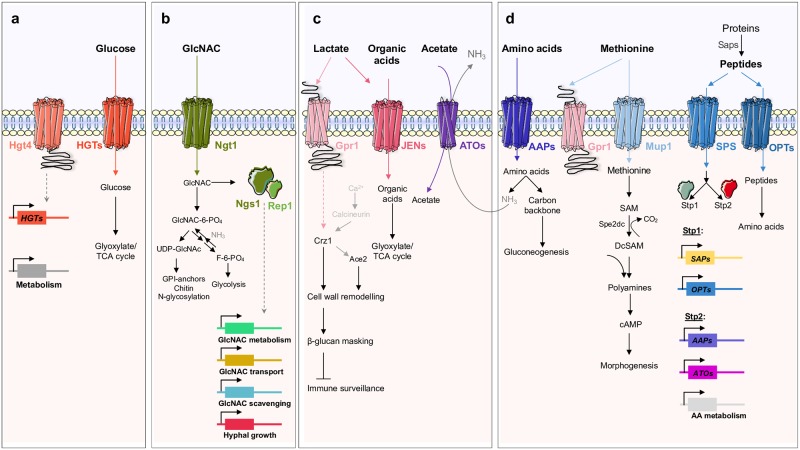
Fig 2. Schematic representation of the main sensing, transport, and transduction systems for the utilization of different host nutrients in Candida species.
(a) In C. albicans, glucose is sensed by Hgt4, generating an intracellular signal that induces the expression of HGTs and other metabolic genes. (b) In C. albicans and C. tropicalis, the uptake of GlcNAc occurs through the Ngt1 transporter. (c) The uptake of carboxylic acids is facilitated by the Jen (in C. albicans) and Ato transporters (in C. albicans and C. glabrata). In C. albicans, Gpr1 is reported to be a lactate and methionine sensor. In the presence of lactate, Gpr1 is thought to activate Crz1 in a calcineurin-independent manner and, together with Ace2, regulates a polygenic response that leads to β-glucan masking. (d) Peptides and amino acids are sensed by the SPS complex, which induces the expression of Opts, Aaps, and Ato transporters, as well as SAPs and amino acid catabolic genes. Intracellular ammonia resulting from the catabolism of GlcNAc or amino acids is exported via Ato transporters. In the presence of methionine, and in low glucose conditions, the methionine-induced morphogenesis is activated via Gpr1 sensor and Mup1 transporter. AA, amino acid; Aap, amino acid permease; ATP, adenosine triphosphate; cAMP, cyclic adenosine monophosphate; DcSAM, decarboxylated S-adenosylmethionine; GlcNAc, N-acetylglucosamine; GPI, glycosylphosphatidylinositol; HGT, hexose transporter; Opt, oligopeptide transporter; SAM, S-adenosylmethionine; SAP, secretory aspartyl proteinase; SPS, Ssy1-Ptr3-SSy5; Sp2DC, Sp2 decarboxylase; TCA, tricarboxylic acid cycle; UDP, uridine diphosphate.
In glucose-limiting conditions, other alternative carbon sources, such as N-acetylglucosamine (GlcNAc) and carboxylic acids, are thought to play a critical role to sustain Candida growth. When infecting tissues and organs, Candida up-regulates several pathways involved in the utilization of alternative carbon sources, such as gluconeogenesis, the glyoxylate cycle, and fatty acid β-oxidation, suggesting that glucose levels may not be sufficient to satisfy the energetic requirements of the cells [28,35–37]. In C. albicans and C. tropicalis, GlcNAc, a monosaccharide produced mainly by bacteria in the gastrointestinal tract, enters the cell through the Ngt1 transporter, and is then sensed by the transcription factors, Ngs1 and Rep1, which control the expression of genes involved in the uptake and catabolism of GlcNAc [38–40] (Fig 2b). Depending on the metabolic state of the cells, GlcNAc can either be converted to uridine diphosphate-N-acetylglucosamine (UDP-GlcNAc) or to fructose-6-phosphate, which then enters the glycolytic pathway (Fig 2b). In C. albicans, GlcNAc can also be used as a signal to induce the expression of several virulence genes involved in white-opaque switching [41], hyphal morphogenesis [38–40,42], and cell death [43]. Additionally, GlcNAc metabolism seems to sustain Candida survival when growing inside phagocytic cells. The export of intracellular ammonia, derived from GlcNAc catabolism, has been reported to promote the alkalization of the phagosome, enabling cells to survive and escape from the acidic environment of the phagolysosome [44]. This mechanism is dependent on the transport of GlcNAc and subsequent catabolism through Hxk1, Nag1, and Dac1 enzymes [44]. Hence, mutants lacking the Ngt1 transporter or GlcNAc catabolic enzymes are defective in neutralizing the phagosome [44]. The ability to manipulate ambient pH is reported for all species of the CTG clade, a phylogenetic group that translates the CUG codon into serine instead of leucine [45]. This is in contrast to what is found for the distantly related C. glabrata, whose genome does not appear to encode homologs of GlcNAc transporters or catabolic enzymes [44].
C. albicans can also raise the extracellular pH by metabolizing carboxylic acids [46]. This phenomenon is physiologically and genetically distinct from the GlcNAc-driven mechanism, as the metabolism of carboxylic acids, when used as the sole carbon source, does not generate ammonia or promote hyphal morphogenesis [44,46]. Physiologically relevant carboxylic acids such as lactate, acetate, succinate, butyrate, and propionate are produced either by host cells or host microbiota [47–49]. Lactate and acetate are particularly abundant in the gut and in vaginal secretions [47,50] but also inside phagocytic cells [51,52]. In C. albicans, the uptake of lactate is mediated by Jen transporters [51,53], while Ato transporters are potentially involved in the transport of acetate in both C. albicans and C. glabrata [52,54] (Fig 2c). These two transporter families are strongly induced after phagocytosis [51,52], and they modulate biofilm formation and resistance to antifungal drugs in both C. albicans and C. glabrata [54–56]. In particular, exposure to lactate has been shown to trigger the masking of β-glucan, a major pathogen-associated molecular pattern (PAMP), in several Candida species [57]. This affects the visibility of these pathogens to host immune defenses, which correlates well with the observed decrease in C. albicans uptake by macrophages and reduced phagocytic recruitment [57,58]. The β-glucan masking phenotype has been proposed to be dependent on Gpr1 and the transcription factor Crz1 [57]. These proteins control the expression of genes associated with the organization of the cell wall, ultimately contributing to the masking effect [57,59]. Therefore, the concomitant exposure of Candida cells to different carboxylic acids potentiates immune evasion and consequently Candida persistence.
The uptake of nitrogen is also critical for Candida survival. Different in vivo studies have demonstrated that genes involved in amino acid uptake and catabolism are strongly up-regulated in C. albicans, especially when phagocytosed by neutrophils and macrophages [36,60–62]. Indeed, several C. albicans and C. glabrata amino acid auxotrophic strains retain full virulence in mice, suggesting that these nutrients are readily available during infection [63–65]. Proteolytic enzymes, namely secretory aspartyl proteinases (SAPs), are of particular importance because they allow Candida to efficiently degrade the complement proteins and host connective tissues [66]. Once available, extracellular amino acids are then sensed by the SPS complex (composed of Ssy1, Ptr3, and Ssy5), which in turn activates the transcription factors, Stp1 and Stp2 (Fig 2d). While Stp1 controls the expression of extracellular proteases and peptide transporters, Stp2 regulates amino acid permeases, Ato transporters, and catabolic enzymes [67,68] (Fig 2d). Along with GlcNAc and carboxylic acids, the catabolism of amino acids represents a third independent mechanism by which Candida rapidly neutralizes acidic microenvironments [52,69]. Previous studies reported that C. albicans mutants lacking STP2 or ATO genes release less ammonia than wild-type controls, failing to efficiently neutralize the acidic phagosome and undergo hyphal morphogenesis, which consequently affects their ability to escape phagocytic cells [52,70]. Recent data, however, suggest that the phagosomal membrane is highly permeable to ammonia, and the observed alkalization is rather a direct consequence of proton leakage induced by hyphal growth [71,72]. The transport of methionine via the high-affinity permease Mup1 and its subsequent metabolism have been also shown to induce morphogenesis in a process that is dependent on Gpr1 and the cAMP-PKA (cyclic Adenosine Monophosphate-Protein Kinase A) signaling cascade [73,74]. The methionine-induced morphogenesis pathway triggers the activation of adenylate cyclase by the production of increased levels of polyamines such as spermine and spermidine. These compounds are generated by the intracellular conversion of methionine into S-adenosylmethionine (SAM) and its decarboxylation by Spe2, which donates aminopropyl groups for polyamine synthesis [73] (Fig 2d).
Environmental pH fluctuations shape Candida physiology and pathogenicity
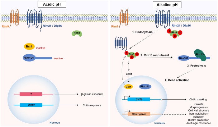
Changes in ambient pH represent an additional stress that Candida and other pathogens face in the human host. While the pH of human blood and tissues is slightly alkaline (pH 7.4), the pH of the oral cavity and the gastrointestinal and genitourinary tracts is acidic (2 < pH < 6). Adaptation to differing ambient pHs is critical for survival and growth in these niches. In fungi, including Candida species, pH signaling is mediated by the Rim pathway [75]. In C. albicans, the external pH is sensed by Rim21/Dfg16, Rim9, and an arrestin-like protein Rim8. Under alkaline pH, Rim8 is hyperphosphorylated, a signal that triggers the endocytosis of the plasma membrane complex and the recruitment of the signaling protease Rim13. This protease then cleaves the C-terminal inhibitory domain of Rim101, resulting in its activation. The activation of Rim101 promotes the expression of target genes involved in morphogenesis [76–79], growth [80], cell-wall remodeling [80], iron metabolism [81,82], adhesion [80], biofilm formation, and antifungal tolerance [75,83,84] (Fig 3).
Fig 3. Candida adaptation to pH fluctuations.
In Candida species, pH adaptation is mediated by the Rim pathway. Under acidic pH, the exposure of both chitin and β-glucan is enhanced and facilitates their recognition by the host innate immune system. Chitin exposure is promoted by the repression of both Rim101 and Bcr1, resulting in reduced expression of CHT2. β-glucan exposure is regulated by a noncanonical signaling pathway. Under alkaline pH, Rim8 is hyperphosphorylated, a signal that induces the endocytosis of the Rim complex and the recruitment of Rim13. The C-terminal proteolysis of Rim101 by Rim13 activates it and promotes the expression of target genes, including CHT2.
On the other hand, the adaptation of C. albicans to acidic environments drives cell-wall remodeling by enhancing the exposure of two key fungal PAMPs (chitin and β-glucan) at the cell surface [85]. While pH-dependent β-glucan exposure is regulated by a noncanonical signaling pathway, the remodeling of chitin is coordinated by several transcription factors, including Rim101, Bcr1, and Efg1 (Fig 3) [85,86]. The exposure of β-glucan at the cell surface hyperactivates the immune system largely through the recognition of the immunostimulatory β-glucan by Dectin-1, which enhances the recruitment of neutrophils and macrophages to the site of the infection [85]. This pH-dependent β-glucan exposure was also observed in C. dubliniensis and C. tropicalis, but not in C. auris or C. glabrata [85,86]. Surprisingly, adaptation to acidic environments induces β-glucan masking in C. krusei, suggesting that the outputs of pH-dependent signal transduction differ between these Candida species [85]. Additionally, the pH-dependent reorganization of the cell wall fluctuates over time in C. albicans, with β-glucan and chitin being masked after an initial period of exposure [86]. While the subsequent β-glucan masking is mediated by farnesol, this quorum-sensing molecule does not trigger the chitin cloaking [86]. These temporal fluctuations suggest dynamic cell-wall responses to environmental pH. Moreover, the early PAMP exposure appears to govern the outcome of the infection because subsequent remasking on the cell wall does not compensate for the initial induction of strong proinflammatory responses [86].
Adaptation to oxygen-limiting niches is critical for Candida virulence
Oxygen levels inside the human host can vary greatly. While some niches are rich in oxygen, such as exposed skin or oral mucosa, others are anoxic or hypoxic, including the gastrointestinal tract [87]. Consequently, Candida cells must adapt to low-oxygen environments, particularly when colonizing the human gut, developing lesions or growing in biofilms [87,88]. Analyses of gene expression profiles of C. albicans cells shifted from normoxia to hypoxic growth conditions revealed the induction of several pathways, including glycolytic gene expression via Tye7 [89–91], fatty acid metabolism [92,93], heme biosynthesis and iron metabolism [89,92,94], cell-wall structure [89,92,94], and sterol biosynthesis via Upc2 [95,96]. In contrast, genes involved in the oxidative respiration were repressed [89,92,94]. Additionally, the Sit4 phosphatase, the Ccr4 mRNA deacetylase, and the Sko1 transcription factor have been identified as potential regulators of an early hypoxic response (10–20 min) [91,94].
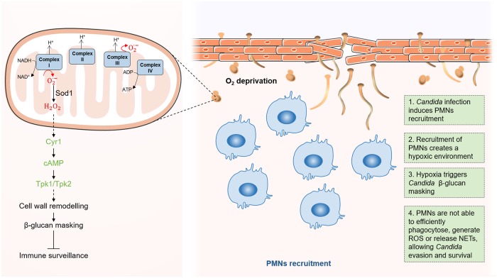
Besides affecting the cellular metabolism and energy homeostasis, adaptation to hypoxia induces hyphal growth in C. albicans [94] and promotes immune evasion by triggering β-glucan masking at the cell surface [97]. β-glucan masking leads to reduced phagocytosis and attenuates local immune responses [97]. In contrast to lactate-induced β-glucan masking, hypoxia-induced masking does not depend on Gpr1 and Crz1. Instead, hypoxia-induced masking is mediated by mitochondrial and cAMP-PKA signaling [57,97]. Hypoxia induces the generation of mitochondrial superoxide [98,99], which is rapidly converted into diffusible hydrogen peroxide by superoxide dismutase 1 (Fig 4). Hydrogen peroxide has been proposed to somehow activate the cAMP-PKA pathway, which, in turn, triggers cell-wall remodeling and β-glucan masking [97]. However, the mechanism by which β-glucan masking is achieved at the cell surface remains unclear.
Fig 4. Candida adaptation to hypoxic host niches.
During C. albicans infections, the recruitment of PMNs creates an hypoxic environment [88]. In the fungus, this oxygen limitation triggers increased formation of ROS, such as superoxide (O2•−), from the electron transport chain [98,99]. Superoxide is then converted into diffusible hydrogen peroxide (H2O2) by the action of Sod1. H2O2 has been proposed to activate adenylyl cyclase (Cyr1) and cAMP-PKA (Tpk1/2) signaling, which in turn triggers cell-wall remodeling and β-glucan masking [97]. This β-glucan masking allows the fungus to evade phagocytosis by the PMNs [88]. cAMP, cyclic Adenosine Monophosphate; NET, neutrophil extracellular trap; PKA, Protein Kinase A; PMN, polymorphonuclear leukocyte; ROS, reactive oxygen species; Sod1, superoxide dismutase 1.
Hypoxia-induced β-glucan masking has been observed for some other pathogenic Candida species, namely C. tropicalis and C. krusei, but not in C. glabrata, C. guilliermondi, or C. parapsilosis [97]. Therefore, during their evolution, hypoxic signaling has become integrated with PAMP masking only in some Candida pathogens. The adaptation to hypoxic environments enhances the ability of these Candida species to colonize the host. For example, it was shown that the recruitment of polymorphonuclear leukocytes (PMNs) to sites of C. albicans infection in mice was the main cause of hypoxia [88] (Fig 4). However, because of the hypoxia-induced β-glucan masking by C. albicans cells, these PMNs are not able to efficiently phagocytose the fungus, generate reactive oxygen species (ROS), or release extracellular DNA traps, allowing C. albicans to survive. Continued exposure to hypoxia leads to accumulation of lactate, prolonging the masking effect. Additionally, it was also observed that the antifungal activity of fluconazole is considerably reduced under hypoxic conditions. We speculate that the molecular mechanism behind this observation might include Upc2, considering its dual role in activating hypoxia-induced β-glucan masking [97] and conferring azole antifungal resistance [100]. In contrast to C. albicans, C. tropicalis is not able to induce β-glucan masking in response to hypoxia, and this species is more susceptible to PMN attack [88]. This is in agreement with the fact that C. tropicalis mainly infects neutropenic patients [101]. The molecular mechanisms allowing hypoxic adaptation are not completely defined. Nevertheless, it is clear that some Candida species take advantage of low-oxygen environments, either generated during infection or imposed by the specific host niche, to thrive by avoiding immune surveillance and escaping from antifungal therapy.
Candida adaptation to temperature shifts is essential for full virulence
The human body temperature is considered to be a potent nonspecific defense against fungal infection, especially in febrile patients, because high temperatures considerably restrict fungal growth [102,103]. The human host presents fever as one of the first responses against a Candida infection, thereby exposing the fungal cells to temperatures ranging from 37 °C to 42 °C. These temperature fluctuations profoundly influence many physiological aspects of C. albicans, including morphology, mating, phenotypic switching, and drug resistance [104].
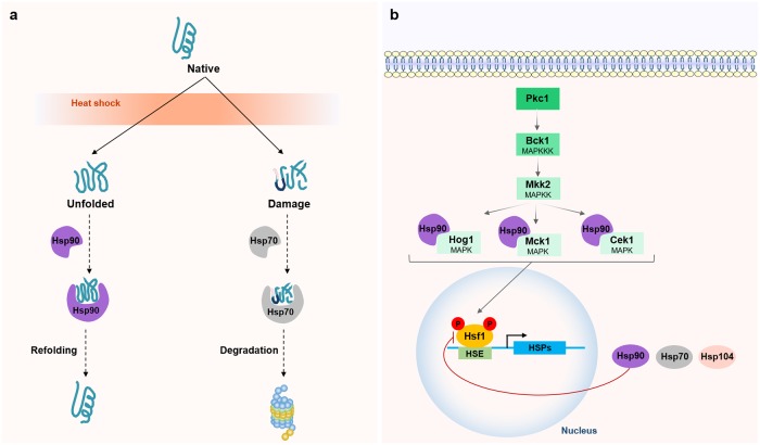
Changes in ambient temperature are sensed by a broad diversity of mechanisms. One of the most studied pathways is the evolutionarily conserved heat shock response, which mediates thermal homeostasis by controlling the levels of heat shock proteins (HSPs) [105]. HSPs are molecular chaperones sequestered in response to heat shock, rescuing proteins from unfolding or targeting damaged proteins for degradation. In C. albicans, the expression of HSP genes is activated by the heat shock transcription factor 1 (Hsf1), which becomes phosphorylated in response to temperature elevations, including thermal transitions that mimic fever [106,107]. After adaptation to the exposed temperature, Hsf1 phosphorylation returns to basal levels and several lines of evidence have suggested the existence of a negative feedback loop, in which Hsp90 negatively regulates Hsf1 [107–109]. Besides Hsf1, Hsp90 also controls the activation of other regulators that mediate long-term thermal adaptation (Fig 5). These include several mitogen-activated protein kinase (MAPK) signaling pathways, particularly the Hog1, Mkc1, and Cek1 pathways, which are intimately associated with cell-wall remodeling [110,111]. Other small HSPs such as Hsp12 and Hsp21 have also been identified as crucial for C. albicans to resist thermal stress [112,113]. HSPs and their associated signaling pathways have been widely implicated in antifungal resistance, emerging as potential antifungal targets to treat Candida infections [114]. Moreover, the activation of the Hsf1 transcriptional program in C. albicans has been associated with increased host cell adhesion, damage, and virulence, reinforcing the importance of this regulon in thermal homeostasis [115,116].
Fig 5. Molecular circuits required for thermal adaptation in C. albicans.
(a) HSPs rescue proteins from unfolding or target damaged proteins for degradation. (b) In response to temperature upshifts, Hsf1 becomes phosphorylated, inducing the expression of HSP genes. After thermal adaptation, Hsf1 returns to basal levels through a negative feedback loop dependent on Hsp90. Long-term adaptation is controlled by Hsp90 through Hog1, Mkc1, and Cek1. HSE, heat shock element; Hsf1, heat shock transcription factor 1; HSP, heat shock protein; MAPK, mitogen-activated protein kinase; MAPKK/MAPKKK, MAPK kinase/ MAPKK kinase.
Candida and host microbiota: Avoiding antagonistic interactions in health and disease
The structure of human microbiota is dynamic, often defined by host and environmental factors and also by physical and metabolic interactions between species. While some of these interactions are cooperative, others are antagonistic, and the latter may represent a major obstacle for Candida. This concept gained experimental support through studies involving the depletion of commensal microbiota by continued use of broad-spectrum antibiotics, which resulted in Candida overgrowth [117,118]. This suggests that some commensal microbial colonizers antagonize Candida spp. (and other exogenous pathogens) in order to maintain a homeostatic balance in the host. Some of these interactions are driven by metabolic competition, while others are mediated by quorum-sensing molecules that influence fungal cell behavior and regulate important virulence traits. Although quorum-sensing systems have been explored in great detail for pathogenic bacteria, they are relatively poorly understood in fungi [119]. The C. albicans molecule farnesol was the first quorum-sensing compound to be identified in an eukaryote [120] and has been the object of intense research. Yet, its precise mode of action remains unclear.
Lactobacillus species and C. albicans are a well-documented example of infectious antagonism [121–123]. Lactobacilli are a dominant species of the microbiota of the gastrointestinal and urogenital tracts, and they actively reduce the amount of fungal microbes by producing many fungicidal compounds [121–123]. Other commensal bacteria such as Bacteroides thetaiotamicron or Blautia producta can antagonize C. albicans by stimulating intestinal cells to produce antimicrobial peptides [124]. The pathogenic bacterium Acinetobacter baumanii has been also reported to interact antagonistically with C. albicans by binding to hyphae to promote apoptosis [125]. The elucidation of these types of interaction is of particular interest in the quest for novel targets for antifungal therapy, as the inhibitory secreted factors produced by these antagonists appear to have high fungicidal activity.
The disruption of commensal interactions through alterations in immune competence, by changes in environmental host conditions, or via antibiotic therapy may favor the outgrowth and overrepresentation of pathogenic microbes, with these growing at the expense of those organisms that fail to adapt. While antagonist interactions might lower the risk of infection, synergistic interactions during dysbiotic states are associated with increased pathogenesis because microbes can also interact to enhance colonization and persistence. An illustrative example is the infectious synergism established between several Candida species (including C. albicans, C. dubliniensis, C. tropicalis, and C. krusei) and the gram-positive bacterium Staphylococcus aureus [126,127]. Candida not only provides a substratum for the attachment and colonization of S. aureus but also facilitates its invasion across mucosal barriers, thereby promoting persistence and systemic infection [128].
Host immune defenses: How Candida species counteract the immune response
Microbial pathogens are constantly surveyed by the innate immune system. Phagocytic cells such as dendritic cells, macrophages, monocytes, and neutrophils play important roles in clearing fungal pathogens from the bloodstream and tissues. Loss of innate immune cells or defects in their antifungal activities have major implications for the host. Candida cells are recognized through key PAMPs, some of which are located in the cell wall; for example, β-glucans, chitin, and mannans. These components are sensed by the multiple pattern-recognition receptors (PRRs) expressed by phagocytic cells or secreted (for example, complement components). PPRs mediate binding of the pathogen to the phagocyte, and the PAMP–PRR interactions trigger intracellular signaling pathways within the immune cells that can induce phagocytosis and the production of proinflammatory cytokines and chemokines. In order to attenuate recognition and escape phagocytosis, Candida cells are able to actively mask cell-wall PAMPs [129] and secrete specific proteases that target complement opsonization [130]. Alternatively, some Candida species can induce their phagocytic uptake into endothelial and epithelial cells and use these cells as “safe houses” by preventing maturation of the phagolysosome and subsequent killing [131]. If none of these strategies is employed, Candida cells are likely to be internalized and subjected to a combination of toxic oxidative and nonoxidative mechanisms that attempt to kill an intra- or extracellular yeast cell. These oxidative mechanisms include the production of reactive oxygen and nitrogen species (ROS and RNS, respectively), while nonoxidative killing mechanisms include the release of antimicrobial peptides and the induction of processes related to micronutrient restriction. Of note, while C. albicans is sensitive to the combinatorial stresses imposed by phagocytes [132], C. glabrata has adapted to survive within the inhospitable environment of the phagosome. This pathogen mounts robust stress responses against the ROS implemented by the phagocytic cell and neutralizes the phagocytic environment, thereby escaping phagocytosis [133].
Oxidative, nitrosative, and osmotic/cationic stresses
Phagocytic cells attempt to kill pathogens in part by employing toxic ROS and RNS, either intracellularly or extracellularly, as a major antimicrobial defense mechanism. ROS are produced by the NADPH oxidase complex, a process known as respiratory burst, and include chemicals such as the superoxide anion (O2•), hydrogen peroxide (H2O2), and the hydroxyl radicle (•OH). Furthermore, ROS production in response to C. albicans infection has been shown to lead to the recruitment of additional phagocytes, creating a toxic oxidative environment for the fungus [134]. Inside phagocytes, ROS can interact with nitric oxide (NO), generating toxic products such as peroxynitrite [135]. These toxic chemicals cause irreversible damage to the pathogen by interacting with proteins, lipids, and nucleic acids.
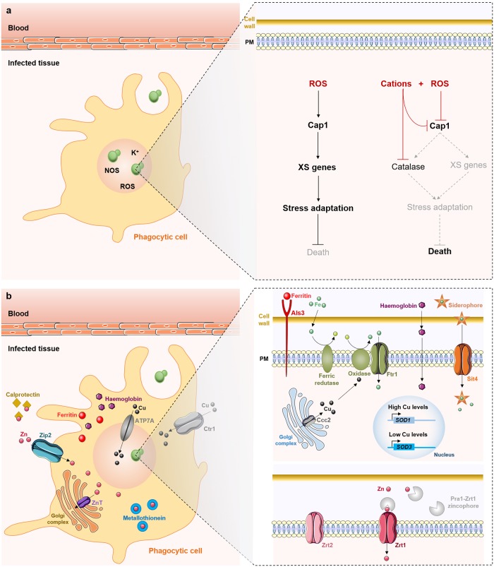
Candida species attempt to counteract these stresses by activating cellular responses that include the activation of genes encoding proteins involved in stress detoxification and reparation. These include catalase, superoxide dismutases, glutathione peroxidases, and thioredoxins (Fig 6a) [136–138]. In C. albicans and C. glabrata, these stress pathways are regulated largely by the Hog1 stress-activated protein kinase [136,139], the transcription factor Cap1 [140–142], and the Rad53 DNA damage checkpoint kinase [143]. Together with the transcription factor Cta4, these signaling pathways play key roles in orchestrating the responses to osmotic, oxidative, and nitrosative stresses in these species [144]. In this way, these regulators promote the fitness of C. albicans during systemic infection. Indeed, mutants lacking these genes display attenuated virulence in mice, as well as impaired tolerance to these stresses in vitro and phagocytic survival [145,146]. Curiously, the oxidative stress response is delayed if the fungus is simultaneously exposed to cationic and oxidative stress [147]. This is thought to contribute to the ability of phagocytic cells to efficiently kill invading pathogens (Fig 6a) [132]. Given the importance of these stress response pathways for Candida survival, key molecular players involved may represent attractive targets for antifungal development.
Fig 6. Host immune defenses and adaptation mechanisms displayed by C. albicans and C. glabrata.
(a) Cap1 plays a key role in the activation of responses to ROS generated by phagocytic cells, leading to the induction of oxidative stress genes (XS genes), including catalase, superoxide dismutases, glutathione peroxidases, and thioredoxins, among others. However, cations inhibit catalase and Cap1, thereby delaying the induction of the oxidative stress response and leading to the death of C. albicans cells. (b) Host-enforced micronutrient restriction results in reduced iron, copper, and zinc availability, but C. albicans responds by up-regulating efficient metal-scavenging strategies. Host phagocytes also exploit the toxicity of copper and zinc by pumping these metals in excess into phagosomes to intoxicate internalized pathogens. NOS, nitric oxide species; PM, plasma membrane; ROS, reactive oxygen species; Sod, superoxide dismutase; XS, oxidative stress.
Host-enforced micronutrient restriction
The limitation of micronutrients such as iron, copper, zinc, or manganese is an effective way of controlling the outgrowth of invading microbes. These micronutrients are essential for the survival of both host and pathogen because they function as cofactors for enzymes, transcription factors, and other proteins that play crucial biochemical and cellular functions. However, our immune system attempts to restrict microbial access to these essential elements via a mechanism known as nutritional immunity [148].
Iron has well-studied implications for Candida pathogenesis, being a crucial micronutrient for Candida growth, survival, and virulence [149]. During systemic candidiasis, the host restricts this metal by increasing the levels of iron-binding proteins, such as ferritin and hemoglobin alpha, and accumulating heme oxygenase (Fig 6b) [150,151]. Both C. albicans and C. glabrata have developed efficient iron-scavenging strategies that can overcome these host mechanisms. This contributes to their ability to survive phagocytosis and replicate inside macrophages by using their intracellular storages of iron [152,153]. C. albicans and C. glabrata cells exploit sophisticated iron-uptake systems to acquire either free iron [154,155] or iron from host iron-binding proteins, including hemoglobin [156], ferritin [82], and transferrin (Fig 6b). Additionally, the utilization of siderophores promotes resistance to macrophage killing: in C. glabrata, the Sit1 siderophore-iron transporter mediates iron acquisition, being critical for the survival of the yeast inside macrophages [152].
Copper is also involved in Candida virulence, both positively and negatively. The fungal reductive iron-uptake pathway includes multicopper oxidases, and hence, iron acquisition and mobilization depends on copper availability [157]. Interestingly, the host also uses copper as a defense mechanism against Candida by pumping excess quantities of this metal into Candida-containing phagosomes (Fig 6b) [158]. However, C. albicans adapts to this potential killing mechanism by differentially modulating the expression of copper- and manganese-dependent SODs (Sod1 and Sod3, respectively) [159]. Sod1 is expressed when copper is in excess, but when copper levels decline, Sod3 is then expressed (Fig 6b) [159]. Thus, during infection, C. albicans is able to adjust copper uptake and management by using it as an enzymatic cofactor for SOD enzymes [159].
Zinc is an abundant micronutrient that has crucial roles in cellular functionality for both host and pathogen. The host attempts to limit zinc availability for the fungus by depleting extracellular zinc levels, mainly via calprotectin, an antimicrobial peptide expressed by neutrophils that binds zinc and manganese with high affinity (Fig 6b) [160]. Calprotectin promotes the antimicrobial activity of neutrophil extracellular traps (NETs), which are released by neutrophils after sensing large microbes such as C. albicans hyphae [161–163]. Zinc depletion also occurs inside immune cells as an antifungal mechanism to kill intracellular pathogens such as C. albicans and C. glabrata [164]. During infection, macrophages deplete intracellular zinc by pumping it into the Golgi apparatus via specific ZnT-type zinc transporters (Fig 6b) and increasing the expression of zinc-binding metallothioneins [165]. Additionally, macrophages up-regulate the zinc importer ZIP2 to increase the intracellular levels of zinc (Fig 6b) [166]. This combination of strategies depletes zinc from the extracellular environment while dealing with the increased metabolic demands associated with microbial clearing [166]. To overcome zinc depletion, C. albicans overexpresses ZRT1 and ZRT2 genes, encoding zinc uptake transporter systems Zrt1 and Zrt2 (Fig 6b). Both transporters are regulated by the zinc finger transcription factor Zap1 (also known as Csr1) [167,168] and by pH [79]. Zinc transporters play important roles in Candida pathogenesis because overexpression of Zrt2 increases C. albicans virulence [169]. In addition to functioning as a zinc transporter, Zrt1 also serves as a receptor for the Pra1 zincophore [79,168], a secreted protein that binds and sequesters zinc from host cells during C. albicans invasion (Fig 6b) [170]. Similarly to copper, zinc has also been reported to be pumped in higher amounts into the phagosome to intoxicate internalized pathogens, constituting an important mechanism of killing (Fig 6b) [171].
Environment-triggered biofilm formation and antifungal resistance
So far, we have described major molecular circuits required by Candida species to counteract several constraints they face in the human host. The ability of Candida to adapt to these stresses imparts the flexibility to colonize diverse host niches. The physiological capacity to respond efficiently to stress and survive hostile environments also endows the fungal cells with the advantage of being better prepared for future insults [172,173]. The generation of biofilms might represent another strategy to resist harsh conditions and persist in the human host.
The Candida species most frequently associated with the formation of biofilms, either in host tissues or implanted medical devices, are C. albicans, C. glabrata, C. tropicalis, and C. parapsilosis [174]. Biofilms represent three-dimensional communities of adherent cells, with distinct biological properties, that are embedded in a self-synthesizing extracellular matrix (ECM) composed predominantly of proteins, glycoproteins, carbohydrates, lipids, and nucleic acids [175]. The ECM helps to maintain the overall structural integrity of the biofilm, and it also acts as a physical barrier to drug penetration. Consequently, biofilm cells can survive drug concentrations more than a thousand times higher than those defined for planktonic cells [176]. This phenotype is partly associated with the sequestration of drugs by the biofilm ECM and partly with the occurrence of a subpopulation of so-called “persister cells”. Persister cells exhibit a dormant-like physiology that has been demonstrated to make them highly resistant to antifungals [177]. These features contribute to the intrinsic resistance of Candida biofilms to conventional antifungal treatments, the host immune system, and other environmental perturbations, making biofilm-based infections a clinical challenge.
Genome-wide transcriptional profiling and proteomic approaches have identified hundreds of genes that are differentially expressed between C. albicans biofilm and planktonic cells. The up-regulation of glycolytic and sulfur amino acid genes, similar to what is observed when cells grow under hypoxia, suggests that Candida biofilms constitute a heterogeneous environment with hypoxic niches [178]. Moreover, more than 50 transcriptional regulators and 101 other genes have functionally validated roles in the formation of Candida biofilms [179–181]. Some of these play important roles in hyphal formation, adhesion, drug resistance, and matrix production (all intrinsic characteristics of biofilms), as well as in stress adaptation. It is not surprising, then, that adaptation to specific environmental niches modulates the ability of cells to form biofilms and, consequently, to resist antifungals [54,55,58,59,182–184].
Final remarks and future perspectives
Candida cells regulate specific sets of genes, including many involved in an array of stresses and metabolic pathways, in order to thrive and persist in the human host. In addition to conferring metabolic flexibility and stress resistance, the physiological reprogramming has been associated with enhanced virulence through impaired immune recognition, increased biofilm formation, and/or acquired antifungal tolerance and resistance. Although remarkable progress has been made in the last few decades in our understanding of the impact of host-derived stresses on Candida physiology and pathogenicity, many details remain unclear. During an infection, Candida cells are exposed to multiple environmental constraints, sometimes imposed consecutively, and at other times imposed simultaneously. Yet, in vitro experiments are predominantly designed to study individual environmental signals, often at single time points, rather than combinatorial stresses over time. Much progress has been achieved using the first approach. While this has given us valuable insights, it rather oversimplifies biological reality. The analysis of combinatorial stresses and of the dynamism of these inputs would mimic host conditions more closely and reveal more detailed views of which stress or stresses prevail and dictate the outcome of different types of infection. The same principle applies to infection and biofilm models, in which usually interactions between only a few different microbial populations have generally been examined. Most knowledge in the field comes from studies of either C. albicans or C. glabrata. Yet, the regulatory circuits required to effectively respond to each constraint, including antifungal treatments, differ considerably between the different Candida species, illustrating how heterogeneous these pathogens are. With the unprecedented emergence of multidrug resistant species such as C. auris, there is an urgent need to develop new effective antifungals. The integration of omics data with in vivo models, which mimic host conditions more closely, is now a powerful strategy to unravel molecular processes underlying adaptive phenotypes. These platforms have already produced novel lines of research and improved the identification of new potential therapeutic targets for vaccine and antifungal drug development, enhancing our ability to develop novel strategies to fight Candida infections.
Funding Statement
Work at CBMA is supported by the Contrato-Programa UIBD/04050/2020 funded by Portuguese national funds through the FCT I.P. RA and CBA are recipients of FCT PhD fellowships (PD/BD/113813/2015 and PD/BD/135208/2017, respectively). Research stay of RA at KU Leuven was supported by Boehringer Ingelheim Fonds. Work at KU Leuven is supported by grants from the Fund for Scientific Research Flanders (FWO grant nr: G0F8519N) and by the Research Council of the KU Leuven (grant nr: C14/17/063). Work at the University of Exeter is funded by a programme grant from the UK Medical Research Council (MRC) [www.mrc.ac.uk: MR/M026663/1], by the MRC Centre for Medical Mycology, University of Exeter [MR/N006364/1], and by the Wellcome Trust [www.wellcome.ac.uk: 097377]. The funders had no role in study design, data collection and analysis, decision to publish, or preparation of the manuscript.
References
- 1.Kumamoto CA. Inflammation and gastrointestinal Candida colonization. Current Opinion in Microbiology. 2011;14(4): 386–391. 10.1016/j.mib.2011.07.015 [DOI] [PMC free article] [PubMed] [Google Scholar]
- 2.Limon JJ, Skalski JH, Underhill DM. Commensal Fungi in Health and Disease. Cell Host Microbe. 2017;22: 156–165. 10.1016/j.chom.2017.07.002 [DOI] [PMC free article] [PubMed] [Google Scholar]
- 3.Cauchie M, Desmet S, Lagrou K. Candida and its dual lifestyle as a commensal and a pathogen. Res Microbiol. 2017;168: 802–810. 10.1016/j.resmic.2017.02.005 [DOI] [PubMed] [Google Scholar]
- 4.Pappas PG, Lionakis MS, Arendrup MC, Ostrosky-Zeichner L, Kullberg BJ. Invasive candidiasis. Nat Rev Dis Prim. 2018;4: 18026 10.1038/nrdp.2018.26 [DOI] [PubMed] [Google Scholar]
- 5.Tsai M-H, Hsu J-F, Yang L-Y, Pan Y-B, Lai M-Y, Chu S-M, et al. Candidemia due to uncommon Candida species in children: new threat and impacts on outcomes. Sci Rep. 2018;8: 15239 10.1038/s41598-018-33662-x [DOI] [PMC free article] [PubMed] [Google Scholar]
- 6.Singh S, Sobel JD, Bhargava P, Boikov D, Vazquez JA. Vaginitis Due to Candida krusei: Epidemiology, Clinical Aspects, and Therapy. Clin Infect Dis. 2002;35(9): 1066–1070. 10.1086/343826 [DOI] [PubMed] [Google Scholar]
- 7.Bougnoux ME, Brun S, Zahar JR. Healthcare-associated fungal outbreaks: new and uncommon species, new molecular tools for investigation and prevention. Antimicrobial Resistance and Infection Control. 2018;7: 45 10.1186/s13756-018-0338-9 [DOI] [PMC free article] [PubMed] [Google Scholar]
- 8.Kohlenberg A, Struelens MJ, Monnet DL, Plachouras D, Apfalter P, Lass-Flörl C, et al. Candida auris: Epidemiological situation, laboratory capacity and preparedness in European Union and European economic area countries, 2013 to 2017. Eurosurveillance. 2018;23(13): 18–00136. 10.2807/1560-7917.ES.2018.23.13.18-00136 [DOI] [PMC free article] [PubMed] [Google Scholar]
- 9.Morrell M, Fraser VJ, Kollef MH. Delaying the empiric treatment of Candida bloodstream infection until positive blood culture results are obtained: a potential risk factor for hospital mortality. Antimicrob Agents Chemother. 2005;49(9): 3640–3645. 10.1128/AAC.49.9.3640-3645.2005 [DOI] [PMC free article] [PubMed] [Google Scholar]
- 10.Perlin DS, Rautemaa-Richardson R, Alastruey-Izquierdo A. The global problem of antifungal resistance: prevalence, mechanisms, and management. The Lancet Infectious Diseases. 2017;17(12): e383–e392. 10.1016/S1473-3099(17)30316-X [DOI] [PubMed] [Google Scholar]
- 11.Pfaller MA, Diekema DJ, Turnidge JD, Castanheira M, Jones RN. Twenty years of the SENTRY Antifungal Surveillance Program: Results for Candida species from 1997–2016. Open Forum Infect Dis. 2019;6(Suppl. 1): S79–S94. 10.1093/ofid/ofy358 [DOI] [PMC free article] [PubMed] [Google Scholar]
- 12.Nobile CJ, Johnson AD. Candida albicans Biofilms and Human Disease. Annu Rev Microbiol. 2015;69: 71–92. 10.1146/annurev-micro-091014-104330 [DOI] [PMC free article] [PubMed] [Google Scholar]
- 13.Demers EG, Biermann AR, Masonjones S, Crocker AW, Ashare A, Stajich JE, et al. Evolution of drug resistance in an antifungal-naive chronic Candida lusitaniae infection. Proc Natl Acad Sci U S A. 2018;115(47): 12040–12045. 10.1073/pnas.1807698115 [DOI] [PMC free article] [PubMed] [Google Scholar]
- 14.Thompson DS, Carlisle PL, Kadosh D. Coevolution of morphology and virulence in Candida species. Eukaryotic Cell. 2011;10(9): 1173–1182. 10.1128/EC.05085-11 [DOI] [PMC free article] [PubMed] [Google Scholar]
- 15.Noble SM, Gianetti BA, Witchley JN. Candida albicans cell-type switching and functional plasticity in the mammalian host. Nature Reviews Microbiology. 2017;15(2): 96–108. 10.1038/nrmicro.2016.157 [DOI] [PMC free article] [PubMed] [Google Scholar]
- 16.Tao L, Du H, Guan G, Dai Y, Nobile CJ, Liang W, et al. Discovery of a “White-Gray-Opaque” Tristable Phenotypic Switching System in Candida albicans: Roles of Non-genetic Diversity in Host Adaptation. PLoS Biol. 2014;12(4): e1001830 10.1371/journal.pbio.1001830 [DOI] [PMC free article] [PubMed] [Google Scholar]
- 17.Pande K, Chen C, Noble SM. Passage through the mammalian gut triggers a phenotypic switch that promotes Candida albicans commensalism. Nat Genet. 2013;45(9): 1088–1091. 10.1038/ng.2710 [DOI] [PMC free article] [PubMed] [Google Scholar]
- 18.Hopke A, Brown AJP, Hall RA, Wheeler RT. Dynamic Fungal Cell Wall Architecture in Stress Adaptation and Immune Evasion. Trends in Microbiology. 2018;26(4): 284–295. 10.1016/j.tim.2018.01.007 [DOI] [PMC free article] [PubMed] [Google Scholar]
- 19.Garcia-Rubio R, de Oliveira HC, Rivera J, Trevijano-Contador N. The Fungal Cell Wall: Candida, Cryptococcus, and Aspergillus Species. Frontiers in Microbiology. 2020;10: 2993 10.3389/fmicb.2019.02993 [DOI] [PMC free article] [PubMed] [Google Scholar]
- 20.Johnston M. Feasting, fasting and fermenting: Glucose sensing in yeast and other cells. Trends in Genetics. 1999;15(1): 29–33. 10.1016/s0168-9525(98)01637-0 [DOI] [PubMed] [Google Scholar]
- 21.Ene IV., Brunke S, Brown AJP, Hube B. Metabolism in fungal pathogenesis. Cold Spring Harb Perspect Med. 2014;4(12): a019695 10.1101/cshperspect.a019695 [DOI] [PMC free article] [PubMed] [Google Scholar]
- 22.Carlson M. Glucose repression in yeast. Curr Opin Microbiol. 1999;2(2): 202–207. 10.1016/S1369-5274(99)80035-6 [DOI] [PubMed] [Google Scholar]
- 23.Yin Z, Smith RJ, Brown AJP. Multiple signalling pathways trigger the exquisite sensitivity of yeast gluconeogenic mRNAs to glucose. Mol Microbiol. 1996;20(4): 751–764. 10.1111/j.1365-2958.1996.tb02514.x [DOI] [PubMed] [Google Scholar]
- 24.López-Boado YS, Herrero P, Gascón S, Moreno F. Catabolite inactivation of isocitrate lyase from Saccharomyces cerevisiae. Arch Microbiol. 1987;147(3): 231–234. 10.1007/BF00463480 [DOI] [PubMed] [Google Scholar]
- 25.Holzer H. Proteolytic catabolite inactivation in Saccharomyces cerevisiae. Revisiones sobre biologia celular: RBC. 1989;21: 305–319. [PubMed] [Google Scholar]
- 26.Sandai D, Yin Z, Selway L, Stead D, Walker J, Leach MD, et al. The evolutionary rewiring of ubiquitination targets has reprogrammed the regulation of carbon assimilation in the pathogenic yeast Candida albicans. mBio. 2012;3(6): e00495–12. 10.1128/mBio.00495-12 [DOI] [PMC free article] [PubMed] [Google Scholar]
- 27.Childers DS, Raziunaite I, Mol Avelar G, Mackie J, Budge S, Stead D, et al. The Rewiring of Ubiquitination Targets in a Pathogenic Yeast Promotes Metabolic Flexibility, Host Colonization and Virulence. PLoS Pathog. 2016;12(4): e1005566 10.1371/journal.ppat.1005566 [DOI] [PMC free article] [PubMed] [Google Scholar]
- 28.Barelle CJ, Priest CL, MacCallum DM, Gow NAR, Odds FC, Brown AJP. Niche-specific regulation of central metabolic pathways in a fungal pathogen. Cell Microbiol. 2006;8: 961–971. 10.1111/j.1462-5822.2005.00676.x [DOI] [PMC free article] [PubMed] [Google Scholar]
- 29.Hudson DA, Sciascia QL, Sanders RJ, Norris GE, Edwards PJB, Sullivan PA, et al. Identification of the dialysable serum inducer of germ-tube formation in Candida albicans. Microbiology. 2004;150(Pt 9): 3041–3049. 10.1099/mic.0.27121-0 [DOI] [PubMed] [Google Scholar]
- 30.Buu LM, Chen YC. Impact of glucose levels on expression of hypha-associated secreted aspartyl proteinases in Candida albicans. J Biomed Sci. 2014;21: 22 10.1186/1423-0127-21-22 [DOI] [PMC free article] [PubMed] [Google Scholar]
- 31.Maidan MM, Thevelein JM, Van Dijck P. Carbon source induced yeast-to-hypha transition in Candida albicans is dependent on the presence of amino acids and on the G-protein-coupled receptor Gpr1. Biochemical Society Transactions. 2005;33(Pt 1): 291–293. 10.1042/BST0330291 [DOI] [PubMed] [Google Scholar]
- 32.Mandal SM, Mahata D, Migliolo L, Parekh A, Addy PS, Mandal M, et al. Glucose directly promotes antifungal resistance in the fungal pathogen, Candida spp. J Biol Chem. 2014;289(37): 25468–25473. 10.1074/jbc.C114.571778 [DOI] [PMC free article] [PubMed] [Google Scholar]
- 33.Rodaki A, Bohovych IM, Enjalbert B, Young T, Odds FC, Gow NAR, et al. Glucose promotes stress resistance in the fungal pathogen Candida albicans. Mol Biol Cell. 2009;20(22): 4845–4855. 10.1091/mbc.E09-01-0002 [DOI] [PMC free article] [PubMed] [Google Scholar]
- 34.Tucey TM, Verma J, Harrison PF, Snelgrove SL, Lo TL, Scherer AK, et al. Glucose Homeostasis Is Important for Immune Cell Viability during Candida Challenge and Host Survival of Systemic Fungal Infection. Cell Metab. 2018;27(5): 988–1006.e7. 10.1016/j.cmet.2018.03.019 [DOI] [PMC free article] [PubMed] [Google Scholar]
- 35.Fradin C, Kretschmar M, Nichterlein T, Gaillardin C, D’Enfert C, Hube B. Stage-specific gene expression of Candida albicans in human blood. Mol Microbiol. 2003;47(6): 1523–1543. 10.1046/j.1365-2958.2003.03396.x [DOI] [PubMed] [Google Scholar]
- 36.Fradin C, De Groot P, MacCallum D, Schaller M, Klis F, Odds FC, et al. Granulocytes govern the transcriptional response, morphology and proliferation of Candida albicans in human blood. Mol Microbiol. 2005;56(2): 397–415. 10.1111/j.1365-2958.2005.04557.x [DOI] [PubMed] [Google Scholar]
- 37.Lorenz MC, Fink GR. The glyoxylate cycle is required for fungal virulence. Nature. 2001;412: 83–86. 10.1038/35083594 [DOI] [PubMed] [Google Scholar]
- 38.Naseem S, Min K, Spitzer D, Gardin J, Konopka JB. Regulation of hyphal growth and N-acetylglucosamine catabolism by two transcription factors in Candida albicans. Genetics. 2017;206(1): 299–314. 10.1534/genetics.117.201491 [DOI] [PMC free article] [PubMed] [Google Scholar]
- 39.Su C, Lu Y, Liu H. N-acetylglucosamine sensing by a GCN5-related N-acetyltransferase induces transcription via chromatin histone acetylation in fungi. Nat Commun. 2016;7: 12916 10.1038/ncomms12916 [DOI] [PMC free article] [PubMed] [Google Scholar]
- 40.Alvarez FJ, Konopka JB. Identification of an N-acetylglucosamine transporter that mediates hyphal induction in Candida albicans. Mol Biol Cell. 2007;18(3):965–975. Epub 27 Dec 2006. 10.1091/mbc.E06-10-0931 [DOI] [PMC free article] [PubMed] [Google Scholar]
- 41.Huang G, Yi S, Sahni N, Daniels KJ, Srikantha T, Soll DR. N-acetylglucosamine induces white to opaque switching, a mating prerequisite in Candida albicans. PLoS Pathog. 2010;6(3): e1000806 10.1371/journal.ppat.1000806 [DOI] [PMC free article] [PubMed] [Google Scholar]
- 42.Simonetti N, Strippoli V, Cassone A. Yeast-mycelial conversion induced by N-acetyl-D-glucosamine in Candida albicans. Nature. 1974;250(464): 344–346. 10.1038/250344a0 [DOI] [PubMed] [Google Scholar]
- 43.Du H, Guan G, Li X, Gulati M, Tao L, Cao C, et al. N-Acetylglucosamine-induced cell death in Candida albicans and its implications for adaptive mechanisms of nutrient sensing in yeasts. MBio. 2015;6(5): e01376–15. 10.1128/mBio.01376-15 [DOI] [PMC free article] [PubMed] [Google Scholar]
- 44.Vesely EM, Williams RB, Konopka JB, Lorenz MC. N-Acetylglucosamine Metabolism Promotes Survival of Candida albicans in the Phagosome. mSphere. 2017;2(5): e00357–17. 10.1128/mSphere.00357-17 [DOI] [PMC free article] [PubMed] [Google Scholar]
- 45.Butler G, Rasmussen MD, Lin MF, Santos MAS, Sakthikumar S, Munro CA, et al. Evolution of pathogenicity and sexual reproduction in eight Candida genomes. Nature. 2009;459(7247): 657–662. 10.1038/nature08064 [DOI] [PMC free article] [PubMed] [Google Scholar]
- 46.Danhof HA, Vylkova S, Vesely EM, Ford AE, Gonzalez-Garay M, Lorenz MC. Robust Extracellular pH Modulation by Candida albicans during Growth in Carboxylic Acids. mBio. 2016;7(6): e01646–16. 10.1128/mBio.01646-16 [DOI] [PMC free article] [PubMed] [Google Scholar]
- 47.Rowland I, Gibson G, Heinken A, Scott K, Swann J, Thiele I, et al. Gut microbiota functions: metabolism of nutrients and other food components. European Journal of Nutrition. 2018;57(1): 1–24. Epub 9 Apr 2017. 10.1007/s00394-017-1445-8 [DOI] [PMC free article] [PubMed] [Google Scholar]
- 48.Morrison DJ, Preston T. Formation of short chain fatty acids by the gut microbiota and their impact on human metabolism. Gut Microbes. 2016;7(3): 189–200. 10.1080/19490976.2015.1134082 [DOI] [PMC free article] [PubMed] [Google Scholar]
- 49.Valdes AM, Walter J, Segal E, Spector TD. Role of the gut microbiota in nutrition and health. BMJ. 2018;361: k2179 10.1136/bmj.k2179 [DOI] [PMC free article] [PubMed] [Google Scholar]
- 50.Owen DH, Katz DF. A vaginal fluid simulant. Contraception. 1999;59: 91–95. 10.1016/s0010-7824(99)00010-4 [DOI] [PubMed] [Google Scholar]
- 51.Vieira N, Casal M, Johansson B, MacCallum DM, Brown AJP, Paiva S. Functional specialization and differential regulation of short-chain carboxylic acid transporters in the pathogen Candida albicans. Mol Microbiol. 2010;75: 1337–1354. 10.1111/j.1365-2958.2009.07003.x [DOI] [PMC free article] [PubMed] [Google Scholar]
- 52.Danhof HA, Lorenz MC. The Candida albicans ATO Gene Family Promotes Neutralization of the Macrophage Phagolysosome. Infect Immun. 2015;83(11): 4416–4426. 10.1128/IAI.00984-15 [DOI] [PMC free article] [PubMed] [Google Scholar]
- 53.Soares-Silva I, Paiva S, Kötter P, Entian K-D, Casal M. The disruption of JEN1 from Candida albicans impairs the transport of lactate. Mol Membr Biol. 2004;21: 403–411. 10.1080/09687860400011373 [DOI] [PubMed] [Google Scholar]
- 54.Mota S, Alves R, Carneiro C, Silva S, Brown AJ, Istel F, et al. Candida glabrata susceptibility to antifungals and phagocytosis is modulated by acetate. Front Microbiol. 2015;6: 919 10.3389/fmicb.2015.00919 [DOI] [PMC free article] [PubMed] [Google Scholar]
- 55.Alves R, Mota S, Silva S, Rodrigues C F., Alistair AJ, Henriques M, et al. The carboxylic acid transporters Jen1 and Jen2 affect the architecture and fluconazole susceptibility of Candida albicans biofilm in the presence of lactate. Biofouling. 2017;33: 943–954. 10.1080/08927014.2017.1392514 [DOI] [PubMed] [Google Scholar]
- 56.Alves R, Kastora SL, Gomes-Gonçalves A, Azevedo N, Rodrigues CF, Silva S, et al. Transcriptional responses of Candida glabrata biofilm cells to fluconazole are modulated by the carbon source. NPJ Biofilms Microbiomes. 2020;6: 4 10.1038/s41522-020-0114-5 [DOI] [PMC free article] [PubMed] [Google Scholar]
- 57.Ballou ER, Avelar GM, Childers DS, Mackie J, Bain JM, Wagener J, et al. Lactate signalling regulates fungal β-glucan masking and immune evasion. Nat Microbiol. 2016;2: 16238 10.1038/nmicrobiol.2016.238 [DOI] [PMC free article] [PubMed] [Google Scholar]
- 58.Ene I V., Adya AK, Wehmeier S, Brand AC, Maccallum DM, Gow NAR, et al. Host carbon sources modulate cell wall architecture, drug resistance and virulence in a fungal pathogen. Cell Microbiol. 2012;14: 1319–1335. 10.1111/j.1462-5822.2012.01813.x [DOI] [PMC free article] [PubMed] [Google Scholar]
- 59.Ene I V., Heilmann CJ, Sorgo AG, Walker LA, De Koster CG, Munro CA, et al. Carbon source-induced reprogramming of the cell wall proteome and secretome modulates the adherence and drug resistance of the fungal pathogen Candida albicans. Proteomics. 2012;12: 3164–3179. 10.1002/pmic.201200228 [DOI] [PMC free article] [PubMed] [Google Scholar]
- 60.Zakikhany K, Naglik JR, Schmidt-westhausen A, Holland G, Schaller M, Hube B. In vivo transcript profiling of Candida albicans identifies a gene essential for interepithelial dissemination. Cell Microbiol. 2007;9(12): 2938–2954. 10.1111/j.1462-5822.2007.01009.x [DOI] [PubMed] [Google Scholar]
- 61.Lorenz MC, Bender JA, Fink GR. Transcriptional response of Candida albicans upon internalization by macrophages. Eukaryot Cell. 2004;3: 1076–1087. 10.1128/EC.3.5.1076-1087.2004 [DOI] [PMC free article] [PubMed] [Google Scholar]
- 62.Rubin-Bejerano I, Fraser I, Grisafi P, Fink GR. Phagocytosis by neutrophils induces an amino acid deprivation response in Saccharomyces cerevisiae and Candida albicans. Proc Natl Acad Sci U S A. 2003. 10.1073/pnas.1834481100 [DOI] [PMC free article] [PubMed] [Google Scholar]
- 63.Jacobsen ID, Brunke S, Seider K, Schwarzmüller T, Firon A, D’Enfért C, et al. Candida glabrata persistence in mice does not depend on host immunosuppression and is unaffected by fungal amino acid auxotrophy. Infect Immun. 2010;78(3): 1066–1077. 10.1128/IAI.01244-09 [DOI] [PMC free article] [PubMed] [Google Scholar]
- 64.Kirsch DR, Whitney RR. Pathogenicity of Candida albicans auxotrophic mutants in experimental infections. Infect Immun. 1991;59(9): 3297–3300. [DOI] [PMC free article] [PubMed] [Google Scholar]
- 65.Alonso-Monge R, Navarro-García F, Molero G, Diez-Orejas R, Gustin M, Pla J, et al. Role of the mitogen-activated protein kinase hog1p in morphogenesis and virulence of Candida albicans. J Bacteriol. 1999;181(10): 3058–3068. [DOI] [PMC free article] [PubMed] [Google Scholar]
- 66.Gropp K, Schild L, Schindler S, Hube B, Zipfel PF, Skerka C. The yeast Candida albicans evades human complement attack by secretion of aspartic proteases. Mol Immunol. 2009;47(2–3): 465–475. 10.1016/j.molimm.2009.08.019 [DOI] [PubMed] [Google Scholar]
- 67.Martínez P, Ljungdahl PO. An ER packaging chaperone determines the amino acid uptake capacity and virulence of Candida albicans. Mol Microbiol. 2004;51(2): 371–384. 10.1046/j.1365-2958.2003.03845.x [DOI] [PubMed] [Google Scholar]
- 68.Martinez P, Ljungdahl PO. Divergence of Stp1 and Stp2 Transcription Factors in Candida albicans Places Virulence Factors Required for Proper Nutrient Acquisition under Amino Acid Control. Mol Cell Biol. 2005;25(21): 9435–9446. 10.1128/MCB.25.21.9435-9446.2005 [DOI] [PMC free article] [PubMed] [Google Scholar]
- 69.Vylkova S, Lorenz MC. Modulation of Phagosomal pH by Candida albicans Promotes Hyphal Morphogenesis and Requires Stp2p, a Regulator of Amino Acid Transport. PLoS Pathog. 2014;10(3): e1003995 10.1371/journal.ppat.1003995 [DOI] [PMC free article] [PubMed] [Google Scholar]
- 70.Vylkova S, Carman AJ, Danhof HA, Collette JR, Zhou H, Lorenz MC. The fungal pathogen Candida albicans autoinduces hyphal morphogenesis by raising extracellular pH. mBio. 2011;2(3): e00055–11. 10.1128/mBio.00055-11 [DOI] [PMC free article] [PubMed] [Google Scholar]
- 71.Westman J, Moran G, Mogavero S, Hube B, Grinstein S. Candida albicans hyphal expansion causes phagosomal membrane damage and luminal alkalinization. mBio. 2018;9(5): e01226–18. 10.1128/mBio.01226-18 [DOI] [PMC free article] [PubMed] [Google Scholar]
- 72.May RC, Casadevall A. In Fungal Intracellular Pathogenesis, Form Determines Fate. mBio. 2018;9(5): e02092–18. 10.1128/mBio.02092-18 [DOI] [PMC free article] [PubMed] [Google Scholar]
- 73.Schrevens S, Van Zeebroeck G, Riedelberger M, Tournu H, Kuchler K, Van Dijck P. Methionine is required for cAMP-PKA-mediated morphogenesis and virulence of Candida albicans. Mol Microbiol;108(3): 258–275. 2018. 10.1111/mmi.13933 [DOI] [PubMed] [Google Scholar]
- 74.Miwa T, Takagi Y, Shinozaki M, Yun CW, Schell WA, Perfect JR, et al. Gpr1, a putative G-protein-coupled receptor, regulates morphogenesis and hypha formation in the pathogenic fungus Candida albicans. Eukaryot Cell. 2004;3(4): 919–931. 10.1128/EC.3.4.919-931.2004 [DOI] [PMC free article] [PubMed] [Google Scholar]
- 75.Cornet M, Gaillardin C. pH signaling in human fungal pathogens: A new target for antifungal strategies. Eukaryotic Cell. 2014;13(3): 342–352. 10.1128/EC.00313-13 [DOI] [PMC free article] [PubMed] [Google Scholar]
- 76.Ramon AM, Porta A, Fonzi WA. Effect of environmental pH on morphological development of Candida albicans is mediated via the PacC-related transcription factor encoded by PRR2. J Bacteriol. 1999;181(24): 7524–7530. [DOI] [PMC free article] [PubMed] [Google Scholar]
- 77.Porta A, Ramon AM, Fonzi WA. PRR1, a homolog of Aspergillus nidulans palF, controls pH-dependent gene expression and filamentation in Candida albicans. J Bacteriol. 1999;181(24): 7516–7523. [DOI] [PMC free article] [PubMed] [Google Scholar]
- 78.Davis D, Wilson RB, Mitchell AP. RIM101-Dependent and -Independent Pathways Govern pH Responses in Candida albicans. Mol Cell Biol. 2000;20(3): 971–978. 10.1128/mcb.20.3.971-978.2000 [DOI] [PMC free article] [PubMed] [Google Scholar]
- 79.Bensen ES, Martin SJ, Li M, Berman J, Davis DA. Transcriptional profiling in Candida albicans reveals new adaptive responses to extracellular pH and functions for Rim101p. Mol Microbiol. 2004;54(5): 1335–1351. 10.1111/j.1365-2958.2004.04350.x [DOI] [PubMed] [Google Scholar]
- 80.Nobile CJ, Solis N, Myers CL, Fay AJ, Deneault JS, Nantel A, et al. Candida albicans transcription factor Rim101 mediates pathogenic interactions through cell wall functions. Cell Microbiol. 2008;10(11): 2180–2196. 10.1111/j.1462-5822.2008.01198.x [DOI] [PMC free article] [PubMed] [Google Scholar]
- 81.Baek YU, Li M, Davis DA. Candida albicans ferric reductases are differentially regulated in response to distinct forms of iron limitation by the Rim101 and CBF transcription factors. Eukaryot Cell. 2008;7(7): 1168–1179. 10.1128/EC.00108-08 [DOI] [PMC free article] [PubMed] [Google Scholar]
- 82.Almeida RS, Brunke S, Albrecht A, Thewes S, Laue M, Edwards JE, et al. The hyphal-associated adhesin and invasin Als3 of Candida albicans mediates iron acquisition from host ferritin. PLoS Pathog. 2008;4(11): e1000217 10.1371/journal.ppat.1000217 [DOI] [PMC free article] [PubMed] [Google Scholar]
- 83.Garnaud C, García-Oliver E, Wang Y, Maubon D, Bailly S, Despinasse Q, et al. The rim pathway mediates antifungal tolerance in Candida albicans through newly identified Rim 101 transcriptional targets, including Hsp90 and Ipt1. Antimicrob Agents Chemother. 2018;62(3): e01785–17. 10.1128/AAC.01785-17 [DOI] [PMC free article] [PubMed] [Google Scholar]
- 84.Marr KA, Rustad TR, John H R, White TC. The trailing end point phenotype in antifungal susceptibility testing is pH dependent. Antimicrob Agents Chemother. 1999;43(6):1383–1386. 10.1128/aac.43.6.1383 [DOI] [PMC free article] [PubMed] [Google Scholar]
- 85.Sherrington SL, Sorsby E, Mahtey N, Kumwenda P, Lenardon MD, Brown I, et al. Adaptation of Candida albicans to environmental pH induces cell wall remodelling and enhances innate immune recognition. PLoS Pathog. 2017;13(5): e1006403 10.1371/journal.ppat.1006403 [DOI] [PMC free article] [PubMed] [Google Scholar]
- 86.Cottier F, Sherrington S, Cockerill S, del Olmo Toledo V, Kissane S, Tournu H, et al. Remasking of Candida albicans β-Glucan in Response to Environmental pH Is Regulated by Quorum Sensing. Alspaugh JA, editor. mBio. 2019;10: e02347–19. 10.1128/mBio.02347-19 [DOI] [PMC free article] [PubMed] [Google Scholar]
- 87.Taylor CT. Hypoxia in the Gut. Cell Mol. Gasteroenterol. Hepatol. 2018;5(1): 61–62. 10.1016/j.jcmgh.2017.09.005 [DOI] [PMC free article] [PubMed] [Google Scholar]
- 88.Lopes JP, Stylianou M, Backman E, Holmberg S, Jass J, Claesson R, et al. Evasion of Immune Surveillance in Low Oxygen Environments Enhances Candida albicans Virulence. mBio. 2018;9(6): e02120–18. 10.1128/mBio.02120-18 [DOI] [PMC free article] [PubMed] [Google Scholar]
- 89.Askew C, Sellam A, Epp E, Hogues H, Mullick A, Nantel A, et al. Transcriptional regulation of carbohydrate metabolism in the human pathogen Candida albicans. PLoS Pathog. 2009;5(10): e1000612 10.1371/journal.ppat.1000612 [DOI] [PMC free article] [PubMed] [Google Scholar]
- 90.Bonhomme J, Chauvel M, Goyard S, Roux P, Rossignol T, D’Enfert C. Contribution of the glycolytic flux and hypoxia adaptation to efficient biofilm formation by Candida albicans. Mol Microbiol. 2011;80(4): 995–1013. 10.1111/j.1365-2958.2011.07626.x [DOI] [PubMed] [Google Scholar]
- 91.Sellam A, van het Hoog M, Tebbji F, Beaurepaire C, Whiteway M, Nantelc A. Modeling the transcriptional regulatory network that controls the early hypoxic response in Candida albicans. Eukaryot Cell. 2014;13(5): 675–690. 10.1128/EC.00292-13 [DOI] [PMC free article] [PubMed] [Google Scholar]
- 92.Setiadi ER, Doedt T, Cottier F, Noffz C, Ernst JF. Transcriptional Response of Candida albicans to Hypoxia: Linkage of Oxygen Sensing and Efg1p-regulatory Networks. J Mol Biol. 2006;361(3): 399–411. 10.1016/j.jmb.2006.06.040 [DOI] [PubMed] [Google Scholar]
- 93.Stichternoth C, Ernst JF. Hypoxic adaptation by Efg1 regulates biofilm formation by Candida albicans. Appl Environ Microbiol. 2009;75(11): 3663–3672. 10.1128/AEM.00098-09 [DOI] [PMC free article] [PubMed] [Google Scholar]
- 94.Synnott JM, Guida A, Mulhern-Haughey S, Higgins DG, Butler G. Regulation of the Hypoxic Response in Candida albicans. Eukaryot Cell. 2010;9(11): 1734–1746. 10.1128/EC.00159-10 [DOI] [PMC free article] [PubMed] [Google Scholar]
- 95.MacPherson S, Akache B, Weber S, De Deken X, Raymond M, Turcotte B. Candida albicans zinc cluster protein Upc2p confers resistance to antifungal drugs and is an activator of ergosterol biosynthetic genes. Antimicrob Agents Chemother. 2005;49(5): 1745–1752. 10.1128/AAC.49.5.1745-1752.2005 [DOI] [PMC free article] [PubMed] [Google Scholar]
- 96.Znaidi S, Weber S, Al-Abdin OZ, Bomme P, Saidane S, Drouin S, et al. Genomewide location analysis of Candida albicans Upc2p, a regulator of sterol metabolism and azole drug resistance. Eukaryot Cell. 2008;7(5): 836–847. 10.1128/EC.00070-08 [DOI] [PMC free article] [PubMed] [Google Scholar]
- 97.Pradhan A, Avelar GM, Bain JM, Childers DS, Larcombe DE, Netea MG, et al. Hypoxia Promotes Immune Evasion by Triggering β-Glucan Masking on the Candida albicans Cell Surface via Mitochondrial and cAMP-Protein Kinase A Signaling. mBio. 2018;9(6): e01318–18. 10.1128/mBio.01318-18 [DOI] [PMC free article] [PubMed] [Google Scholar]
- 98.Hamanaka RB, Chandel NS. Mitochondrial reactive oxygen species regulate hypoxic signaling. Current Opinion in Cell Biology. 2009;21(6): 894–899. 10.1016/j.ceb.2009.08.005 [DOI] [PMC free article] [PubMed] [Google Scholar]
- 99.Waypa GB, Smith KA, Schumacker PT. O2 sensing, mitochondria and ROS signaling: The fog is lifting. Molecular Aspects of Medicine. 2016;47–48: 76–89. 10.1016/j.mam.2016.01.002 [DOI] [PMC free article] [PubMed] [Google Scholar]
- 100.Vasicek EM, Berkow EL, Flowers SA, Barker KS, Rogers PD. UPC2 is Universally Essential for Azole Antifungal Resistance in Candida albicans. Eukaryot Cell. 2014;13(7):933–946. 10.1128/EC.00221-13 [DOI] [PMC free article] [PubMed] [Google Scholar]
- 101.Kontoyiannis DP, Vaziri I, Hanna HA, Boktour M, Thornby J, Hachem R, et al. Risk Factors for Candida tropicalis Fungemia in Patients with Cancer. Clin Infect Dis. 2001;33(10): 1676–1681. 10.1086/323812 [DOI] [PubMed] [Google Scholar]
- 102.Robert VA, Casadevall A. Vertebrate Endothermy Restricts Most Fungi as Potential Pathogens. J Infect Dis. 2009;200(10): 1623–1626. 10.1086/644642 [DOI] [PubMed] [Google Scholar]
- 103.Casadevall A. Thermal Restriction as an Antimicrobial Function of Fever. PLoS Pathogens. 2016;12(5): e1005577 10.1371/journal.ppat.1005577 [DOI] [PMC free article] [PubMed] [Google Scholar]
- 104.Shapiro RS, Robbins N, Cowen LE. Regulatory Circuitry Governing Fungal Development, Drug Resistance, and Disease. Microbiol Mol Biol Rev. 2011;75(2): 213–267. 10.1128/MMBR.00045-10 [DOI] [PMC free article] [PubMed] [Google Scholar]
- 105.Lindquist S. Heat-shock proteins and stress tolerance in microorganisms. Curr Opin Genet Dev. 1992;2(5): 748–755. 10.1016/s0959-437x(05)80135-2 [DOI] [PubMed] [Google Scholar]
- 106.Nicholls S, Leach MD, Priest CL, Brown AJP. Role of the heat shock transcription factor, Hsf1, in a major fungal pathogen that is obligately associated with warm-blooded animals. Mol Microbiol. 2009;74(4): 844–861. 10.1111/j.1365-2958.2009.06883.x [DOI] [PMC free article] [PubMed] [Google Scholar]
- 107.Leach MD, Tyc KM, Brown AJP, Klipp E. Modelling the regulation of thermal adaptation in Candida albicans, a major fungal pathogen of humans. PLoS ONE. 2012;7(3): e32467 10.1371/journal.pone.0032467 [DOI] [PMC free article] [PubMed] [Google Scholar]
- 108.Duina AA, Kalton HM, Gaber RF. Requirement for Hsp90 and a CyP-40-type cyclophilin in negative regulation of the heat shock response. J Biol Chem. 1998;273(30): 18974–18978. 10.1074/jbc.273.30.18974 [DOI] [PubMed] [Google Scholar]
- 109.Zou J, Guo Y, Guettouche T, Smith DF, Voellmy R. Repression of heat shock transcription factor HSF1 activation by HSP90 (HSP90 complex) that forms a stress-sensitive complex with HSF1. Cell. 1998;94(4): 471–480. 10.1016/s0092-8674(00)81588-3 [DOI] [PubMed] [Google Scholar]
- 110.Leach MD, Budge S, Walker L, Munro C, Cowen LE, Brown AJP. Hsp90 Orchestrates Transcriptional Regulation by Hsf1 and Cell Wall Remodelling by MAPK Signalling during Thermal Adaptation in a Pathogenic Yeast. PLoS Pathog. 2012;8(12): e1003069 10.1371/journal.ppat.1003069 [DOI] [PMC free article] [PubMed] [Google Scholar]
- 111.Ene I V., Walker LA, Schiavone M, Lee KK, Martin-Yken H, Dague E, et al. Cell wall remodeling enzymes modulate fungal cell wall elasticity and osmotic stress resistance. mBio. 2015;6(4): e00986 10.1128/mBio.00986-15 [DOI] [PMC free article] [PubMed] [Google Scholar]
- 112.Mayer FL, Wilson D, Jacobsen ID, Miramón P, Slesiona S, Bohovych IM, et al. Small but crucial: The novel small heat shock protein Hsp21 mediates stress adaptation and virulence in Candida albicans. PLoS ONE. 2012;7(6); e38584 10.1371/journal.pone.0038584 [DOI] [PMC free article] [PubMed] [Google Scholar]
- 113.Fu MS, de Sordi L, Mühlschlegel FA. Functional characterization of the small heat shock protein Hsp12p from Candida albicans. PLoS ONE. 2012;7(8); e42894 10.1371/journal.pone.0042894 [DOI] [PMC free article] [PubMed] [Google Scholar]
- 114.Gong Y, Li T, Yu C, Sun S. Candida albicans Heat Shock Proteins and Hsps-Associated Signaling Pathways as Potential Antifungal Targets. Front Cell Infect Microbiol. 2017;7: 520 10.3389/fcimb.2017.00520 [DOI] [PMC free article] [PubMed] [Google Scholar]
- 115.Leach MD, Farrer RA, Tan K, Miao Z, Walker LA, Cuomo CA, et al. Hsf1 and Hsp90 orchestrate temperature-dependent global transcriptional remodelling and chromatin architecture in Candida albicans. Nat Commun. 2016;7: 11704 10.1038/ncomms11704 [DOI] [PMC free article] [PubMed] [Google Scholar]
- 116.Nicholls S, MacCallum DM, Kaffarnik FAR, Selway L, Peck SC, Brown AJP. Activation of the heat shock transcription factor Hsf1 is essential for the full virulence of the fungal pathogen Candida albicans. Fungal Genet Biol. 2011;48(3): 297–305. Epub 9 Sep 2010. 10.1016/j.fgb.2010.08.010 [DOI] [PMC free article] [PubMed] [Google Scholar]
- 117.Mason KL, Downward JRE, Falkowski NR, Young VB, Kao JY, Huffnagle GB. Interplay between the gastric bacterial microbiota and Candida albicans during postantibiotic recolonization and gastritis. Infect Immun. 2012;80(1): 150–158. Epub 10 Oct 2011. 10.1128/IAI.05162-11 [DOI] [PMC free article] [PubMed] [Google Scholar]
- 118.Mason KL, Downward JRE, Mason KD, Falkowski NR, Eaton KA, Kao JY, et al. Candida albicans and bacterial microbiota interactions in the cecum during recolonization following broad-spectrum antibiotic therapy. Infect Immun. 2012;80(10): 3371–3380. 10.1128/IAI.00449-12 [DOI] [PMC free article] [PubMed] [Google Scholar]
- 119.Polke M, Jacobsen ID. Quorum sensing by farnesol revisited. Current Genetics. 2017;63(5): 791–797. 10.1007/s00294-017-0683-x [DOI] [PubMed] [Google Scholar]
- 120.Hornby JM, Jensen EC, Lisec AD, Tasto JJ, Jahnke B, Shoemaker R, et al. Quorum Sensing in the Dimorphic Fungus Candida albicans Is Mediated by Farnesol. Appl Environ Microbiol. 2001;67(7): 2982–2992. 10.1128/AEM.67.7.2982-2992.2001 [DOI] [PMC free article] [PubMed] [Google Scholar]
- 121.Jang SJ, Lee K, Kwon B, You HJ, Ko GP. Vaginal lactobacilli inhibit growth and hyphae formation of Candida albicans. Sci Rep. 2019;9(1): 8121 10.1038/s41598-019-44579-4 [DOI] [PMC free article] [PubMed] [Google Scholar]
- 122.Itapary dos Santos C, Ramos França Y, Duarte Lima Campos C, Quaresma Bomfim MR, Oliveira Melo B, Assunção Holanda R, et al. Antifungal and Antivirulence Activity of Vaginal Lactobacillus Spp. Products against Candida Vaginal Isolates. Pathogens. 2019;8(3): E150 10.3390/pathogens8030150 [DOI] [PMC free article] [PubMed] [Google Scholar]
- 123.Graf K, Last A, Gratz R, Allert S, Linde S, Westermann M, et al. Keeping Candida commensal: How lactobacilli antagonize pathogenicity of Candida albicans in an in vitro gut model. DMM Dis Model Mech. 2019;12(9): dmm039719 10.1242/dmm.039719 [DOI] [PMC free article] [PubMed] [Google Scholar]
- 124.Fan D, Coughlin LA, Neubauer MM, Kim J, Kim MS, Zhan X, et al. Activation of HIF-1α and LL-37 by commensal bacteria inhibits Candida albicans colonization. Nature Medicine. 2015;21(7): 808–814. 10.1038/nm.3871 [DOI] [PMC free article] [PubMed] [Google Scholar]
- 125.Gaddy JA, Tomaras AP, Actis LA. The Acinetobacter baumannii 19606 OmpA protein plays a role in biofilm formation on abiotic surfaces and in the interaction of this pathogen with eukaryotic cells. Infect Immun. 2009;77(8): 3150–3160. 10.1128/IAI.00096-09 [DOI] [PMC free article] [PubMed] [Google Scholar]
- 126.Nash EE, Peters BM, Fidel PL, Noverr MC. Morphology-independent virulence of Candida species during polymicrobial intra-abdominal infections with Staphylococcus aureus. Infect Immun. 2015;84(1): 90–98. 10.1128/IAI.01059-15 [DOI] [PMC free article] [PubMed] [Google Scholar]
- 127.Todd OA, Fidel PL, Harro JM, Hilliard JJ, Tkaczyk C, Sellman BR, et al. Candida albicans augments Staphylococcus aureus virulence by engaging the staphylococcal agr quorum sensing system. mBio. 2019;10(3): e00910–19. 10.1128/mBio.00910-19 [DOI] [PMC free article] [PubMed] [Google Scholar]
- 128.Schlecht LM, Peters BM, Krom BP, Freiberg JA, Hänsch GM, Filler SG, et al. Systemic Staphylococcus aureus infection mediated by Candida albicans hyphal invasion of mucosal tissue. Microbiol (United Kingdom). 2015;161(Pt 1): 168–181. 10.1099/mic.0.083485-0 [DOI] [PMC free article] [PubMed] [Google Scholar]
- 129.Childers DS, Avelar GM, Bain JM, Larcombe DE, Pradhan A, Budge S, et al. Impact of the Environment upon the Candida albicans Cell Wall and Resultant Effects upon Immune Surveillance Curr Top Microbiol Immunol Springer, Berlin, Heidelb; 2019. Forthcoming 2020. 10.1007/82_2019_182 [DOI] [PubMed] [Google Scholar]
- 130.Speth C, Rambach G, Würzner R, Lass-Flörl C. Complement and fungal pathogens: An update. Mycoses. 2008;51(6): 477–496. 10.1111/j.1439-0507.2008.01597.x [DOI] [PubMed] [Google Scholar]
- 131.Filler SG, Sheppard DC. Fungal invasion of normally non-phagocytic host cells. PLoS Pathog. 2006;2(12): e129 10.1371/journal.ppat.0020129 [DOI] [PMC free article] [PubMed] [Google Scholar]
- 132.Kaloriti D, Jacobsen M, Yin Z, Patterson M, Tillmann A, Smith DA, et al. Mechanisms underlying the exquisite sensitivity of Candida albicans to combinatorial cationic and oxidative stress that enhances the potent fungicidal activity of phagocytes. mBio. 2014;5(4): e01334–14. 10.1128/mBio.01334-14 [DOI] [PMC free article] [PubMed] [Google Scholar]
- 133.Seider K, Brunke S, Schild L, Jablonowski N, Wilson D, Majer O, et al. The Facultative Intracellular Pathogen Candida glabrata Subverts Macrophage Cytokine Production and Phagolysosome Maturation. J Immunol. 2011;187(6): 3072–3086. 10.4049/jimmunol.1003730 [DOI] [PubMed] [Google Scholar]
- 134.Brothers KM, Gratacap RL, Barker SE, Newman ZR, Norum A, Wheeler RT. NADPH Oxidase-Driven Phagocyte Recruitment Controls Candida albicans Filamentous Growth and Prevents Mortality. PLoS Pathog. 2013;9(10): e1003634 10.1371/journal.ppat.1003634 [DOI] [PMC free article] [PubMed] [Google Scholar]
- 135.Prolo C, Álvarez MN, Radi R. Peroxynitrite, a potent macrophage-derived oxidizing cytotoxin to combat invading pathogens. BioFactors. 2014;40(2): 215–225. Epub 26 Nov 2013. 10.1002/biof.1150 [DOI] [PMC free article] [PubMed] [Google Scholar]
- 136.Enjalbert B, Smith DA, Cornell MJ, Alam I, Nicholls S, Brown AJP, et al. Role of the Hog1 stress-activated protein kinase in the global transcriptional response to stress in the fungal pathogen Candida albicans. Mol Biol Cell. 2006;17(2): 1018–1032. Epub 7 Dec 2005. 10.1091/mbc.E05-06-0501 [DOI] [PMC free article] [PubMed] [Google Scholar]
- 137.Enjalbert B, MacCallum DM, Odds FC, Brown AJP. Niche-specific activation of the oxidative stress response by the pathogenic fungus Candida albicans. Infect Immun. 2007;75(5): 2143–2151. 10.1128/IAI.01680-06 [DOI] [PMC free article] [PubMed] [Google Scholar]
- 138.Pradhan A, Herrero-de-Dios C, Belmonte R, Budge S, Lopez Garcia A, Kolmogorova A, et al. Elevated catalase expression in a fungal pathogen is a double-edged sword of iron. PLoS Pathog. 2017;13(5): e1006405 10.1371/journal.ppat.1006405 [DOI] [PMC free article] [PubMed] [Google Scholar]
- 139.Smith DA, Nicholls S, Morgan BA, Brown AJP, Quinn J. A conserved stress-activated protein kinase regulates a core stress response in the human pathogen Candida albicans. Mol Biol Cell. 2004;15(9): 4179–4190. 10.1091/mbc.E04-03-0181 [DOI] [PMC free article] [PubMed] [Google Scholar]
- 140.Alarco AM, Raymond M. The bZip transcription factor Cap1p is involved in multidrug resistance and oxidative stress response in Candida albicans. J Bacteriol. 1999;181(3): 700–708. [DOI] [PMC free article] [PubMed] [Google Scholar]
- 141.Znaidi S, Barker KS, Weber S, Alarco AM, Liu TT, Boucher G, et al. Identification of the Candida albicans Cap1p regulon. Eukaryot Cell. 2009;8(6): 806–820. 10.1128/EC.00002-09 [DOI] [PMC free article] [PubMed] [Google Scholar]
- 142.Zhang X, De Micheli M, Coleman ST, Sanglard D, Moye-Rowley WS. Analysis of the oxidative stress regulation of the Candida albicans transcription factor, Cap1p. Mol Microbiol. 2000;36(3):618–629. 10.1046/j.1365-2958.2000.01877.x [DOI] [PubMed] [Google Scholar]
- 143.da Silva Dantas A, Patterson MJ, Smith DA, MacCallum DM, Erwig LP, Morgan BA, et al. Thioredoxin Regulates Multiple Hydrogen Peroxide-Induced Signaling Pathways in Candida albicans. Mol Cell Biol. 2010;30(19): 4550–4563. 10.1128/MCB.00313-10 [DOI] [PMC free article] [PubMed] [Google Scholar]
- 144.Chiranand W, McLeod I, Zhou H, Lynn JJ, Vega LA, Myers H, et al. CTA4 transcription factor mediates induction of nitrosative stress response in Candida albicans. Eukaryot Cell. 2008;7(2): 268–278. Epub 14 Dec 2007. 10.1128/EC.00240-07 [DOI] [PMC free article] [PubMed] [Google Scholar]
- 145.Chaves GM, Bates S, MacCallum DM, Odds FC. Candida albicans GRX2, encoding a putative glutaredoxin, is required for virulence in a murine model. Genetics and Molecular Research. 2007;6(4): 1051–1063. [PubMed] [Google Scholar]
- 146.Hwang CS, Rhie GE, Oh JH, Huh WK, Yim HS, Kang SO. Copper- and zinc-containing superoxide dismutase (Cu/ZnSOD) is required for the protection of Candida albicans against oxidative stresses and the expression of its full virulence. Microbiology. 2002;148(Pt 11): 3705–3713. 10.1099/00221287-148-11-3705 [DOI] [PubMed] [Google Scholar]
- 147.Kos I, Patterson MJ, Znaidi S, Kaloriti D, da Silva Dantas A, Herrero-de-Dios CM, et al. Mechanisms underlying the delayed activation of the cap1 transcription factor in Candida albicans following combinatorial oxidative and cationic stress important for phagocytic potency. mBio. 2016;7(2): e00331 10.1128/mBio.00331-16 [DOI] [PMC free article] [PubMed] [Google Scholar]
- 148.Hood MI, Skaar EP. Nutritional immunity: Transition metals at the pathogen-host interface. Nature Reviews Microbiology. 2012;10(8): 525–537. 10.1038/nrmicro2836 [DOI] [PMC free article] [PubMed] [Google Scholar]
- 149.Ramanan N, Wang Y. A high-affinity iron permease essential for Candida albicans virulence. Science. 2000;288(5468): 1062–1064. 10.1126/science.288.5468.1062 [DOI] [PubMed] [Google Scholar]
- 150.Potrykus J, Stead D, MacCallum DM, Urgast DS, Raab A, van Rooijen N, et al. Fungal Iron Availability during Deep Seated Candidiasis Is Defined by a Complex Interplay Involving Systemic and Local Events. PLoS Pathog. 2013;9(10): e1003676 10.1371/journal.ppat.1003676 [DOI] [PMC free article] [PubMed] [Google Scholar]
- 151.Potrykus J, Ballou ER, Childers DS, Brown AJP. Conflicting Interests in the Pathogen-Host Tug of War: Fungal Micronutrient Scavenging Versus Mammalian Nutritional Immunity. PLoS Pathog. 2014;10(3): e1003910 10.1371/journal.ppat.1003910 [DOI] [PMC free article] [PubMed] [Google Scholar]
- 152.Nevitt T, Thiele DJ. Host iron withholding demands siderophore utilization for Candida glabrata to survive macrophage killing. PLoS Pathog. 2011;7(3): e1001322 10.1371/journal.ppat.1001322 [DOI] [PMC free article] [PubMed] [Google Scholar]
- 153.Seider K, Gerwien F, Kasper L, Allert S, Brunke S, Jablonowski N, et al. Immune evasion, stress resistance, and efficient nutrient acquisition are crucial for intracellular survival of Candida glabrata within macrophages. Eukaryot Cell. 2014;13(1): 170–183. Epub 20 Dec 2013. 10.1128/EC.00262-13 [DOI] [PMC free article] [PubMed] [Google Scholar]
- 154.Knight SAB, Lesuisse E, Stearman R, Klausner RD, Dancis A. Reductive iron uptake by Candida albicans: Role of copper, iron and the TUP1 regulator. Microbiology. 2002;148(Pt 1): 29–40. 10.1099/00221287-148-1-29 [DOI] [PubMed] [Google Scholar]
- 155.Knight SAB, Vilaire G, Lesuisse E, Dancis A. Iron acquisition from transferrin by Candida albicans depends on the reductive pathway. Infect Immun. 2005;73(9): 5482–5492. 10.1128/IAI.73.9.5482-5492.2005 [DOI] [PMC free article] [PubMed] [Google Scholar]
- 156.Kuznets G, Vigonsky E, Weissman Z, Lalli D, Gildor T, Kauffman SJ, et al. A Relay Network of Extracellular Heme-Binding Proteins Drives C. albicans Iron Acquisition from Hemoglobin. PLoS Pathog. 2014;10(10): e1004407 10.1371/journal.ppat.1004407 [DOI] [PMC free article] [PubMed] [Google Scholar]
- 157.Mackie J, Szabo EK, Urgast DS, Ballou ER, Childers DS, MacCallum DM, et al. Host-imposed copper poisoning impacts fungal micronutrient acquisition during systemic Candida albicans infections. PLoS ONE. 2016;11(6): e0158683 10.1371/journal.pone.0158683 [DOI] [PMC free article] [PubMed] [Google Scholar]
- 158.Ballou ER, Wilson D. The roles of zinc and copper sensing in fungal pathogenesis. Current Opinion in Microbiology. 2016;32: 128–134. 10.1016/j.mib.2016.05.013 [DOI] [PMC free article] [PubMed] [Google Scholar]
- 159.Li CX, Gleason JE, Zhang SX, Bruno VM, Cormack BP, Culotta VC. Candida albicans adapts to host copper during infection by swapping metal cofactors for superoxide dismutase. Proc Natl Acad Sci. 2015;112(38): E5336–E5342. 10.1073/pnas.1513447112 [DOI] [PMC free article] [PubMed] [Google Scholar]
- 160.Edgeworth J, Gorman M, Bennett R, Freemont P, Hogg N. Identification of p8,14 as a highly abundant heterodimeric calcium binding protein complex of myeloid cells. J Biol Chem. 1991;266(12): 7706–7713. [PubMed] [Google Scholar]
- 161.Urban CF, Ermert D, Schmid M, Abu-Abed U, Goosmann C, Nacken W, et al. Neutrophil extracellular traps contain calprotectin, a cytosolic protein complex involved in host defense against Candida albicans. PLoS Pathog. 2009;5(10): e1000639 10.1371/journal.ppat.1000639 [DOI] [PMC free article] [PubMed] [Google Scholar]
- 162.Urban CF, Reichard U, Brinkmann V, Zychlinsky A. Neutrophil extracellular traps capture and kill Candida albicans and hyphal forms. Cell Microbiol. 2006;8(4): 668–676. 10.1111/j.1462-5822.2005.00659.x [DOI] [PubMed] [Google Scholar]
- 163.Branzk N, Lubojemska A, Hardison SE, Wang Q, Gutierrez MG, Brown GD, et al. Neutrophils sense microbe size and selectively release neutrophil extracellular traps in response to large pathogens. Nat Immunol. 2014;15(11): 1017–1025. 10.1038/ni.2987 [DOI] [PMC free article] [PubMed] [Google Scholar]
- 164.Miramón P, Kasper L, Hube B. Thriving within the host: Candida spp. interactions with phagocytic cells. Medical Microbiology and Immunology. 2013;202(3): 183–195. 10.1007/s00430-013-0288-z [DOI] [PubMed] [Google Scholar]
- 165.Vignesh KS, Landero Figueroa JA, Porollo A, Caruso JA, Deepe GS. Zinc Sequestration: Arming Phagocyte Defense against Fungal Attack. PLoS Pathog. 2013;9(12): e1003815 10.1371/journal.ppat.1003815 [DOI] [PMC free article] [PubMed] [Google Scholar]
- 166.Subramanian Vignesh K, Landero Figueroa JA, Porollo A, Caruso JA, Deepe GS. Granulocyte macrophage-colony stimulating factor induced Zn sequestration enhances macrophage superoxide and limits intracellular pathogen survival. Immunity. 2013;39(4): 697–710. 10.1016/j.immuni.2013.09.006 [DOI] [PMC free article] [PubMed] [Google Scholar]
- 167.Kim MJ, Kil M, Jung JH, Kim J. Roles of zinc-responsive transcription factor Csr1 in filamentous growth of the pathogenic yeast Candida albicans. J Microbiol Biotechnol. 2008;18(2): 242–247. [PubMed] [Google Scholar]
- 168.Nobile CJ, Nett JE, Hernday AD, Homann OR, Deneault JS, Nantel A, et al. Biofilm matrix regulation by Candida albicans Zap1. PLoS Biol. 2009;7(6): e1000133 10.1371/journal.pbio.1000133 [DOI] [PMC free article] [PubMed] [Google Scholar]
- 169.Xu W, Solis N V., Ehrlich RL, Woolford CA, Filler SG, Mitchell AP. Activation and Alliance of Regulatory Pathways in C. albicans during Mammalian Infection. PLoS Biol. 2015;13(2): e1002076 10.1371/journal.pbio.1002076 [DOI] [PMC free article] [PubMed] [Google Scholar]
- 170.Citiulo F, Jacobsen ID, Miramón P, Schild L, Brunke S, Zipfel P, et al. Candida albicans scavenges host zinc via Pra1 during endothelial invasion. PLoS Pathog. 2012;8(6): e1002777 10.1371/journal.ppat.1002777 [DOI] [PMC free article] [PubMed] [Google Scholar]
- 171.Sheldon JR, Skaar EP. Metals as phagocyte antimicrobial effectors. Current Opinion in Immunology. 2019;60: 1–9. 10.1016/j.coi.2019.04.002 [DOI] [PMC free article] [PubMed] [Google Scholar]
- 172.Brown AJP, Budge S, Kaloriti D, Tillmann A, Jacobsen MD, Yin Z, et al. Stress adaptation in a pathogenic fungus. Journal of Experimental Biology. 2014;217(Pt 1): 144–155. 10.1242/jeb.088930 [DOI] [PMC free article] [PubMed] [Google Scholar]
- 173.Brown AJP, Gow NAR, Warris A, Brown GD. Memory in Fungal Pathogens Promotes Immune Evasion, Colonisation, and Infection. Trends in Microbiology. 2019;27(3): 219–230. Epub 30 Nov 2018. 10.1016/j.tim.2018.11.001 [DOI] [PubMed] [Google Scholar]
- 174.Cavalheiro M, Teixeira MC. Candida Biofilms: Threats, Challenges, and Promising Strategies. Front Med. 2018;5: 28 10.3389/fmed.2018.00028 [DOI] [PMC free article] [PubMed] [Google Scholar]
- 175.Soll DR, Daniels KJ. Plasticity of Candida albicans Biofilms. Microbiol Mol Biol Rev. 2016;80(3): 565–595. 10.1128/MMBR.00068-15 [DOI] [PMC free article] [PubMed] [Google Scholar]
- 176.Taff HT, Mitchell KF, Edward JA, Andes DR. Mechanisms of Candida biofilm drug resistance. Future Microbiology. 2013;8(10): 1325–1337. 10.2217/fmb.13.101 [DOI] [PMC free article] [PubMed] [Google Scholar]
- 177.Wuyts J, Van Dijck P, Holtappels M. Fungal persister cells: The basis for recalcitrant infections? PLoS Pathog. 2018;14(10): e1007301 10.1371/journal.ppat.1007301 [DOI] [PMC free article] [PubMed] [Google Scholar]
- 178.Rossignol T, Ding C, Guida A, D’Enfert C, Higgins DG, Butler G. Correlation between biofilm formation and the hypoxic response in Candida parapsilosis. Eukaryot Cell. 2009;8(4): 550–559. 10.1128/EC.00350-08 [DOI] [PMC free article] [PubMed] [Google Scholar]
- 179.Zarnowski R, Westler WM, de Lacmbouh GA, Marita JM, Bothe JR, Bernhardt J, et al. Novel entries in a fungal biofilm matrix encyclopedia. mBio. 2014;5: e01333–14. 10.1128/mBio.01333-14 [DOI] [PMC free article] [PubMed] [Google Scholar]
- 180.Nobile CJ, Fox EP, Nett JE, Sorrells TR, Mitrovich QM, Hernday AD, et al. A recently evolved transcriptional network controls biofilm development in Candida albicans. Cell. 2012;148: 126–138. 10.1016/j.cell.2011.10.048 [DOI] [PMC free article] [PubMed] [Google Scholar]
- 181.Martínez-Gomariz M, Perumal P, Mekala S, Nombela C, Chaffin WLJ, Gil C. Proteomic analysis of cytoplasmic and surface proteins from yeast cells, hyphae, and biofilms of Candida albicans. Proteomics. 2009;9(8): 2230–2252. 10.1002/pmic.200700594 [DOI] [PMC free article] [PubMed] [Google Scholar]
- 182.Ene IV., Cheng SC, Netea MG, Brown AJP. Growth of Candida albicans cells on the physiologically relevant carbon source lactate affects their recognition and phagocytosis by immune cells. Infect Immun. 2013;81: 238–248. 10.1128/IAI.01092-12 [DOI] [PMC free article] [PubMed] [Google Scholar]
- 183.Chew SY, Ho KL, Cheah YK, Ng TS, Sandai D, Brown AJP, et al. Glyoxylate cycle gene ICL1 is essential for the metabolic flexibility and virulence of Candida glabrata. Sci Rep. 2019;9(1): 2843 10.1038/s41598-019-39117-1 [DOI] [PMC free article] [PubMed] [Google Scholar]
- 184.Oliveira-Pacheco J, Alves R, Costa-Barbosa A, Cerqueira-Rodrigues B, Pereira-Silva P, Paiva S, et al. The role of Candida albicans transcription factor RLM1 in response to carbon adaptation. Front Microbiol. 2018;9: 1127 10.3389/fmicb.2018.01127 [DOI] [PMC free article] [PubMed] [Google Scholar]


