Capsule Summary:
Alpha-gal-containing glycolipids activate basophils sensitized with plasma from alpha-gal allergic subjects in an IgE-dependent manner suggesting a role for glycolipid in the effector phase of IgE-mediated food allergy.
Keywords: alpha-gal, glycolipid, meat, food allergy, basophil
TO THE EDITOR:
Alpha-gal mammalian meat allergy is a novel food allergy associated with tick bites and specific (s)IgE antibodies to the oligosaccharide galactose-α-1,3-galactose (alpha-gal)(1). Alpha-gal food allergy challenges the current paradigm for food allergy in several ways. First, reactions are typically delayed, appearing 3–6 hours after eating mammalian meat. Second, alpha-gal allergy is associated with IgE antibodies against a carbohydrate moiety rather than a protein. Third, patients can develop alpha-gal food allergy in late adulthood after a clear period of immunologic tolerance. Our understanding of the pathogenesis of alpha-gal allergy is poor. Immunomodulatory properties of ticks appear to play an important role in its development. In the US, serum IgE antibodies to alpha-gal rise after tick bites from the lone star tick, Amblyomma americanum(1).
Allergic reactions to alpha-gal may not occur with every allergen exposure. This variability depends on the amount of allergen ingested and the biological macromolecules within the alpha-gal-containing food. During food challenges, we found that lipid-rich mammalian meats were associated with more consistent, severe reactions(2). Thus, we hypothesized that glycolipid could activate allergic effector cells in alpha-gal allergy. A role for lipids in enhancing allergenic potency has been described in peach(3) and cow’s milk allergies(4). We now demonstrate that alpha-gal-sIgE binds mammalian glycolipids and that alpha-gal-containing glycolipids can activate basophils sensitized with alpha-gal-sIgE, highlighting a potential role for glycolipid in alpha-gal meat allergy.
To determine whether alpha-gal-sIgE could bind glycolipid containing alpha-gal, we took advantage of the ability of the MHC I-like antigen presenting molecule CD1d to bind glycolipid in order to fix the alpha-gal-containing glycolipids onto a solid phase (ImmunoCAP). The hydrophobic groove of CD1d can bind a diversity of lipids (5). Lipids bound by CD1d include glycolipids with beta-anomeric glyosidic linkages, including isoglobotrihexosylceramide (iGb3) and globotrihexosylceramide (Gb3) (6) as well as the synthetic Gb3 analog PBS-112 and iGb3 analog PBS-113 (7). The sugar groups of beta-linked glycolipids protrude outward from the CD1d binding cleft (6) providing an opportunity for recognition by antibodies specific for the relevant oligosaccharide. The polar heads of Gb3, iGB3, PBS-112, and PBS-113 contain alpha-gal moieties, but notably, iGb3 and PBS-113 contain the galactose-α-1,3-galactose alpha-gal moiety (from here on, referred to as “alpha-gal”) implicated in alpha-gal mammalian meat allergy (7), while PBS-112 and Gb3 contain galactose-α-1,4-galactose moieties (Figure E1, A–C, in this article’s Online Repository at www.jacionline.org). Experimental procedures are detailed in the Methods section in this article’s Online Repository. Briefly, human (h)CD1d monomers loaded with glycolipids Gb3 and iGb3 were biotinylated as described (8) and attached to the streptavidin solid phase of the ImmunoCAP (Phadia) (Figure E1, D, in this article’s Online Repository). Plasma IgE from mammalian-meat allergic subjects bound glycolipid presented by hCD1d and did so with increased specificity to glycolipids containing alpha-gal (Figure E1, E, in this article’s Online Repository). Unloaded, biotinylated hCD1d monomers served as a negative control (Figure E1, E, in this article’s Online Repository). Additionally, plasma from 2 non-alpha-gal allergic control subjects without alpha-gal-sIgE (<0.10kU/L) did not bind Gb3, iGb3, and PBS-113 (not shown).
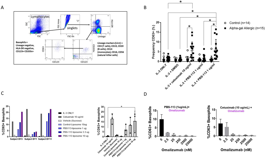
To determine whether alpha-gal-containing glycolipid could functionally trigger basophil degranulation, we used an indirect basophil activation test (BAT) and measured upregulation in the frequency of CD63+ basophils as a marker of basophil activation. Donor basophils within peripheral blood mononuclear cells (PBMCs) from a non-alpha-gal allergic subject were stripped of IgE with cold lactic acid and then sensitized with plasma from control or alpha-gal allergic (AGA) subjects. Lactic acid stripping of primary human basophils resulted in a 25-fold decrease in surface-bound IgE, but did not affect surface expression of the human FcεRIα (Figure E2 in this article’s Online Repository). Sensitized cells were exposed to IL-3 alone or IL-3 and one of the following stimuli for 30 minutes: dimethylsulfoxide (DMSO) carrier, crosslinking anti-IgE antibodies, the alpha-gal-containing glycoprotein cetuximab, alpha-gal-containing glycolipid PBS-113, or glycolipid PBS-112 (contains galactose-α-1,4-galactose). As we had limited access to the naturally occurring glycolipids Gb3 and iGb3, we used their synthetic analogs, PBS-112 and PBS-113, in these experiments. Glycolipids were not loaded onto CD1d, but were added directly to the media. CD63 expression on Lineage−HLA-DR−CD123+CD203c+ basophils was assessed by flow cytometry (Figure 1A). We found that the frequency of CD63+ basophils increased significantly following sensitization with AGA plasma and stimulation with alpha-gal-containing glycolipid (p<0.05, Figure 1B). PBS-112, which does not contain galactose-α-1,3-galactose alpha-gal moieties, did not affect CD63 expression (Figure 1B). We have previously shown that when alpha-gal allergic subjects undergo mammalian meat challenge, the frequency of circulating basophils that upregulate cell-surface expression of CD63 peaks at 4 hours after challenge. Notably, the frequency of CD63+ basophils also increased in some non-alpha-gal allergic controls following meat challenge (1). Similarly, in our indirect BAT we did see modest increases in the frequency of CD63+ donor basophils sensitized with control plasma and challenged with alpha-gal-containing compounds. We speculate that this could be due to signaling through activating Fcγ receptors on basophils bound to alpha-gal-specific IgG. However, taken in aggregate, activation of donor basophils by alpha-gal-containing compounds was more robust when donor basophils were sensitized with alpha-gal allergic plasma (Figure 1B).
Figure 1. Glycolipid-Mediated Basophil Activation in Alpha-Gal Allergy.
(A) Flow cytometry gating strategy used to identify activated Lineage−HLA-DR−CD123+CD203c+CD63+ basophils. (B) Frequency of CD63+ activated donor basophils increases when PBMCs are sensitized with alpha-gal allergic plasma and stimulated with alpha-gal-containing glycolipid PBS-113. Glycolipids were not loaded onto CD1d, but added directly to media. Controls = open circles, n=14; Alpha-gal allergic = black squares, n=15. *p<0.05 by Mann-Whitney Test. (C) Frequency of CD63+ activated donor basophils increases when PBMCs are sensitized with alpha-gal allergic plasma and stimulated with increasing doses of alpha-gal-containing liposome (PBS113-Liposome). Individual subject responses are shown on the left and pooled results using plasma from 3 alpha-gal allergic subjects are shown on the right. *p<0.05 by 1-way ANOVA followed by Dunnett’s multiple comparison’s test. (D) Basophil activation mediated by alpha-gal-containing glycolipid (PBS-113) and glycoprotein (cetuximab) is impaired in the presence of omalizumab, a monoclonal antibody against IgE. Pooled results from 2 alpha-gal allergic subjects.
Surprisingly, using this in vitro system, we did not observe a significant correlation between the frequency of CD63+ cells and alpha-gal-sIgE levels or alpha-gal-sIgE and total IgE ratio despite the broad range of alpha-gal-sIgE in the plasma samples used (0.27 to 96 IU/ml, Figure E3, A–D, in this article’s Online Repository). However, a significant practical limitation to this in vitro system is the variability in donor leukocyte preparations, in native IgE stripping and IgE loading, and in cellular response to IL-3 priming across the pooled experiments. Taken together, these factors likely explain why we did not observe a correlation.
The logarithm of a compound’s partition coeeficient between n-octanol and water (c-log P) is an established means of measuring a compound’s hydrophilicity, with low hydrophilicity corresponding to high c-log P values. The c-log P of PBS-113 is 13.1, indicating that it will not be monomeric in aqueous solution, but will be associated with other lipids, most stably in lipid bilayers. This suggests that when alpha-gal-containing glycolipids are added directly to aqueous media containing sensitized, primary human basophils, they are likely to aggregate into lipid bilayers that could serve as multivalent antigen sources that could crosslink alpha-gal-specific IgE-FcεRI complexes. To explore this hypothesis, we generated blank control liposomes and liposomes containing PBS-113 (PBS113-liposome, Table E2, in this article’s Online Repository), both approximately 200 nm in size, and used these particles to stimulate basophils sensitized with plasma containing alpha-gal-specific IgE. We found that compared to stimulation with control liposomes or the sucrose vehicle, the frequency of CD63+ basophils increased following stimulation with PBS113-liposome in a dose-dependent manner (Figure 1C). This demonstrates that alpha-gal-containing glycolipid incorporated into stable lipid bilayers (PBS113-liposome) served as a multivalent source of glycolipid antigen with the ability to crosslink surface-bound IgE on human basophils. Notably, the hapten dinitrophenyl incorporated into phospholipid liposomes has been shown to induce degranulation of rat basophil leukemia cells sensitized dinitrophenyl-specific IgE(9). Our data expand and reinforce this earlier observation, but within the context of glycolipid-mediated activation of primary human basophils sensitized with a glycan-specific IgE.
Basophil stimulation took place in 75% RPMI media which contains no lipids or proteins and 25% human plasma vol/vol. The protein concentration of this hydrophilic stimulation environment is lower than in whole blood. The structures PBS-113 forms that allow it to stimulate appropriately sensitized basophils in this low protein environment may also form in the higher protein environment of plasma or whole blood. However, PBS-113 stimulation in this setting may be muted due to decreased alpha-gal glycolipid multimerization and increased binding of alpha-gal glycolipids to plasma proteins present at higher concentrations.
To investigate whether basophil activation was IgE-dependent, donor basophils were stripped of IgE and incubated with plasma from alpha-gal allergic subjects in the presence of increasing concentrations of the monoclonal blocking anti-IgE antibody omalizumab. We found that the frequency of CD63+ basophils declined as the concentration of omalizumab increased (Figure 1D). Moreover, omalizumab blocked basophil activation induced by alpha-gal-containing glycolipid (PBS-113) or glycoprotein (cetuximab) antigens (Figure 1D). These data suggest that a 30-minute incubation with alpha-gal-containing compounds activate appropriately sensitized basophils in an IgE-dependent fashion and that this effect is independent of the form in which the glycan antigen is presented.
To our knowledge, this is the first demonstration that mammalian glycolipid can activate allergic effector cells via surface-bound specific IgE. Given the delayed reaction onset after mammalian meat ingestion in alpha-gal allergic individuals, perhaps failure of antigen to appear rapidly in circulation or packaging of immunogenic lipids with plasma proteins or CD1d glycolipid antigen-presenting molecules delays allergic effector cell activation, possibly explaining the delay in allergic symptoms. These results suggest a unique role for glycolipid rarely described in IgE-mediated food allergy.
Supplementary Material
Acknowledgments:
We thank Drs. Michael Kulis, Andrew Spector and Erin Steinbach for critical review of this manuscript. We thank Dr. Kelly Orgel, Dr. Ping Ye, Lisa J. Workman, Gerald F.M. Watts and the UNC Flow Cytometry Core Facility for technical assistance. We thank the NIH Tetramer Core Facility for providing CD1d monomers.
Funding: This work was supported by NIH grant UM1AI30936 to the UNC Food Allergy Initiative and R01AI135049. Dr. Iweala was supported by NIH grant T32AI007062 and a 2019 Thurston Arthritis Research Center Pilot Award. Dr. Brennan was supported by the NIH grant K08AI102945 and generous support from the Goodfellow and Karol families. The UNC Flow Cytometry Core Facility is supported in part by NIH grant P30CA016086 Cancer Center Core Support Grant to the UNC Lineberger Comprehensive Cancer Center. Research reported in this publication was supported in part by the North Carolina Biotech Center Institutional Support Grant 2017-IDG-1025 and by the National Institutes of Health 1UM2AI30836-01. The funders had no role in study design, data collection and analysis, decision to publish, or preparation of the manuscript. The content is solely the responsibility of the authors and does not necessarily represent the official views of the National Institutes of Health.
Footnotes
Conflicts of Interest: The authors declare that we have no relevant conflicts of interest.
Publisher's Disclaimer: This is a PDF file of an unedited manuscript that has been accepted for publication. As a service to our customers we are providing this early version of the manuscript. The manuscript will undergo copyediting, typesetting, and review of the resulting proof before it is published in its final form. Please note that during the production process errors may be discovered which could affect the content, and all legal disclaimers that apply to the journal pertain.
References:
- 1.Commins SP, James HR, Stevens W, Pochan SL, Land MH, King C, et al. Delayed clinical and ex vivo response to mammalian meat in patients with IgE to galactose-alpha-1,3-galactose. J Allergy Clin Immunol. 2014;134(1):108–15. [DOI] [PMC free article] [PubMed] [Google Scholar]
- 2.Steinke JW, Pochan SL, James HR, Platts-Mills TAE, Commins SP. Altered metabolic profile in patients with IgE to galactose-alpha-1,3-galactose following in vivo food challenge. J Allergy Clin Immunol. 2016;138(5):1465–7 e8. [DOI] [PMC free article] [PubMed] [Google Scholar]
- 3.Tordesillas L, Cubells-Baeza N, Gomez-Casado C, Berin C, Esteban V, Barcik W, et al. Mechanisms underlying induction of allergic sensitization by Pru p 3 Clin Exp Allergy. 2017. [DOI] [PMC free article] [PubMed] [Google Scholar]
- 4.Jyonouchi S, Abraham V, Orange JS, Spergel JM, Gober L, Dudek E, et al. Invariant natural killer T cells from children with versus without food allergy exhibit differential responsiveness to milk-derived sphingomyelin. J Allergy Clin Immunol. 2011;128(1):102–9 e13. [DOI] [PMC free article] [PubMed] [Google Scholar]
- 5.Cox D, Fox L, Tian R, Bardet W, Skaley M, Mojsilovic D, et al. Determination of cellular lipids bound to human CD1d molecules. PLoS One. 2009;4(5):e5325. [DOI] [PMC free article] [PubMed] [Google Scholar]
- 6.Brennan PJ, Brigl M, Brenner MB. Invariant natural killer T cells: an innate activation scheme linked to diverse effector functions. Nat Rev Immunol. 2013;13(2):101–17. [DOI] [PubMed] [Google Scholar]
- 7.Yin N, Long X, Goff RD, Zhou D, Cantu C 3rd, Mattner J, et al. Alpha anomers of iGb3 and Gb3 stimulate cytokine production by natural killer T cells. ACS Chem Biol. 2009;4(3):199–208. [DOI] [PMC free article] [PubMed] [Google Scholar]
- 8.Erwin EA, Custis NJ, Satinover SM, Perzanowski MS, Woodfolk JA, Crane J, et al. Quantitative measurement of IgE antibodies to purified allergens using streptavidin linked to a high-capacity solid phase. J Allergy Clin Immunol. 2005;115(5):1029–35. [DOI] [PubMed] [Google Scholar]
- 9.Cooper AD, Balakrishnan K, McConnell HM. Mobile haptens in liposomes stimulate serotonin release by rat basophil leukemia cells in the presence of specific immunoglobulin E. J Biol Chem. 1981;256(18):9379–81. [PubMed] [Google Scholar]
Associated Data
This section collects any data citations, data availability statements, or supplementary materials included in this article.


