PLOS ONE logoLink to PLOS ONE
. 2021 Mar 9;16(3):e0246244. doi: 10.1371/journal.pone.0246244

EZH2 inhibition decreases neuroblastoma proliferation and in vivo tumor growth

Laura V Bownes 1,#, Adele P Williams 1,#, Raoud Marayati 1, Laura L Stafman 1, Hooper Markert 1, Colin H Quinn 1, Nikita Wadhwani 1, Jamie M Aye 2, Jerry E Stewart 1, Karina J Yoon 3, Elizabeth Mroczek-Musulman 4, Elizabeth A Beierle 1,*
Editor: Joe W Ramos5
PMCID: PMC7942994  PMID: 33690617

Abstract

Investigation of the mechanisms responsible for aggressive neuroblastoma and its poor prognosis is critical to identify novel therapeutic targets and improve survival. Enhancer of Zeste Homolog 2 (EZH2) is known to play a key role in supporting the malignant phenotype in several cancer types and knockdown of EZH2 has been shown to decrease tumorigenesis in neuroblastoma cells. We hypothesized that the EZH2 inhibitor, GSK343, would affect cell proliferation and viability in human neuroblastoma. We utilized four long-term passage neuroblastoma cell lines and two patient-derived xenolines (PDX) to investigate the effects of the EZH2 inhibitor, GSK343, on viability, motility, stemness and in vivo tumor growth. Immunoblotting confirmed target knockdown. Treatment with GSK343 led to significantly decreased neuroblastoma cell viability, migration and invasion, and stemness. GSK343 treatment of mice bearing SK-N-BE(2) neuroblastoma tumors resulted in a significant decrease in tumor growth compared to vehicle-treated animals. GSK343 decreased viability, and motility in long-term passage neuroblastoma cell lines and decreased stemness in neuroblastoma PDX cells. These data demonstrate that further investigation into the mechanisms responsible for the anti-tumor effects seen with EZH2 inhibitors in neuroblastoma cells is warranted.

Introduction

Neuroblastoma, a neural crest tumor, continues to be responsible for over 15% of all pediatric cancer deaths [1]. Children with high-risk disease fare the most poorly, and minimal advances have been made in improving their outcomes [2]. Novel pathways and targets must be investigated to provide innovative therapeutic interventions for these children.

Enhancer of Zeste Homolog 2 (EZH2), a SET domain-containing histone methyltransferase, has become a target of interest in numerous malignancies including glioblastoma, leukemia, ovary, lung, colon, and breast cancers [3]. EZH2 belongs to the catalytic subunit of the polycomb repressive complex 2 (PRC2) along with two additional proteins, embryonic ectoderm development (EED) and suppressor of zeste 12 (SUZ12). PRC2 mediates gene silencing primarily by regulating chromatin structure, and consequently, gene expression. PRC2 catalyzes trimethylation by removing a methyl group from S-adenosyl methionine (SAM) to histone H3 lysine 27 (H3K27me3), which is an important epigenetic factor determining stem cell differentiation [3,4].

The oncogenic role of EZH2 is defined by the methylation of H3K27 of tumor suppressor genes, and EZH2 has been implicated in other tumorigenic pathways including β-catenin, Ras, and nuclear factor kappa B (NF-κB) [3,5]. Overexpression of EZH2 has been shown to be a marker of advanced disease in prostate and breast cancers while inactivating mutations suggest a worse prognosis in myeloid neoplasms [3,6]. In neuroblastoma, it has been shown that high expression of EZH2 correlated with poor prognosis [7]. Other investigators demonstrated that suppression of PRC2 subunits in neuroblastoma decreased tumor growth in vitro and in vivo [8]. EZH2 upregulation has been demonstrated to activate the Src kinase pathway in cancer, in addition to various other kinase dependent pathways [3]. Due to the evidence for the role of EZH2 in tumorigenesis, several EZH2 inhibitors have been developed and are in various stages of evaluation, including GSK343, which is a SAM-competitive inhibitor of EZH2 [5].

Based on the findings supporting the role of EZH2 in promoting tumorigenesis and its correlation with poor outcome in neuroblastoma, we hypothesized that blocking EZH2 function in neuroblastoma cell lines would result in decreased proliferation and motility in vitro and impede tumor growth in vivo. Further, EZH2 has been shown to activate Src, a protein kinase upstream from focal adhesion kinase (FAK). FAK, a nonreceptor protein tyrosine kinase, has been shown to play an important role in neuroblastoma tumorigenesis [911]. We further hypothesized that EZH2 inhibition with GSK343 would affect FAK expression.

Materials and methods

Cells and cell culture

The human neuroblastoma cell lines SK-N-AS (CRL-2137) and SK-N-BE(2) (CRL-2271) were obtained from American Type Culture Collection (ATCC, Manassas, VA). The isogenic MYCN- SH-EP and MYCN+ WAC(2) cell lines were a generous gift from Dr. M. Schwab (Deutsches Krebsforschungszentrum, Heidelberg, Germany), and have been described previously in detail [12]. Cell lines were maintained under standard culture conditions at 37°C and 5% CO2 in the following media: Dulbecco’s modified Eagle’s medium (DMEM, 30–2601, ATCC) containing 10% fetal bovine serum (FBS, Hyclone, Suwanee, GA), 4 mM L-glutamine (Thermo Fisher Scientific Inc., Waltham, MA), 1 μM non-essential amino acids, and 1 μg/mL penicillin/streptomycin (Gibco, Carlsbad, CA) for SK-N-AS cells; and a 1:1 mixture of Eagle’s minimum essential medium (30–2003, ATCC) and Ham’s F-12 medium (30–2004, ATCC) with 10% fetal bovine serum (Hyclone), 2 mM L-glutamine (Thermo Fisher Scientific), 1 μM non-essential amino acids, and 1 μg/mL penicillin/streptomycin (Gibco) for SK-N-BE(2) cells. SH-EP and WAC(2) cell lines were maintained in RPMI 1640 medium (Corning Inc., Corning, NY) supplemented with 10% fetal bovine serum (Hyclone) and 1 μg/mL penicillin/streptomycin (Gibco). All four cell lines were verified within the last 12 months using short tandem repeat analysis [University of Alabama at Birmingham (UAB) Genomics Core, Birmingham, AL], and deemed free of Mycoplasma infection. The human neuroblastoma patient-derived xenolines (PDXs), COA3 and COA6, were utilized. These PDXs have been previously described in detail [9]. Briefly, individual PDX cells were obtained by dissociating the xenograft tumors using a Tumor Dissociation Kit (Miltenyi Biotec, San Diego, CA) per manufacturer’s protocol, and resuspending in neurobasal medium (NB, Life Technologies, Carlsbad, CA) supplemented with B-27 without Vitamin A (Life Technologies), N2 (Life Technologies), L-glutamine (2 mM), epidermal growth factor (10 ng/mL, Miltenyi Biotec), fibroblast growth factor (10 ng/mL, Miltenyi Biotec), amphotericin B (250 μg/mL), and gentamicin (50 μg/mL). Following dissociation, cells were maintained at 37°C with 5% CO2 overnight prior to use for experimentation. Real-time PCR was performed to assess the percentage of human and mouse DNA contained in the PDXs to ensure that the tumors did not harbor excessive murine contamination and had not undergone a transformation to a murine tumor (TRENDD RNA/DNA Isolation and TaqMan QPCR/Genotyping Core Facility, UAB, Birmingham, AL). PDX cells were also verified within the last 12 months using short tandem repeat analysis (UAB Genomics Core). Details regarding patient demographics are provided in S1 Table.

Establishing patient-derived xenolines

The PDX program was approved by the University of Alabama at Birmingham (UAB) Institutional Review Board (X130627006), and the studies were conducted at the University of Alabama at Birmingham, Children’s Hospital of Alabama. From November 2013 through January 2014, pediatric patients (0–21 years) with tumors suspected to be neuroblastoma were identified in the pediatric hematology/oncology or surgery clinics or after admission to the Children’s Hospital of Alabama. Parents of the children were approached, and the study was thoroughly explained to them. Written informed consent was obtained from the patients’ parents prior to collection of tumor tissue. When appropriate, written assent was also obtained from the patients. Consents and assents were witnesses by adults who were independent from the research team and not involved in the studies. There were no specific inclusion/exclusion criteria for the study. COA3 was a MYCN amplified, high-risk primary tumor originating in a female child and COA6 was a MYCN amplified, high-risk primary tumor originating in a male child (S1).

For the PDX studies, all animal experiments were approved by the University of Alabama at Birmingham (UAB) Institutional Animal Care and Use Committee (IACUC-09186) and were conducted within institutional, national and NIH guidelines. Neuroblastoma tumor tissue was obtained fresh from patients with primary tumors and kept in Roswell Park Memorial Institute (RPMI) 1640 medium on ice for transport. Tumor chunks were transplanted subcutaneously into the flank of female NOD SCID mice (Envigo, Prattville, AL). Tumor volumes were measured with calipers and calculated with the standard formula (width2 × length)/2, where the width was the smallest measurement. When tumors reached 2000 mm3, they were harvested, chopped, and sequentially implanted from animal to animal for xenoline expansion. Separate portions of the tumor were dissociated for experiments.

Reagents and antibodies

GSK343 (S7164) was purchased from Selleckchem (Houston, TX). Primary antibodies used for Western blotting included the following: rabbit anti-EZH2 (5246S), anti-H3 (4499S, clone D1H2), anti-FAK (71433S) and anti-H3K27me3 (9733S, clone C36B11) from Cell Signaling (Danvers, MA); anti-FAK(C-20) (sc-558) from Santa Cruz (Santa Cruz Biotechnology, Dallas, TX); anti-FAK (05–537, clone 4.47) from Millipore (EMD Millipore, Burlington, MA); and mouse anti-β-actin from Sigma (A1978, Sigma Aldrich, St. Louis, MO), anti-GAPDH (MAB374, clone 6C5) from Millipore and anti-MYCN from Santa Cruz (sc-53993).

Immunoblotting

Following treatment, cells were lysed on ice in a buffer consisting of 50 mM Tris-HCl (pH 7.4), 150 mM NaCl, 1 mM EDTA, 1% Triton x-100, 1% sodium deoxcycholate, 0.1% SDS, phosphatase inhibitor (P5726, Sigma Aldrich), protease inhibitor (P8340, Sigma Aldrich), and phenylmethylsulfonyl fluoride (PMSF, P7626, Sigma Aldrich) for 30 minutes. Lysates were centrifuged at 14 000 rpm for 30 minutes at 4°C. Protein concentrations were determined using a Micro BCA Protein Assay Kit (Thermo Fisher Scientific). Proteins were separated on SDS-PAGE gels by electrophoresis and transferred to Immobilon®-P polyvinylidene fluoride (PVDF) transfer membrane (EMD Millipore). Precision Plus Protein Kaleidoscope Standards (161–0375, Bio-Rad, Hercules, CA) molecular weight markers were used to confirm expected size of target proteins. Antibodies were used per the manufacturers’ recommended protocol. Samples were visualized by enhanced chemiluminescence (ECL) using Luminata Classico or Luminata Crescendo Western horseradish peroxidase (HPR) substrates (EMD Millipore). Anti-β-actin or vinculin served as an internal control to ensure equal protein loading.

Cell proliferation and viability assays

Proliferation was assessed using the CellTiter 96® Aqueous One Solution Cell Proliferation assay (Promega, Madison, WI). Cells were treated with increasing concentrations of GSK343 (0, 5, 15, 25 μM) for 24 hours and plated (5 × 10³ cells/well) onto 96-well plates. After 24 hours, CellTiter 96® dye (10 μL) was added to each well and the absorbance was measured at 490 nm using a microplate reader (Epoch Microplate Spectrophotometer, BioTek Instruments, Winooski, VT). Viability was evaluated using an alamarBlue® assay (Thermo Fisher Scientific). Cells were treated with increasing concentrations of GSK343 (0, 5, 15, 25 μM) for 24 hours, plated (1.5 × 10³ cells) onto 96-well plates and after 24 hours, 10 μL of alamarBlue® dye was added to each well. The plates were read at using a microplate reader (Epoch Microplate Spectrophotometer) to detect the absorbance at 570 nm, using 600 nm as a reference wavelength. Proliferation and viability experiments were completed with at least three biologic replicates and data reported as fold change ± standard error of the mean (SEM).

Monolayer wound healing (scratch) assay

Effects of GSK343 on SK-N-AS and SK-N-BE(2) cell migration was evaluated utilizing a monolayer wound healing assay. Cells were treated for 24 hours with GSK343 (0, 15 μM) then plated and allowed to reach 80% confluence. A sterile 200 μL pipette tip was employed to make a standard scratch in the well. Images were obtained of the scratch wound at 0, 12, and 24 hours. The area of the open wound in pixels was quantified using the ImageJ MRI Wound Healing Tool [13]. Data were reported as fold change scratch area ± SEM and compared between groups. Monolayer wounding assays were not completed on COA6 cells as they do not attach in cell culture.

Animal statement

Animal experiments were approved by the University of Alabama at Birmingham (UAB) Institutional Animal Care and Use Committee (IACUC-09355) and were conducted within institutional, national, and NIH guidelines. The Department of Comparative Medicine through the Animal Resources Program (ARP) at UAB manages a fully accredited (AAALAC) animal laboratory. 6-week-old female athymic nude mice (Envigo, Prattville, AL) were maintained in a pathogen-free facility with 12-hour light/dark cycles, static conventional housing, and ad libitum access to Harlan Rodent Diet® Teklad 4% fat mouse/rat chow (Envigo) and water. Mice were provided cardboard tubes and wood chew sticks for environmental enrichment. Mice were humanely euthanized by a two-step method by CO2 inhalation followed by cervical dislocation. The in vivo tumors were measured three times per week, and weights measured weekly. After tumor cell injections, the animals were monitored on a daily basis by both our laboratory staff and the animal welfare veterinary staff. Animals that were humanely euthanized with the method listed above were done so for the following IACUC criteria: (i) body condition score less than 2; (ii) weight loss greater than 10% of their body weight; (iii) loss of grooming behavior; (iv) cessation of eating or drinking; (v) tumor size over 2 cm3; (vi) ulcerated tumor; (vii) tumor hindering ambulation. The animals did not undergo any surgical intervention prior to euthanasia for tumor harvest. We utilized 14 animals for the in vivo tumor growth study outlined below and 21 animals for propagating the xenolines.

In vivo tumor growth

SK-N-BE(2) (1.8 × 10⁶) cells in 25% Matrigel (Corning Inc.) were injected into the right flank of 6-week-old, female, athymic nude mice (n = 7 per group) (Envigo). Tumors were measured twice weekly and tumor volumes calculated with the formula (width2 × length)/2, with width being the smallest measurement. Once tumors were palpable (100 mm3), the animals were randomized to receive either 100 μL of sterile phosphate buffered saline (PBS, vehicle) or GSK343 (10 mg/kg/day in 100 μL PBS) once daily via intraperitoneal (IP) injection for 21 days. The GSK343 dosing was based on previously published animal data [4,14]. Flank tumors were measured three times per week, and tumor volumes calculated as described above. The animals were humanely euthanized after 21 days of treatment or when IACUC parameters were met.

Cell migration and invasion assays

Cell migration and invasion assays were performed with COA3 and COA6 human neuroblastoma PDX cells using 6.5 mm Transwell® inserts with 8 μM pore polycarbonate membrane (Corning Inc.). For both assays, the bottoms of the inserts were coated with collagen Type I (10 mg/mL, 50 μL) for 4 hours at 37°C. Additionally, for invasion assays, the top sides of the inserts were coated with Matrigel (1 mg/mL, 50 μL, Corning Inc.) for 4 hours at 37°C. Cells were pretreated with GSK343 (0, 10 μM) for 24 hours and then 4.0 × 10⁴ cells plated into the top portion of the insert. The bottom well contained 10% FBS as a chemo-attractant. Cells were allowed to migrate for 72 hours and invade for 1 week. The inserts were then fixed in 3% paraformaldehyde and stained with crystal violet. SPOT Basic 5.2 (Diagnostic Instruments Inc., Sterling Heights, MI) imaging software was used to photograph the inserts at predetermined locations with a microscope at 40 × and cells quantified using ImageJ software (Ver 1.49, available online at http://imagej.nih.gov/ij). Experiments were repeated in triplicate and migration and invasion reported as fold change ± SEM.

In vitro limiting dilution tumorsphere assay

To determine if GSK343 disrupted the stem cell-like phenotype, tumorsphere forming ability was assessed with in vitro limiting dilution assays [15]. Conditioned COA3 and COA6 media was harvested from untreated cells in culture. Single cells were plated in conditioned media onto 96-well low attachment plates using serial dilutions with 5000, 1000, 100, 50, 20, 10, and 1 cell per well. Cells were then treated with GSK343 (0, 5 μM). A week later, the number of wells containing tumorspheres in each row was counted by a single researcher and the results were analyzed using the extreme limiting dilution analysis software (http://bioinf.wehi.edu.au/software/elda/).

Real-time PCR (qPCR)

The effects of GSK343 on mRNA abundance of known stemness markers were assessed using qPCR. COA6 cells were treated with GSK343 (0, 5 μM) for 72 hours and total cellular RNA was extracted using the RNeasy kit (Qiagen) according to the manufacturer’s protocol. For synthesis of cDNA, 1 μg of RNA was used in a 20-μl reaction mixture utilizing an iScript cDNA Synthesis kit (Bio-Rad) according to the supplier’s instructions. Resulting reverse transcription products were stored at -20°C until further use. For qPCR, SsoAdvanced SYBR® Green Supermix (Bio-Rad) was utilized according to manufacturer’s protocol. Probes specific for the stemness markers Oct4, Nanog, and Sox2, as well as for actin B were obtained (Applied Biosystems, Foster City, CA). qPCR was performed with 10 ng cDNA in 20 μL reaction volume. Amplification was done using an Applied Biosystems 7900HT cycler (Applied Biosystems). Cycling conditions were 95°C for 2 min, followed by 39-cycle amplification at 95°C for 5 secs and 60°C for 30 sec. Experiments were repeated with at least three biologic replicates, and samples were analyzed in triplicate with actin B utilized as an internal control. Data were calculated utilizing the ΔΔCt method [16] and are reported as mean fold change ± SEM.

Immunofluorescence

Cells were plated on glass chamber slides and allowed to attach for 24 hours, treated for 24 hours, then fixed with 4% paraformaldehyde. Cells were permeablized with 0.15% Triton X-100, and the first primary antibody (anti-EZH2, 5246S, Cell Signaling) was added and incubated at room temperature (RT) for 1 hour followed by the addition of the second primary antibody (anti-FAK 4.47, EMD Millipore) that was also incubated for 1 hour at RT. The Alexa Fluor 488 secondary antibody (goat anti-rabbit, A-11034, Invitrogen) was added for 45 minutes at RT. After washing, the second secondary antibody, Alexa Fluor 594 (goat anti-mouse, A-11005, Invitrogen), was added and incubated as above. Prolong® Gold antifade reagent with DAPI (P36931, Invitrogen) was used for mounting. Imaging was performed with a Zeiss LSM 710 Confocal Scanning Microscope with Zen 2008 software (Carl Zeiss MicroImaging, LLC, Thornwood, NY) using a 63× objective with a zoom of 0.9. Fifteen cells per sample were analyzed with 20 images each. Manders overlap coefficients were calculated [17]. Manders coefficients have a value between 0 and 1, with 0 = no overlap and 1 = perfect overlap. These coefficients provide the proportion of overlap of each channel with the other.

Immunoprecipitation

Whole cell lysates were subjected to immunoprecipitation followed by immunoblotting. Briefly, cell lysates were added to Protein A/G agarose beads (Santa Cruz Biotechnology), incubated at 4°C for 1 hour and then spun in the cold to pre-clear the cell lysate. The primary antibody was added to the supernatant and incubated overnight at 4°C. Protein A/G agarose beads were added and incubated for 3 hours and washed on ice. The samples were heated to 100°C for 5 minutes and centrifuged for 1 minute at 14 000 g. Samples (500 μg protein) were loaded onto SDS-PAGE gels and electrophoresed as described above. Rabbit IgG was included on the immunoblots as a control.

Statistical analyses

In vitro experiments were performed at a minimum of three biologic replicates. Data reported as the mean ± SEM. Parametric data between groups was compared using an analysis of variance (ANOVA) or Student’s t-test as appropriate. Non-parametric data were analyzed with Mann-Whitney U test (Wilcoxon Rank Sum Test). Statistical significance was defined as p ≤ 0.05.

Results

EZH2 inhibitor, GSK343, decreased tri-methylation of Histone 3 at Lysine 27

Four long-term passage neuroblastoma cell lines, SK-N-AS, SK-N-BE(2), SH-EP and WAC(2) and two human neuroblastoma PDXs, COA 3 and COA6, were utilized for experimentation. Documentation of expression of EZH2 and the downstream target, tri-methylation of Histone 3 at Lysine 27 (H3K27me3) was necessary prior to further investigation. Immunoblotting revealed that EZH2 and H3K27me3 were both present in both the long-term passage neuroblastoma cell lines (Fig 1A) and the human neuroblastoma PDX cells (Fig 1B and 1C). Treatment with increasing concentrations of GSK343 for 24 hours decreased the tri-methylation of Histone 3 at Lysine 27 and the expression of EZH2 in the PDXs (Fig 1B and 1C), but did not change H3 expression (Fig 1A–1C).

Fig 1. EZH2 inhibitor, GSK343, decreased tri-methylation of Histone 3 at Lysine 27.

Fig 1

(A) Immunoblotting revealed EZH2 and H3K27me3 expression in long-term passage neuroblastoma cell lines. Treatment with increasing concentrations of GSK343 for 24 hours led to a decrease in EZH2 expression with increasing doses. Increasing doses of GSK343 decreased tri-methylation of the EZH2 downstream target, Histone 3 at Lysine 27 (H3K27me3). H3 expression was unchanged. (B, C) Similar to long-term passage cell lines, immunoblotting of the human neuroblastoma PDXs demonstrated a decrease in EZH2 expression with the inhibitor GSK343 as well as a decrease in tri-methylation of Histone 3 at Lysine 27, but H3 was not significantly changed.

GSK343 decreased neuroblastoma proliferation, viability, and motility

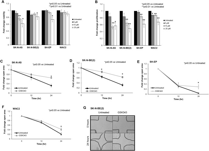
We proceeded to examine the effects of GSK343 on the phenotype of neuroblastoma cells. Viability and proliferation were evaluated with alamarBlue® and CellTiter96®, respectively. Inhibition of EZH2 with GSK343 affected viability in SK-N-AS, SK-N-BE(2) and SH-EP neuroblastoma cell lines (Fig 2A). GSK343 significantly decreased neuroblastoma proliferation (Fig 2B). Information on median lethal dose provided in supplemental S2 Table.

Fig 2. GSK343 decreased neuroblastoma viability, proliferation, and motility.

Fig 2

(A) SK-N-AS, SK-N-BE(2), SH-EP and WAC(2) cells (1.5 × 10³ cells) were treated with increasing concentrations of GSK343 (0, 5, 15, 25 μM) for 24 hours and viability was measured using alamarBlue® assay. GSK343 treatment resulted in decreased viability in SK-N-AS, SK-N-BE(2), and WAC(2) cells. (B) SK-N-AS, SK-N-BE(2), SH-EP and WAC(2) cells (1.5 × 10³ cells) were treated with increasing concentrations of GSK343 (0, 5, 15, 25 μM) for 24 hours and proliferation was measured with using CellTiter® assay. Proliferation was significantly decreased following treatment with GSK343 in all cell lines. (C) SK-N-AS, (D) SK-N-BE(2), (E) SH-EP and (F) WAC(2) cells were treated for 24 hours with GSK343 (15 μM) then plated and allowed to reach 80% confluence. A standard scratch was made in each well and images of the scratch were obtained at 0, 12, and 24 hours. The area of the open wound in pixels was quantified using the ImageJ MRI Wound Healing Tool. By 24 hours, there was a significant decrease in the area of the scratch healed (indicating decreased motility) in cells treated with GSK343 compared to untreated cells. Data were reported as fold change scratch area ± SEM and compared between groups. (G) Representative photomicrographs of SK-N-BE(2) cell wounding assays. GSK343 treated cells (right panel) demonstrated significant reduction in ability to heal the scratch compared to untreated cells (left panel). Data represent at least three biologic replicates.

EZH2 has been shown to affect cell migration and invasion in other malignancies [1820], leading us to examine the role of EZH2 inhibition in neuroblastoma cell motility. Cell motility was evaluated with a monolayer wound healing (scratch assay) in SK-N-AS (Fig 2C), SK-N-BE(2) (Fig 2D and 2G), SH-EP (Fig 2E) and WAC(2) (Fig 2F). There was a significant decrease in the area of the scratch closed, indicating decreased cell motility in cells treated with GSK343. These findings showed that EZH2 affected neuroblastoma viability, proliferation, and motility.

GSK343 decreased tumor growth in vivo

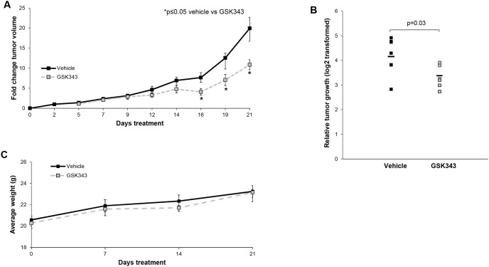
To demonstrate that GSK343 inhibition of EZH2 was relevant in vivo, we proceeded to an animal model. SK-N-BE(2) cells were injected into the right flank of athymic nude mice and animals were monitored for tumor growth. Tumor volumes were measured three times per week. Fold change in tumor volume was significantly smaller in the GSK343 treated animals when compared vehicle treated controls (Fig 3A). Further, relative tumor growth was significantly decreased in mice treated with GSK343 compared to those treated with vehicle (Fig 3B). The GSK343 treatment was well tolerated as demonstrated by constant weight gain in the treated animals (Fig 3C).

Fig 3. GSK343 treatment decreased in vivo tumor volume and growth.

Fig 3

SK-N-BE(2) (1.8 × 10⁶) cells were injected subcutaneously into the right flank of 6-week-old, female, athymic nude mice. Once tumors were palpable (100 mm3), mice were randomized to receive either 100 μL of sterile PBS (vehicle, n = 7) or GSK343 (10 mg/kg/day in 100 μL PBS, n = 7) once daily via intraperitoneal (IP) injection for 21 days. Tumor volumes were monitored three times per week. (A) Animals treated with GSK343 had significantly decreased fold change in tumor volumes compared to mice treated with vehicle. (B) Animals treated with GSK had significantly decreased relative tumor growth compared to those treated with vehicle. (C) Mice were weighed at the beginning of the experiment, weekly, and at the time of euthanasia. There was no significant difference in weights between animals that received vehicle compared to those that received GSK343.

GSK343 decreased neuroblastoma PDX viability, proliferation, and motility

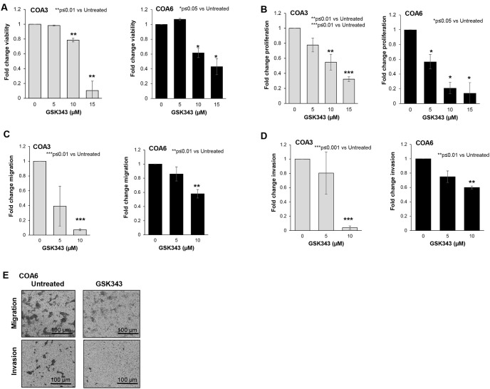
Since long-term passage cell lines may not completely reflect the actual clinical condition [21], we employed human neuroblastoma PDX cells to further study the effects of EZH2 inhibition on neuroblastoma. We again examined viability and proliferation. Viability (Fig 4A) and proliferation (Fig 4B) were significantly decreased following treatment with GSK343. Information on median lethal dose provided in S2. To investigate whether GSK343 affected cell motility, modified Boyden chamber assays were utilized to examine migration and invasion in COA 3 and COA6 PDX cells. There was a significant decrease in cell migration (Fig 4C) and invasion (Fig 4D) with GSK343 treatment. Representative photomicrographs of migration and invasion inserts for COA6 cells are presented in Fig 4E. Black scale bars represent 100 μm.

Fig 4. GSK343 decreased viability and proliferation in human neuroblastoma PDX cells.

Fig 4

(A) COA3 and COA6 (1.5 × 10³) cells were treated with increasing concentrations of GSK343 (0, 5, 15, 25 μM) for 24 hours and viability was measured with alamarBlue® assay. GSK343 treatment resulted in significantly decreased viability. (B) COA3 and COA6 (1.5 × 10³) cells were treated with increasing concentrations of GSK343 (0, 5, 15, 25 μM) for 24 hours and proliferation was measured using CellTiter® assay. Proliferation was significantly decreased following treatment with GSK343.(C) COA3 and COA6 cells were treated for 24 hours with increasing doses of GSK343 (0, 5, 10 μM) and plated (4.0 × 10⁴ cells) in Transwell® inserts. Cells were allowed to migrate for 72 hours. Treatment with GSK343 resulted in significantly decreased migration. (D) After 24 hours of treatment with increasing concentrations of GSK343 (0, 5, 10 μM), COA3 and COA6 cells (4.0 × 10⁴) were plated in Transwell® inserts and allowed to invade for 1 week through a Matrigel layer. Invasion was significantly decreased in the cells treated with GSK343. (E) Representative photomicrographs of migration and invasion inserts for untreated (left panel) and GSK343 treated (right panel) COA6 cells. Scale bars represent 100 μm. Migration was reported as mean percent area positive ± SEM and invasion reported as mean number of cells ± SEM. Experiments were repeated with at least three biologic replicates.

GSK343 decreased stemness in neuroblastoma PDX cells

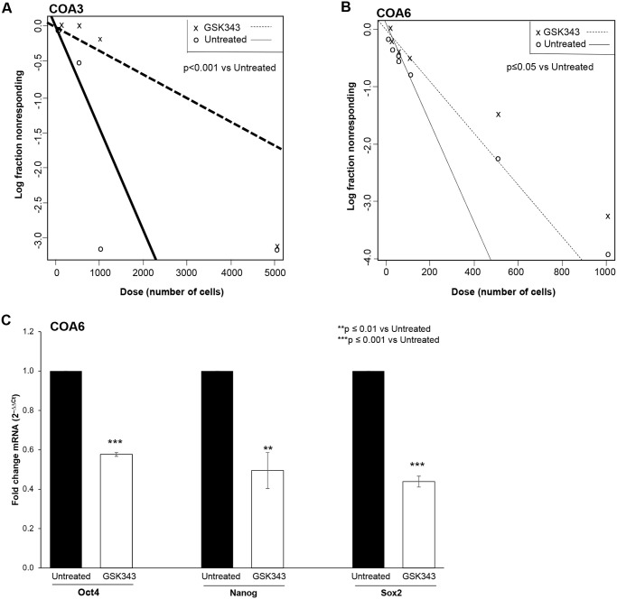
EZH2 has been shown to play a role in maintaining the cancer stem cell-like phenotype in other cancer types [3]. We wished to determine if GSK343-induced EZH2 inhibition reduced the stem cell-like phenotype in the neuroblastoma PDXs, COA3 and COA6. An extreme limiting dilution assay was used to investigate the effect of GSK343 on the ability of PDX cells to form tumorspheres, a marker of cancer cell stemness. When compared to untreated cells, PDX cells treated with GSK343 (0, 5 μM) had significantly decreased sphere forming ability (Fig 5A and 5B), indicating that the cells were less stem cell-like. The mRNA abundance of the stem cell makers, Oct4, Nanog, and Sox2 was examined with qPCR in the COA6 cells treated with GSK343 (0, 5 μM). At 72 hours, GSK343 resulted in a significant decrease in the mRNA abundance of all three markers (Fig 5C). These data provided evidence that GSK343 decreased the cancer stem cell-like phenotype in these human neuroblastoma PDX cells.

Fig 5. GSK343 decreased tumor stemness.

Fig 5

(A) COA3 and (B) COA6 PDX cells were plated as single cell suspension in serum-free, conditioned media onto 96-well low attachment plates using serial dilutions with 5000, 1000, 100, 50, 20, 10, and 1 cell per well. Cells were treated with GSK343 (0, 5 μM). After one week, the number of wells containing tumorspheres in each row was counted by a single researcher and the results were analyzed using the extreme limiting dilution analysis software (http://bioinf.wehi.edu.au/software/elda/). Treatment with GSK343 significantly decreased tumorsphere formation by both the COA3 (A) and COA6 (B) cells, representing a decrease in stem cell-like phenotype. (C) COA6 cells were treated with GSK343 (0, 5 μM) for 72 hours. qPCR demonstrated a significant decrease in mRNA abundance of known stemness markers Oct4, Nanog, and Sox2 following GSK343 treatment compared to untreated cells. Data presented represent results from at least three biologic replicates.

Inhibition of EZH2 led to decreased FAK expression

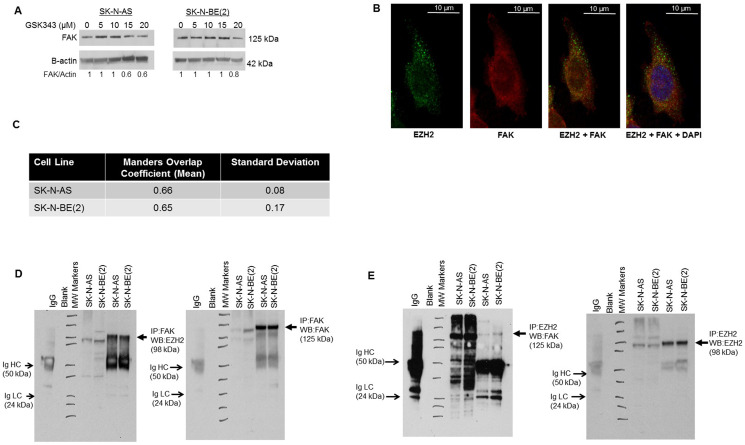
EZH2 has been shown to be associated with Src kinase [3,22,23]. Src activation resulted in increased EZH2 expression, and Src inhibition decreased EZH2 expression in mammary malignancies [3]. We hypothesized that FAK, a Src downstream kinase, may also be affected by GKS343. FAK has been shown to correlate with aggressive neuroblastoma [24], and FAK inhibition resulted in decreased neuroblastoma tumor growth in vivo [25]. Immunoblotting revealed a decrease in FAK expression with increasing doses of GSK343 in long-term passage neuroblastoma cell lines (Fig 6A). Based on these results, dual immunofluorescence staining and confocal microscopy was employed to investigate EZH2 and FAK interaction in neuroblastoma cell lines. Confocal microscopy demonstrated overlap between the two proteins indicating an interaction (Fig 6B and 6C). The Mander’s overlap coefficient [17] for SK-N-AS cells was 0.66, for SK-N-BE(2) cells 0.65 (Fig 6C). These values were greater than 0, indicating staining overlap. Co-immunoprecipitation with EZH2 and FAK antibodies was used to further demonstrate interaction between EZH2 and FAK. Immunoprecipitation with EZH2 followed by immunoblotting for FAK and the reverse, immunoprecipitation with FAK and immunoblotting for EZH2, revealed an interaction between the two proteins (Fig 6D and 6E). These results indicated that EZH2 inhibition led to decreased FAK expression, and EZH2 and FAK interact.

Fig 6. EZH2 and FAK association in neuroblastoma.

Fig 6

(A) SK-N-AS and SK-N-BE(2) cells were treated with increasing doses of GSK343 for 24 hours. Whole cell lysates were examined with immunoblotting. There was a dose dependent decrease in FAK expression. (B) Immunofluorescence staining followed by confocal microscopy was utilized to investigate EZH2 and FAK interaction in SK-N-AS and SK-N-BE(2) cell lines. Representative photographs of SK-N-AS cells are presented. There was an overlap in staining between EZH2 (green, first panel) and FAK (red, second panel), depicted as yellow color (third panel), indicating an interaction. DAPI staining was completed to identify the nucleus (fourth panel). White scale bars represent 10 μm. (C) To quantitate overlap, Manders overlap coefficients were calculated for SK-N-AS (0.66) and SK-N-BE(2) (0.65) cells. These values were greater than 0, providing evidence of an interaction between EZH2 and FAK. (D) Immunoprecipitation with FAK followed by Western blotting for EZH2 was used to demonstrate an interaction between FAK and EZH2 (lanes 6, 7, closed arrows). (E) The reverse, immunoprecipitation for EZH2 followed by Western blotting for FAK revealed an interaction between EZH2 and FAK (lanes 6, 7, closed arrows). IgG served as a control. IgG heavy chain (HC) and light chain (LC) bands are indicated on the blots (open arrows). Whole cell lysates without IP were included on each of the blots (lanes 4, 5).

Discussion

EZH2, a subunit of PCR2, utilizes SAM as a methyl group donor, leading to trimethylation of H3K27 and subsequent transcriptional silencing of target genes. Investigators have found EZH2 to be mutated and/or overexpressed in several malignancies, including prostate, lung, breast, and hepatocellular carcinoma [6], and its overexpression has been shown to be associated with a worse prognosis in neuroblastoma [7]. GSK343 is a competitive inhibitor of SAM. Verma and colleagues found that GSK343 was highly selective for EZH2 over most other methyltransferases tested, with selectivity greater than 1000-fold, with the exception of the highly homologous EZH1, where GSK343 was 60-fold more selective [26]. As such, GSK343 would not be expected to affect the expression of EZH2. In the current study, we found GSK343 treatment in neuroblastoma led to an expected decrease in the EZH2 target, tri-methylation of Histone 3 at Lysine 27 (H3K27me3), but also a decrease in EZH2 expression. Other investigators have documented this finding in various cancer cell lines including cervical cancer [27], colorectal cancer [28], gliomas [4,29], triple negative breast cancer [30], and osteosarcoma [31]. Investigators have hypothesized that changes in c-Myc may be responsible for the effects seen on EZH2. C-Myc has been shown to bind to the EZH2 promoter and regulate EZH2 expression [32]. Xiong and colleagues found that GSK343 decreased expression of c-Myc in osteosarcoma cells [31], leading them to postulate that the effects of GSK343 on c-Myc were responsible for the decreased expression of EZH2 seen with GSK343 treatment [31]. We found MYCN, an important regulator of neuroblastoma, was decreased with increasing concentrations of GSK343 in SH-EP cells (supplemental information, S3). Corvetta et al demonstrated MYCN binding to EZH2 [33], so perhaps a similar mechanism may be in play in the current study and would be an avenue for further research.

The initial phenotypic changes that we explored showed that GSK343 significantly affected proliferation of neuroblastoma cells. Other investigators likewise noted these findings. EZH2 inhibitors were effective at decreasing proliferation of ovarian [34] and cervical cancer cells [27] and of glioma cells [4]. We found that GSK343 impaired neuroblastoma proliferation more than viability in the neuroblastoma cell lines. Other published studies have demonstrated similar findings. Yu and colleagues noted a significant decrease in glioma cell motility after treatment with 5 μM GSK343, but cell viability was not affected until much higher concentrations were used [4]. Similar results were seen in the study of triple negative breast cancer cells where viability never reached 50%, but proliferation was markedly diminished at 2 μM GSK343 [30]. This finding suggests that combining the anti-proliferative effects of GSK343 with a cytotoxic agent could have synergistic or additive effects. Yu et al. investigated GSK343 in combination with adriamycin, a chemotherapy agent, for triple negative breast cancer. The combination therapy resulted in decreased viability in vitro. They also found decreased tumor growth in vivo with the combination treatment when compared to either single agent alone [30]. Stazi and colleagues investigated EZH2 inhibition in combination with standard chemotherapy in glioblastoma with two EZH2 inhibitors and temozolomide. There was decreased viability with the combination treatment compared to EZH2 inhibition alone, suggesting a benefit to adding EZH2 inhibition to cytotoxic chemotherapy [35]. Combining EZH2 inhibition with cytotoxic agents in neuroblastoma could provide an exciting avenue for future studies.

In addition to decreasing cell proliferation and viability, EZH2 affected cell motility in other malignancies [5,28]. Anwar et al. demonstrated that breast cancer metastasis depended upon the phosphorylation of EZH2 at T367. Not only was phosphorylated EZH2 increased in metastatic breast cancer specimens, but they found phosphorylated EZH2 enhanced cell motility and invasion, whereas EZH2 that was not phosphorylated decreased cell proliferation [36]. EZH2 also regulated gain-of-function (GOF) mutations of p53 (mtp53) that promote cancer metastasis. In prostate cancer, the knockdown of EZH2 resulted in a decrease of the protein levels of cancer specific GOF mutations (R273H and R248W) but also decreased cell invasion [37]. Similarly, in metastatic pancreatic cancer, EZH2 increased R248W expression as well as cellular invasion. In our study, the treatment of neuroblastoma cells with GSK343 at concentrations well below the LD50 for the compound significantly decreased neuroblastoma migration and invasion, suggesting that EZH2 may play a role in neuroblastoma metastasis that is independent from simple decreased cell viability.

It is important to note that the SK-N-AS cell line is derived from a MYCN non-amplified tumor while SK-N-BE(2), COA3 and COA6 are MYCN amplified. Further, SH-EP and WAC(2) cell lines are isogenic for MYCN [12] with SH-EP being non-amplified and WAC(2) having MYCN overexpression. MYCN is a proto-oncogene that is associated with high-risk neuroblastoma and, when amplified, is the most important negative prognostic factor in neuroblastoma [10]. Chen et al. showed that MYCN amplified neuroblastoma cells expressed increased levels of EZH2 when compared to non-amplified neuroblastoma cells [8] and showed that treatment with two EZH2 inhibitors, GSK126 and JQEZ5, as well as shRNA knockdown of EZH2 led to decreased tumor growth in MYCN amplified long-term passage cell lines, including SK-N-BE(2) [8]. This group also found SK-N-AS, to be sensitive to EZH2 inhibition and proposed a concurrent oncogenic mutation, NRAS, as potentially driving these effects [8]. Our findings demonstrated EZH2 inhibition with GSK343 decreased the malignant phenotype in MYCN amplified and non-amplified cell lines. The finding that EZH2 inhibition affected both amplified and non-amplified cell lines similarly makes the continued investigation of EZH2 as a therapeutic target for high-risk neuroblastoma crucial and exciting, since the majority of children with high-risk disease does not have MYCN amplified tumors.

In the original studies looking at pharmacokinetics of GSK343 in rodents, it was felt that GSK343 displayed too high a clearance and might not be suitable for in vivo studies [26]. However, our study and those of other investigators have shown in vivo efficacy. In a flank murine model of cervical cancer (SiHa cells), GSK343 treatment resulted in significant reduction in tumor growth [27]. Other investigators have used GSK343 inhibition in studies of neural tumors. Stazi and colleagues utilized GSK343 and showed decreased glioblastoma tumor growth in vivo [35]. Yu et al demonstrated a significant increase in animal survival in mice bearing intracranial U87 glioma tumors when treated with GSK343 compared to vehicle treated animals [4]. In the current study, we found that animals treated with GSK343 had significantly decreased tumor growth, supporting the role for EZH2 inhibition in neuroblastoma. Importantly, we did not see changes in animal weights with GSK343 treatment compared to those treated with vehicle, indicating that decreased animal growth was not the etiology of decreased tumor size.

Cancer cell stemness is a crucial area of investigation due to the thought that these cells may be responsible for cancer cell self-renewal, and thereby recurrent and refractory disease [38,39]. EZH2 may promote stemness by increasing the trimethylation of H3K27 of stemness regulators, thereby decreasing these regulators at the transcriptional level. Zhang found that EZH2 repressed the stemness regulator ATOH8 in hepatocellular carcinoma [40]. In addition, EZH2 interacted with NF-κB, promoting glioma cell proliferation, and the loss of this interaction repressed the self-renewal ability of the glioma cells [41]. Based on these findings, we hypothesized that treatment with GSK343 would lead to reduced neuroblastoma stemness. We showed the effects of GSK343 on stemness by observing decreased tumorsphere formation and decreased mRNA abundance of known stemness markers.

Utilizing confocal microscopy and immunoprecipitation, we demonstrated an interaction between FAK and EZH2. The concept of FAK binding to other proteins is not unfounded. Other investigators have shown interactions between FAK and growth factors, tumor suppressors, and other proteins affecting cancer growth. We have previously shown a direct association between FAK and VEGFR3 [42] and FAK and p53 in neuroblastoma [43]. Sieg showed that FAK associated with PDGF and EGF receptors to promote cancer cell motility [44], and Liu and others demonstrated an association of FAK with IGF-1R in pancreatic cancer cell lines leading to increased cell survival [45]. One of the first investigations of FAK and EZH2’s interaction found both proteins overexpressed in endometrial carcinoma, suggesting that these proteins play a role in a more aggressive tumor phenotype and worse prognosis [22]. Gnani demonstrated an association between FAK and EZH2 by showing that FAK knockdown downregulated EZH2 and the overexpression of FAK increased EZH2 in hepatocellular carcinoma [46]. In light of these previous studies, it would follow that FAK may interact with EZH2 to affect the neuroblastoma cell phenotype.

There are some discrepancies regarding the role of FAK in neuroblastoma. Examination of the Kocak (R2) database indicated that greater PTK2 (FAK) gene abundance is associated with better overall survival in neuroblastoma [47]. However, the studies examining FAK protein expression indicated that higher protein expression was associated with worse disease including MYCN amplification and metastasis [4850]. We have a few explanations for these discrepancies. First, gene expression does not always translate and equate with protein expression due to translational and post-translational modifications of gene products. Second, most of the protein data in the literature indicate a relation between high FAK expression and patients with high-risk disease or amplification of the MYCN oncogene, but these studies focused on MYCN amplified or high-risk disease. Since the gene expression datasets include all comers for the disease including very low, low and intermediate risk patients, there may be factors related to disease stratification that may contribute to the conflicting findings. In fact, in one of the earliest publications investigating FAK protein expression with immunohistochemistry in 70 neuroblastoma patient samples, patient overall or event free survival did not relate to FAK staining in a statistically significant manner. Positive IHC staining for FAK was, however, associated with MYCN amplified disease in high-risk patients.

Conclusion

In the current study, we showed that EZH2 inhibition with GSK343 in neuroblastoma decreased proliferation, viability, motility, and tumor growth in vivo. The data presented also support an association between EZH2 and FAK in neuroblastoma. These findings provide evidence that EZH2 inhibitors and the mechanisms driving their anti-tumor effects warrant further investigation in neuroblastoma.

Supporting information

S1 Fig

(TIF)

S1 Table

(TIF)

S2 Table

(TIF)

S1 Raw images

(PDF)

Acknowledgments

The authors wish to thank Dr. Anita Hjemeland’s laboratory for their assistance with the qPCR. The funding sources had no role in study design, analysis or interpretation of the data, the writing of the manuscript, or in the decision for publication submission.

Data Availability

All relevant data are within the manuscript and its Supporting information files.

Funding Statement

This work was partially funded by institutional grants from the National Cancer Institute including T32 CA229102 (https://grantome.com/grant/NIH/T32-CA229102-01)(LV Bownes, R Marayati), T32 CA183926 (https://grantome.com/grant/NIH/T32-CA183926-01)(AP Williams), T32 CA091078 (https://grantome.com/grant/NIH/T32-CA091078-14) (LL Stafman), 5T32BM00836 (CH Quinn), and P30 AR048311 and P30 AI27667 to the University of Alabama at Birmingham, Flow Cytometry Core (https://www.uab.edu/medicine/cfar/core-facilities/basic-research-core/flow-cytometry-core). Funding was also provided by the Lombardi Cancer Research Fund/Starr Children’s Fund (https://www.lombardifoundation.org/home-annual-report), Sid Strong Foundation (https://sidstrongfound.org/), Elaine Roberts Foundation (https://www.elainerobertsfoundation.org/), and Open Hearts Overflowing Hands (https://openhandsoverflowinghearts.org/) (EA Beierle, JM Aye). The funders had no role in study design, data collection and analysis, decision to publish, or preparation of the manuscript.

References

  • 1.Maris JM. The biologic basis for neuroblastoma heterogeneity and risk stratification. Curr Opin Pediatr. 2005;17(1):7–13. Epub 2005/01/22. 10.1097/01.mop.0000150631.60571.89 [DOI] [PubMed] [Google Scholar]
  • 2.Newman EA, Nuchtern JG. Recent biologic and genetic advances in neuroblastoma: Implications for diagnostic, risk stratification, and treatment strategies. Semin Pediatr Surg. 2016;25(5):257–64. Epub 2016/12/14. 10.1053/j.sempedsurg.2016.09.007 [DOI] [PubMed] [Google Scholar]
  • 3.Han Li C, Chen Y. Targeting EZH2 for cancer therapy: progress and perspective. Curr Protein Pept Sci. 2015;16(6):559–70. Epub 2015/04/10. 10.2174/1389203716666150409100233 [DOI] [PMC free article] [PubMed] [Google Scholar]
  • 4.Yu T, Wang Y, Hu Q, Wu W, Wu Y, Wei W, et al. The EZH2 inhibitor GSK343 suppresses cancer stem-like phenotypes and reverses mesenchymal transition in glioma cells. Oncotarget. 2017;8(58):98348–59. Epub 2017/12/13. 10.18632/oncotarget.21311 [DOI] [PMC free article] [PubMed] [Google Scholar]
  • 5.Kim KH, Roberts CW. Targeting EZH2 in cancer. Nat Med. 2016;22(2):128–34. Epub 2016/02/06. 10.1038/nm.4036 [DOI] [PMC free article] [PubMed] [Google Scholar]
  • 6.Chase A, Cross NC. Aberrations of EZH2 in cancer. Clin Cancer Res. 2011;17(9):2613–8. Epub 2011/03/04. 10.1158/1078-0432.CCR-10-2156 [DOI] [PubMed] [Google Scholar]
  • 7.Li Z, Takenobu H, Setyawati AN, Akita N, Haruta M, Satoh S, et al. EZH2 regulates neuroblastoma cell differentiation via NTRK1 promoter epigenetic modifications. Oncogene. 2018;37(20):2714–27. Epub 2018/03/07. 10.1038/s41388-018-0133-3 [DOI] [PMC free article] [PubMed] [Google Scholar]
  • 8.Chen L, Alexe G, Dharia NV, Ross L, Iniguez AB, Conway AS, et al. CRISPR-Cas9 screen reveals a MYCN-amplified neuroblastoma dependency on EZH2. J Clin Invest. 2018;128(1):446–62. Epub 2017/12/05. 10.1172/JCI90793 [DOI] [PMC free article] [PubMed] [Google Scholar]
  • 9.Stafman LL, Williams AP, Marayati R, Aye JM, Markert HR, Garner EF, et al. Focal Adhesion Kinase Inhibition Contributes to Tumor Cell Survival and Motility in Neuroblastoma Patient-Derived Xenografts. Sci Rep. 2019;9(1):13259. Epub 2019/09/15. 10.1038/s41598-019-49853-z [DOI] [PMC free article] [PubMed] [Google Scholar]
  • 10.Megison ML, Stewart JE, Nabers HC, Gillory LA, Beierle EA. FAK inhibition decreases cell invasion, migration and metastasis in MYCN amplified neuroblastoma. Clin Exp Metastasis. 2013;30(5):555–68. Epub 2012/12/05. 10.1007/s10585-012-9560-7 [DOI] [PMC free article] [PubMed] [Google Scholar]
  • 11.Beierle EA. MYCN, neuroblastoma and focal adhesion kinase (FAK). Front Biosci (Elite Ed). 2011;3:421–33. Epub 2011/01/05. [DOI] [PMC free article] [PubMed] [Google Scholar]
  • 12.Schweigerer L, Breit S, Wenzel A, Tsunamoto K, Ludwig R, Schwab M. Augmented MYCN Expression Advances the Malignant Phenotype of Human Neuroblastoma Cells: Evidence for Induction of Autocrine Growth Factor Activity. Cancer Research. 1990;50(14):4411–6. [PubMed] [Google Scholar]
  • 13.https://imagej.nih.gov/ij/.
  • 14.Zhou J, Huang S, Wang Z, Huang J, Xu L, Tang X, et al. Targeting EZH2 histone methyltransferase activity alleviates experimental intestinal inflammation. Nat Commun. 2019;10(1):2427. Epub 2019/06/05. 10.1038/s41467-019-10176-2 [DOI] [PMC free article] [PubMed] [Google Scholar]
  • 15.Hu Y, Smyth GK. ELDA: extreme limiting dilution analysis for comparing depleted and enriched populations in stem cell and other assays. J Immunol Methods. 2009;347(1–2):70–8. Epub 2009/07/02. 10.1016/j.jim.2009.06.008 [DOI] [PubMed] [Google Scholar]
  • 16.Winer J, Jung CK, Shackel I, Williams PM. Development and validation of real-time quantitative reverse transcriptase-polymerase chain reaction for monitoring gene expression in cardiac myocytes in vitro. Anal Biochem. 1999;270(1):41–9. Epub 1999/05/18. 10.1006/abio.1999.4085 [DOI] [PubMed] [Google Scholar]
  • 17.Manders E, Verbeek F, Aten J. Measurement of co-localization of objects in dual-colour confocal images. Journal of Microscopy. 1993;169(3):375–82. [DOI] [PubMed] [Google Scholar]
  • 18.Suva ML, Riggi N, Janiszewska M, Radovanovic I, Provero P, Stehle JC, et al. EZH2 is essential for glioblastoma cancer stem cell maintenance. Cancer Res. 2009;69(24):9211–8. Epub 2009/11/26. 10.1158/0008-5472.CAN-09-1622 [DOI] [PubMed] [Google Scholar]
  • 19.Ferraro A, Boni T, Pintzas A. EZH2 regulates cofilin activity and colon cancer cell migration by targeting ITGA2 gene. PLoS One. 2014;9(12):e115276. Epub 2014/12/31. 10.1371/journal.pone.0115276 [DOI] [PMC free article] [PubMed] [Google Scholar]
  • 20.Yi X, Guo J, Guo J, Sun S, Yang P, Wang J, et al. EZH2-mediated epigenetic silencing of TIMP2 promotes ovarian cancer migration and invasion. Sci Rep. 2017;7(1):3568. Epub 2017/06/18. 10.1038/s41598-017-03362-z [DOI] [PMC free article] [PubMed] [Google Scholar]
  • 21.Jung J, Seol HS, Chang S. The Generation and Application of Patient-Derived Xenograft Model for Cancer Research. Cancer Res Treat. 2018;50(1):1–10. 10.4143/crt.2017.307 [DOI] [PMC free article] [PubMed] [Google Scholar]
  • 22.Zhou J, Roh JW, Bandyopadhyay S, Chen Z, Munkarah AR, Hussein Y, et al. Overexpression of enhancer of zeste homolog 2 (EZH2) and focal adhesion kinase (FAK) in high grade endometrial carcinoma. Gynecol Oncol. 2013;128(2):344–8. Epub 2012/08/09. 10.1016/j.ygyno.2012.07.128 [DOI] [PubMed] [Google Scholar]
  • 23.Smith HW, Hirukawa A, Sanguin-Gendreau V, Nandi I, Dufour CR, Zuo D, et al. An ErbB2/c-Src axis links bioenergetics with PRC2 translation to drive epigenetic reprogramming and mammary tumorigenesis. Nat Commun. 2019;10(1):2901. Epub 2019/07/03. 10.1038/s41467-019-10681-4 [DOI] [PMC free article] [PubMed] [Google Scholar]
  • 24.Beierle EA, Massoll NA, Hartwich J, Kurenova EV, Golubovskaya VM, Cance WG, et al. Focal adhesion kinase expression in human neuroblastoma: immunohistochemical and real-time PCR analyses. Clin Cancer Res. 2008;14(11):3299–305. Epub 2008/06/04. 10.1158/1078-0432.CCR-07-1511 [DOI] [PubMed] [Google Scholar]
  • 25.Beierle EA, Ma X, Stewart J, Nyberg C, Trujillo A, Cance WG, et al. Inhibition of focal adhesion kinase decreases tumor growth in human neuroblastoma. Cell Cycle. 2010;9(5):1005–15. Epub 2010/02/18. 10.4161/cc.9.5.10936 [DOI] [PMC free article] [PubMed] [Google Scholar]
  • 26.Verma SK, Tian X, LaFrance LV, Duquenne C, Suarez DP, Newlander KA, et al. Identification of Potent, Selective, Cell-Active Inhibitors of the Histone Lysine Methyltransferase EZH2. ACS Med Chem Lett. 2012;3(12):1091–6. Epub 2012/12/13. 10.1021/ml3003346 [DOI] [PMC free article] [PubMed] [Google Scholar]
  • 27.Ding M, Zhang H, Li Z, Wang C, Chen J, Shi L, et al. The polycomb group protein enhancer of zeste 2 is a novel therapeutic target for cervical cancer. Clin Exp Pharmacol Physiol. 2015;42(5):458–64. Epub 2015/03/06. 10.1111/1440-1681.12382 [DOI] [PubMed] [Google Scholar]
  • 28.Ying L, Yan F, Williams BR, Xu P, Li X, Zhao Y, et al. (-)-Epigallocatechin-3-gallate and EZH2 inhibitor GSK343 have similar inhibitory effects and mechanisms of action on colorectal cancer cells. Clin Exp Pharmacol Physiol. 2018;45(1):58–67. Epub 2017/09/20. 10.1111/1440-1681.12854 [DOI] [PubMed] [Google Scholar]
  • 29.Mohammad F, Weissmann S, Leblanc B, Pandey DP, Højfeldt JW, Comet I, et al. EZH2 is a potential therapeutic target for H3K27M-mutant pediatric gliomas. Nature Medicine. 2017;23(4):483–92. 10.1038/nm.4293 [DOI] [PubMed] [Google Scholar]
  • 30.Yu Y, Qi J, Xiong J, Jiang L, Cui D, He J, et al. Epigenetic Co-Deregulation of EZH2/TET1 is a Senescence-Countering, Actionable Vulnerability in Triple-Negative Breast Cancer. Theranostics. 2019;9(3):761–77. Epub 2019/02/28. 10.7150/thno.29520 [DOI] [PMC free article] [PubMed] [Google Scholar]
  • 31.Xiong X, Zhang J, Liang W, Cao W, Qin S, Dai L, et al. Fuse-binding protein 1 is a target of the EZH2 inhibitor GSK343, in osteosarcoma cells. Int J Oncol. 2016;49(2):623–8. Epub 2016/06/10. 10.3892/ijo.2016.3541 [DOI] [PubMed] [Google Scholar]
  • 32.Kaur M, Cole MD. MYC acts via the PTEN tumor suppressor to elicit autoregulation and genome-wide gene repression by activation of the Ezh2 methyltransferase. Cancer Res. 2013;73(2):695–705. Epub 2012/11/09. 10.1158/0008-5472.CAN-12-2522 [DOI] [PMC free article] [PubMed] [Google Scholar]
  • 33.Corvetta D, Chayka O, Gherardi S, D’Acunto CW, Cantilena S, Valli E, et al. Physical interaction between MYCN oncogene and polycomb repressive complex 2 (PRC2) in neuroblastoma: functional and therapeutic implications. J Biol Chem. 2013;288(12):8332–41. Epub 2013/01/31. 10.1074/jbc.M113.454280 [DOI] [PMC free article] [PubMed] [Google Scholar]
  • 34.Amatangelo MD, Garipov A, Li H, Conejo-Garcia JR, Speicher DW, Zhang R. Three-dimensional culture sensitizes epithelial ovarian cancer cells to EZH2 methyltransferase inhibition. Cell Cycle. 2013;12(13):2113–9. Epub 2013/06/14. 10.4161/cc.25163 [DOI] [PMC free article] [PubMed] [Google Scholar]
  • 35.Stazi G, Taglieri L, Nicolai A, Romanelli A, Fioravanti R, Morrone S, et al. Dissecting the role of novel EZH2 inhibitors in primary glioblastoma cell cultures: effects on proliferation, epithelial-mesenchymal transition, migration, and on the pro-inflammatory phenotype. Clin Epigenetics. 2019;11(1):173. Epub 2019/12/04. 10.1186/s13148-019-0763-5 [DOI] [PMC free article] [PubMed] [Google Scholar] [Retracted]
  • 36.Anwar T, Arellano-Garcia C, Ropa J, Chen YC, Kim HS, Yoon E, et al. p38-mediated phosphorylation at T367 induces EZH2 cytoplasmic localization to promote breast cancer metastasis. Nat Commun. 2018;9(1):2801. Epub 2018/07/20. 10.1038/s41467-018-05078-8 [DOI] [PMC free article] [PubMed] [Google Scholar]
  • 37.Zhao Y, Ding L, Wang D, Ye Z, He Y, Ma L, et al. EZH2 cooperates with gain-of-function p53 mutants to promote cancer growth and metastasis. Embo j. 2019;38(5). Epub 2019/02/07. 10.15252/embj.201899599 [DOI] [PMC free article] [PubMed] [Google Scholar]
  • 38.Codd AS, Kanaseki T, Torigo T, Tabi Z. Cancer stem cells as targets for immunotherapy. Immunology. 2018;153(3):304–14. Epub 2017/11/19. 10.1111/imm.12866 [DOI] [PMC free article] [PubMed] [Google Scholar]
  • 39.Alisi A, Cho WC, Locatelli F, Fruci D. Multidrug resistance and cancer stem cells in neuroblastoma and hepatoblastoma. Int J Mol Sci. 2013;14(12):24706–25. Epub 2013/12/20. 10.3390/ijms141224706 [DOI] [PMC free article] [PubMed] [Google Scholar]
  • 40.Zhang Y, Tang B, Song J, Yu S, Li Y, Su H, et al. Lnc-PDZD7 contributes to stemness properties and chemosensitivity in hepatocellular carcinoma through EZH2-mediated ATOH8 transcriptional repression. J Exp Clin Cancer Res. 2019;38(1):92. Epub 2019/02/23. 10.1186/s13046-019-1106-2 [DOI] [PMC free article] [PubMed] [Google Scholar]
  • 41.Liu H, Sun Y, Qi X, Gordon RE, O’Brien JA, Yuan H, et al. EZH2 Phosphorylation Promotes Self-Renewal of Glioma Stem-Like Cells Through NF-κB Methylation. Front Oncol. 2019;9:641. Epub 2019/08/06. 10.3389/fonc.2019.00641 [DOI] [PMC free article] [PubMed] [Google Scholar]
  • 42.Stewart JE, Ma X, Megison M, Nabers H, Cance WG, Kurenova EV, et al. Inhibition of FAK and VEGFR-3 binding decreases tumorigenicity in neuroblastoma. Mol Carcinog. 2015;54(1):9–23. Epub 2013/07/23. 10.1002/mc.22070 [DOI] [PMC free article] [PubMed] [Google Scholar]
  • 43.Gillory LA, Stewart JE, Megison ML, Waters AM, Beierle EA. Focal adhesion kinase and p53 synergistically decrease neuroblastoma cell survival. J Surg Res. 2015;196(2):339–49. 10.1016/j.jss.2015.03.021 [DOI] [PMC free article] [PubMed] [Google Scholar]
  • 44.Sieg DJ, Hauck CR, Ilic D, Klingbeil CK, Schaefer E, Damsky CH, et al. FAK integrates growth-factor and integrin signals to promote cell migration. Nat Cell Biol. 2000;2(5):249–56. Epub 2000/05/12. 10.1038/35010517 [DOI] [PubMed] [Google Scholar]
  • 45.Liu W, Bloom DA, Cance WG, Kurenova EV, Golubovskaya VM, Hochwald SN. FAK and IGF-IR interact to provide survival signals in human pancreatic adenocarcinoma cells. Carcinogenesis. 2008;29(6):1096–107. Epub 2008/02/12. 10.1093/carcin/bgn026 [DOI] [PMC free article] [PubMed] [Google Scholar]
  • 46.Gnani D, Romito I, Artuso S, Chierici M, De Stefanis C, Panera N, et al. Focal adhesion kinase depletion reduces human hepatocellular carcinoma growth by repressing enhancer of zeste homolog 2. Cell Death Differ. 2017;24(5):889–902. Epub 2017/03/25. 10.1038/cdd.2017.34 [DOI] [PMC free article] [PubMed] [Google Scholar]
  • 47.R2: Genomics Analysis and Visualization Platform http://r2.amc.nl.
  • 48.Lee S, Qiao J, Paul P, O’Connor KL, Evers MB, Chung DH. FAK is a critical regulator of neuroblastoma liver metastasis. Oncotarget. 2012;3(12):1576–87. Epub 2012/12/06. 10.18632/oncotarget.732 [DOI] [PMC free article] [PubMed] [Google Scholar]
  • 49.Kratimenos P, Koutroulis I, Syriopoulou V, Michailidi C, Delivoria-Papadopoulos M, Klijanienko J, et al. FAK-Src-paxillin system expression and disease outcome in human neuroblastoma. Pediatr Hematol Oncol. 2017;34(4):221–30. Epub 2017/10/19. 10.1080/08880018.2017.1360969 [DOI] [PubMed] [Google Scholar]
  • 50.Beierle EA, Massoll NA, Hartwich J, Kurenova EV, Golubovskaya VM, Cance WG, et al. Focal Adhesion Kinase Expression in Human Neuroblastoma: Immunohistochemical and Real-time PCR Analyses. Clinical Cancer Research. 2008;14(11):3299–305. 10.1158/1078-0432.CCR-07-1511 [DOI] [PubMed] [Google Scholar]

Decision Letter 0

Joe W Ramos

26 Aug 2020

PONE-D-20-19538

EZH2 inhibition decreases neuroblastoma proliferation and in vivo tumor growth

PLOS ONE

Dear Dr. Beierle,

Thank you for submitting your manuscript to PLOS ONE. After careful consideration, we feel that it has merit but does not fully meet PLOS ONE’s publication criteria as it currently stands. Therefore, we invite you to submit a revised version of the manuscript that addresses the points raised during the review process.

Please see below the comments for the reviewers. There are a number of very significant issues. Significantly you will need to address whether the effects on cell invasion and migration are due to effects in viability. Dead cells do not move. Also there are several questions related to the nature of the cell lines used (xenolines rather than PDX), clarification of why these specific ones were chosen and the limited number of cell lines tested was also a concern. Questions related to the inhibitor concentration and potential off-target effects are also raised and should be addressed.

Please submit your revised manuscript by Oct 10 2020 11:59PM. If you will need more time than this to complete your revisions, please reply to this message or contact the journal office at plosone@plos.org. When you're ready to submit your revision, log on to https://www.editorialmanager.com/pone/ and select the 'Submissions Needing Revision' folder to locate your manuscript file.

Please include the following items when submitting your revised manuscript:

  • A rebuttal letter that responds to each point raised by the academic editor and reviewer(s). You should upload this letter as a separate file labeled 'Response to Reviewers'.

  • A marked-up copy of your manuscript that highlights changes made to the original version. You should upload this as a separate file labeled 'Revised Manuscript with Track Changes'.

  • An unmarked version of your revised paper without tracked changes. You should upload this as a separate file labeled 'Manuscript'.

If you would like to make changes to your financial disclosure, please include your updated statement in your cover letter. Guidelines for resubmitting your figure files are available below the reviewer comments at the end of this letter.

If applicable, we recommend that you deposit your laboratory protocols in protocols.io to enhance the reproducibility of your results. Protocols.io assigns your protocol its own identifier (DOI) so that it can be cited independently in the future. For instructions see: http://journals.plos.org/plosone/s/submission-guidelines#loc-laboratory-protocols

We look forward to receiving your revised manuscript.

Kind regards,

Joe W. Ramos, Ph.D.

Academic Editor

PLOS ONE

Journal Requirements:

When submitting your revision, we need you to address these additional requirements.

1. Please ensure that your manuscript meets PLOS ONE's style requirements, including those for file naming. The PLOS ONE style templates can be found at

https://journals.plos.org/plosone/s/file?id=wjVg/PLOSOne_formatting_sample_main_body.pdf and

https://journals.plos.org/plosone/s/file?id=ba62/PLOSOne_formatting_sample_title_authors_affiliations.pdf

2. Thank you for including your ethics statement:

'Animal experiments were approved by the Institutional Animal Care and Use Committee (IACUC-09355) and were conducted within institutional, national, and NIH guidelines.'

(a) Please amend your current ethics statement to include the full name of the animal ethics committee that approved your specific study, including the full name of the affilitated institution.

(b) Once you have amended this/these statement(s) in the Methods section of the manuscript, please add the same text to the “Ethics Statement” field of the submission form (via “Edit Submission”).

For additional information about PLOS ONE submissions requirements for ethics oversight of animal work, please refer to http://journals.plos.org/plosone/s/submission-guidelines#loc-animal-research.

3. Please provide additional details regarding participant consent for obtaining the neuroblastoma tumor tissue. In the ethics statement in the Methods and online submission information, please ensure that you have specified (1) whether consent was informed and (2) what type you obtained (for instance, written or verbal, and if verbal, how it was documented and witnessed). If your study included minors, state whether you obtained consent from parents or guardians. If the need for consent was waived by the ethics committee, please include this information.

4. At this time, we request that you  please report additional details in your Methods section regarding animal care, as per our editorial guidelines:

(1) Please state the number of mice used in the both the PDX and cell line xenograft study  

Thank you for your attention to this request.

5. We noticed minor instances of text overlap with the following previous publication(s), which need to be addressed:

(1) https://laacs.org/wp-content/uploads/2019/01/oral-abstracts.pdf

(2) https://doaj.org/article/e301bf95fdb34b2097d8ae0defd2aedc

(2) https://www.sciencedirect.com/science/article/pii/S1936523318303711?via%3Dihub  

The text that needs to be addressed involves the (1, 2) Abstract and (3) Introduction section (first paragraph).

In your revision please ensure you cite all your sources (including your own works), and quote or rephrase any duplicated text outside the methods section. Further consideration is dependent on these concerns being addressed.

6. In your Methods section, please provide additional information about the participant recruitment method and the demographic details of your participants for the collection of neuroblastoma tumor tissue. Please ensure you have provided sufficient details to replicate the analyses such as: a) the recruitment date range (month and year), b) a description of any inclusion/exclusion criteria that were applied to participant recruitment, c) a table of relevant demographic details, d) a description of how participants were recruited, and e) descriptions of where participants were recruited and where the research took place.

7. At this time, we ask that you please provide scale bars on the microscopy images presented in Figure 4F and 6B and refer to the scale bar in the corresponding Figure legend.

8. PLOS ONE now requires that authors provide the original uncropped and unadjusted images underlying all blot or gel results reported in a submission’s figures or Supporting Information files. This policy and the journal’s other requirements for blot/gel reporting and figure preparation are described in detail at https://journals.plos.org/plosone/s/figures#loc-blot-and-gel-reporting-requirements and https://journals.plos.org/plosone/s/figures#loc-preparing-figures-from-image-files. When you submit your revised manuscript, please ensure that your figures adhere fully to these guidelines and provide the original underlying images for all blot or gel data reported in your submission. See the following link for instructions on providing the original image data: https://journals.plos.org/plosone/s/figures#loc-original-images-for-blots-and-gels.

In your cover letter, please note whether your blot/gel image data are in Supporting Information or posted at a public data repository, provide the repository URL if relevant, and provide specific details as to which raw blot/gel images, if any, are not available. Email us at plosone@plos.org if you have any questions.

9. Please amend either the abstract on the online submission form (via Edit Submission) or the abstract in the manuscript so that they are identical.

[Note: HTML markup is below. Please do not edit.]

Reviewers' comments:

Reviewer's Responses to Questions

Comments to the Author

1. Is the manuscript technically sound, and do the data support the conclusions?

The manuscript must describe a technically sound piece of scientific research with data that supports the conclusions. Experiments must have been conducted rigorously, with appropriate controls, replication, and sample sizes. The conclusions must be drawn appropriately based on the data presented.

Reviewer #1: Partly

Reviewer #2: Partly

Reviewer #3: Yes

**********

2. Has the statistical analysis been performed appropriately and rigorously?

Reviewer #1: Yes

Reviewer #2: Yes

Reviewer #3: Yes

**********

3. Have the authors made all data underlying the findings in their manuscript fully available?

The PLOS Data policy requires authors to make all data underlying the findings described in their manuscript fully available without restriction, with rare exception (please refer to the Data Availability Statement in the manuscript PDF file). The data should be provided as part of the manuscript or its supporting information, or deposited to a public repository. For example, in addition to summary statistics, the data points behind means, medians and variance measures should be available. If there are restrictions on publicly sharing data—e.g. participant privacy or use of data from a third party—those must be specified.

Reviewer #1: Yes

Reviewer #2: Yes

Reviewer #3: Yes

**********

4. Is the manuscript presented in an intelligible fashion and written in standard English?

PLOS ONE does not copyedit accepted manuscripts, so the language in submitted articles must be clear, correct, and unambiguous. Any typographical or grammatical errors should be corrected at revision, so please note any specific errors here.

Reviewer #1: Yes

Reviewer #2: Yes

Reviewer #3: Yes

**********

5. Review Comments to the Author

Please use the space provided to explain your answers to the questions above. You may also include additional comments for the author, including concerns about dual publication, research ethics, or publication ethics. (Please upload your review as an attachment if it exceeds 20,000 characters)

Reviewer #1: The authors have performed a series of experiments to attempt to evaluate the efficacy of GSK343, an EZH2 inhibitor, in neuroblastoma. The manuscript presents a somewhat limited exploration of the efficacy of GSK343 and its effects on a small panel of neuroblastoma cell lines.

The manuscript is generally well written and summarizes the data well. The manuscript suffers from a few correctable weaknesses, detailed below.

1) The results compare only three neuroblastoma cell lines overall. Data from additional cell lines would provide more clarity about the relative effects and mechanisms

2) the authors should further discuss the potential for off-target effects and the relative specificity of GSK343, particularly at the tested concentrations

3) The concentrations required to demonstrate EZH2 inhibition in cell lines are quite high - are these concentrations achievable in in vivo models without toxicity? The authors should include a comment on the therapeutic window

4) the authors should elaborate on the potential mechanisms by which GSK343 reduces EZH2 expression levels

5) the doses used in the migration assay for SKNAS cells are high enough to result in cell lethality, which would lead to misinterpretation of the migration assays. Repeating assays at lower doses should be used to validate specific effects on migration. As a separate point, the dose used in SKNBE2 cells is not clear

Reviewer #2: The is a tremendous unmet clinical need to develop efficacious and safe therapies for high-risk neuroblastoma (NB). Inhibitors to EZH2 are of great interest in aggressive NB since analysis of clinical NB specimens indicate that EZH2 overexpression is associated with increased risk of relapse. This is an important area of study. Bowness et al use a small panel of NB lines to determine to what extent EZH2 inhibition as a monotherapy can block NB growth, survival, migration and invasion.

Major Comments:

The abstract is incorrectly written. The authors do not use a PDX. They use a xenoline established from the PDX. This needs to be clarified throughout the manuscript. As an example,

the title for Figure 4 legend is not correct. It should read: GSK343 decreased proliferation and survival in a xenoline derived from a human neuroblastoma PDX.

The author should include information on the NB cell lines and the xenoline regarding site of origin and relapse status.

In Figure 1, the authors state that GSK343 decreases EZH2 protein and its downstream effector – trimethylation of H3K27. There is no quantification of the EZH2 protein levels which appears to be quite modest. The antibody (C36B11) detects endogenous levels of histone H3 only when tri-methylated on Lys27. For the authors to definitively say that H3K27me levels were decreased in the presence of the GSK343, the total levels of the H3 protein need to be examined.

Fig. 2 – SK-N-AS cells were treated with 15 uM GSK343 to evaluate motility. There is no discussion if GSK343-mediated inhibition of migration and invasion could be influenced by effects on viability. In Fig. 2B, 15 uM GSK343 decreased viability. These data need to be reconciled.

Fig 2 B – In the SK-N-BE(2) cells, effects on 25 uM GSK343 on viability while evidently statistically significant, are from a biological standpoint very modest. Is this because these cells underwent cell cycle arrest? The concentration of 25 uM is very high and likely off target? Is this in the range of clinically achievable concentrations of GSK343?

In Fig 3, SK-N-BE(2) cells were used for the flank study. Why was this model chosen instead of the SK-N-AS? There is a modest decrease in tumor growth but tumors continue to grow during the dosing period. How was the dose of 10mg/kg/day chosen? There is no data presented that this dose actually decreases H3K27 trimethylation.

It appears that the mice in both groups were taken down at the same time point. The tumor weight data should be included.

Apoptosis was measured by the percentage of cells in sub-G1 phase of the cell cycle. This can include cells that were necrotic as well. Annexin V/PI staining or analysis of caspase 3/7 activation/PARP cleavage should be included to confirm these data.

In Fig 4, the COA6 xenoline was treated for 24 hours with increasing doses of GSK343 (0, 5, 10 μM) and plated in Transwell® inserts and migration monitored for 72 hrs. Treatment with GSK343 resulted in significantly decreased migration, However, 10 uM also significantly decreases viability (Fig 4B). The authors need to reconcile the possible effects of decreased viability on invasion. In the Materials and Methods, it states that the cells were allowed to invade for one week. Please discuss why this long time period was required.

The limiting dilution data in Figure 5A is not clear. Some of the untreated samples appear to associate with the “stem cell” frequency of the GSK343-treated samples.

Minor comments:

The authors state they used real-time PCR to ensure that the PDX does not have murine contamination. PDXs typically have some level of murine stromal components. Assume the authors are referring to the xenoline established from the PDX? That would not have murine contamination.

The GSK343 is characterized as a SAM-competitive inhibitor of PCR2. Are there other examples in the literature of the inhibitor destabilizing EZH2 protein levels? More information on the GSK343 and effects on EZH2 degradation should be discussed.

The authors include the following sentence twice in the MM section:

The PDX program was previously described in detail (10).

It is not clear why different concentrations of GSK343 were used in Fig 5A and 5B.

To determine if GSK343 disrupted the stem cell-like phenotype, tumorsphere forming

ability was assessed with in vitro limiting dilution assays. Conditioned COA6 media was

harvested from untreated cells in culture for the assay. Please discus why the conditioned media was used.

Fig. 6B. Please provide more details on the number of cells analyzed by the Mander’s overlap analysis.

In Fig. 6D and 6E, please denote the heavy and light chains on the westerns.

The authors discuss that the gain-of-function (GOF) mutations of p53 can promote cancer metastasis. For clarity purposed, be sure to mention that you are referring to mtp53 throughout that paragraph (pg. 23 of the discussion).

Reviewer #3: In the present study, authors used two neuroblastoma cell lines (SK-N-AS, SK-N-BE) and one PDX (COA6). The EZH2 inhibitor, GSK343, was employed and knockdown was confirmed with WB. Treatment with GSK343 led to decreased neuroblastoma cell proliferation, viability, migration, and invasion, and decreased stemness. Treatment of mice bearing SK-N-BE(2) neuroblastoma tumors with GSK343 resulted in a significant decrease in tumor growth compared to vehicle-treated animals. GSK343 was found in 2012 (ACS Med Chem Lett. 2012 Oct 19;3(12):1091-6.) and there have been several reports administered GSK343 to several tumor cell lines but not to NB cell lines according to PubMed. The findings by the authors were potentially interesting and the GSK343 effects on NB cells will be informative for NB epigenetic studies. However, several experiments need to be improved to confirm their arguments and for publication in PLOSONE.

Comments:

1. Fig.1, as a loading control, total histone H3 WB is better.

The time of incubation with GSK343 should be mentioned in the legend.

2. Fig.2e: How authors can argue that GSK343 treated cells (right panel) demonstrated significant reduction in ability to heal the scratch compared to untreated cells (left panel)?

Authors should try to quantify the reduction and to present the statistical significance.

3. Fig.3: The representative xenograft photos should be presented for readers.

4. Fig.4: Authors indicated that GSK343 significantly decreased the proliferation and viability of the NB cells. Although they argued that migration and invasion were down-regulated by GSK343, I think their experiments did not address the migration and invasion because of difference of viable cell number by GSK343 treatments.

5. Fig.6B and C: EZH2 mainly locates in nucleus and FAK (PTK2 may be better) mainly locates cytoplasm and nucleus. I can’t distinguish nucleus in Fig.6B because authors did not indicate the single DAPI staining photo. Further, the quality of IHC was not good.

6. Fig.6D and E: The quality of IP-WB experiments were not good because background signals were high. I think direct WB results by using total cell lysates were required for these experiments and MW marker lanes also should be indicated. Further, location of the Ig bands should be indicated in these IP-WBs.

7. R2 database analysis using Kocak database indicated that PTK2 low expression significantly related to the worse prognosis of NB patients. Authors need to discuss the discrepancy.

8. To study the effects of GSK343 on the FAK (PTK2) target molecules and pathways in NB cells will provide the important information for their study.

**********

6. PLOS authors have the option to publish the peer review history of their article (what does this mean?). If published, this will include your full peer review and any attached files.

If you choose “no”, your identity will remain anonymous but your review may still be made public.

Do you want your identity to be public for this peer review? For information about this choice, including consent withdrawal, please see our Privacy Policy.

Reviewer #1: No

Reviewer #2: No

Reviewer #3: No

[NOTE: If reviewer comments were submitted as an attachment file, they will be attached to this email and accessible via the submission site. Please log into your account, locate the manuscript record, and check for the action link "View Attachments". If this link does not appear, there are no attachment files.]

While revising your submission, please upload your figure files to the Preflight Analysis and Conversion Engine (PACE) digital diagnostic tool, https://pacev2.apexcovantage.com/. PACE helps ensure that figures meet PLOS requirements. To use PACE, you must first register as a user. Registration is free. Then, login and navigate to the UPLOAD tab, where you will find detailed instructions on how to use the tool. If you encounter any issues or have any questions when using PACE, please email PLOS at figures@plos.org. Please note that Supporting Information files do not need this step.

PLoS One. 2021 Mar 9;16(3):e0246244. doi: 10.1371/journal.pone.0246244.r002

Author response to Decision Letter 0


21 Nov 2020

Date: Aug 26 2020 04:50AM

To: "Elizabeth A Beierle" elizabeth.beierle@childrensal.org

From: "PLOS ONE" plosone@plos.org

Subject: PLOS ONE Decision: Revision required [PONE-D-20-19538]

PONE-D-20-19538

EZH2 inhibition decreases neuroblastoma proliferation and in vivo tumor growth

PLOS ONE

Dear Dr. Beierle,

Thank you for submitting your manuscript to PLOS ONE. After careful consideration, we feel that it has merit but does not fully meet PLOS ONE’s publication criteria as it currently stands. Therefore, we invite you to submit a revised version of the manuscript that addresses the points raised during the review process.

Please see below the comments for the reviewers. There are a number of very significant issues. Significantly you will need to address whether the effects on cell invasion and migration are due to effects in viability. Dead cells do not move. Also there are several questions related to the nature of the cell lines used (xenolines rather than PDX), clarification of why these specific ones were chosen and the limited number of cell lines tested was also a concern. Questions related to the inhibitor concentration and potential off-target effects are also raised and should be addressed.

Please submit your revised manuscript by Oct 10 2020 11:59PM. If you will need more time than this to complete your revisions, please reply to this message or contact the journal office at plosone@plos.org. When you're ready to submit your revision, log on to https://www.editorialmanager.com/pone/ and select the 'Submissions Needing Revision' folder to locate your manuscript file.

Please include the following items when submitting your revised manuscript:

• A rebuttal letter that responds to each point raised by the academic editor and reviewer(s). You should upload this letter as a separate file labeled 'Response to Reviewers'.

• A marked-up copy of your manuscript that highlights changes made to the original version. You should upload this as a separate file labeled 'Revised Manuscript with Track Changes'.

• An unmarked version of your revised paper without tracked changes. You should upload this as a separate file labeled 'Manuscript'.

If you would like to make changes to your financial disclosure, please include your updated statement in your cover letter. Guidelines for resubmitting your figure files are available below the reviewer comments at the end of this letter.

If applicable, we recommend that you deposit your laboratory protocols in protocols.io to enhance the reproducibility of your results. Protocols.io assigns your protocol its own identifier (DOI) so that it can be cited independently in the future. For instructions see: http://journals.plos.org/plosone/s/submission-guidelines#loc-laboratory-protocols

We look forward to receiving your revised manuscript.

Kind regards,

Joe W. Ramos, Ph.D.

Academic Editor

PLOS ONE

Journal Requirements:

When submitting your revision, we need you to address these additional requirements.

1. Please ensure that your manuscript meets PLOS ONE's style requirements, including those for file naming. The PLOS ONE style templates can be found at

https://journals.plos.org/plosone/s/file?id=wjVg/PLOSOne_formatting_sample_main_body.pdf and

https://journals.plos.org/plosone/s/file?id=ba62/PLOSOne_formatting_sample_title_authors_affiliations.pdf

2. Thank you for including your ethics statement:

'Animal experiments were approved by the Institutional Animal Care and Use Committee (IACUC-09355) and were conducted within institutional, national, and NIH guidelines.'

(a) Please amend your current ethics statement to include the full name of the animal ethics committee that approved your specific study, including the full name of the affiliated institution.

We have completed this request.

(b) Once you have amended this/these statement(s) in the Methods section of the manuscript, please add the same text to the “Ethics Statement” field of the submission form (via “Edit Submission”).

We have completed this request in the manuscript text and the online submission.

For additional information about PLOS ONE submissions requirements for ethics oversight of animal work, please refer to http://journals.plos.org/plosone/s/submission-guidelines#loc-animal-research.

3. Please provide additional details regarding participant consent for obtaining the neuroblastoma tumor tissue. In the ethics statement in the Methods and online submission information, please ensure that you have specified (1) whether consent was informed and (2) what type you obtained (for instance, written or verbal, and if verbal, how it was documented and witnessed). If your study included minors, state whether you obtained consent from parents or guardians. If the need for consent was waived by the ethics committee, please include this information.

We have completed this request and revised the manuscript and the online submission.

4. At this time, we request that you please report additional details in your Methods section regarding animal care, as per our editorial guidelines:

(1) Please state the number of mice used in the both the PDX and cell line xenograft study

We utilized 14 animals for the in vivo tumor growth study and 21 animals for propagating the xenolines. A statement has been added to the methods section.

5. We noticed minor instances of text overlap with the following previous publication(s), which need to be addressed:

(1) https://laacs.org/wp-content/uploads/2019/01/oral-abstracts.pdf

(2) https://doaj.org/article/e301bf95fdb34b2097d8ae0defd2aedc

(2) https://www.sciencedirect.com/science/article/pii/S1936523318303711?via%3Dihub

The text that needs to be addressed involves the (1, 2) Abstract and (3) Introduction section (first paragraph).

These issues have been addressed in the revision. Both sections have been completely re-written.

6. In your Methods section, please provide additional information about the participant recruitment method and the demographic details of your participants for the collection of neuroblastoma tumor tissue. Please ensure you have provided sufficient details to replicate the analyses such as: a) the recruitment date range (month and year), b) a description of any inclusion/exclusion criteria that were applied to participant recruitment, c) a table of relevant demographic details, d) a description of how participants were recruited, and e) descriptions of where participants were recruited and where the research took place.

This requested information has been added to the revised Methods section and the demographics of the patients are provided in Supplemental Table 1.

7. At this time, we ask that you please provide scale bars on the microscopy images presented in Figure 4F and 6B and refer to the scale bar in the corresponding Figure legend.

Scale bars have been added for Figure 4F and 6B.

8. PLOS ONE now requires that authors provide the original uncropped and unadjusted images underlying all blot or gel results reported in a submission’s figures or Supporting Information files. This policy and the journal’s other requirements for blot/gel reporting and figure preparation are described in detail at https://journals.plos.org/plosone/s/figures#loc-blot-and-gel-reporting-requirements and https://journals.plos.org/plosone/s/figures#loc-preparing-figures-from-image-files. When you submit your revised manuscript, please ensure that your figures adhere fully to these guidelines and provide the original underlying images for all blot or gel data reported in your submission. See the following link for instructions on providing the original image data: https://journals.plos.org/plosone/s/figures#loc-original-images-for-blots-and-gels.

In your cover letter, please note whether your blot/gel image data are in Supporting Information or posted at a public data repository, provide the repository URL if relevant, and provide specific details as to which raw blot/gel images, if any, are not available. Email us at plosone@plos.org if you have any questions.

The blots are available in Supporting Information, and this information has been included in the cover letter.

9. Please amend either the abstract on the online submission form (via Edit Submission) or the abstract in the manuscript so that they are identical.

We have amended the abstracts so they are identical.

[Note: HTML markup is below. Please do not edit.]

Reviewers' comments:

Reviewer's Responses to Questions

Comments to the Author

1. Is the manuscript technically sound, and do the data support the conclusions?

The manuscript must describe a technically sound piece of scientific research with data that supports the conclusions. Experiments must have been conducted rigorously, with appropriate controls, replication, and sample sizes. The conclusions must be drawn appropriately based on the data presented.

Reviewer #1: Partly

Reviewer #2: Partly

Reviewer #3: Yes

________________________________________

2. Has the statistical analysis been performed appropriately and rigorously?

Reviewer #1: Yes

Reviewer #2: Yes

Reviewer #3: Yes

________________________________________

3. Have the authors made all data underlying the findings in their manuscript fully available?

The PLOS Data policy requires authors to make all data underlying the findings described in their manuscript fully available without restriction, with rare exception (please refer to the Data Availability Statement in the manuscript PDF file). The data should be provided as part of the manuscript or its supporting information, or deposited to a public repository. For example, in addition to summary statistics, the data points behind means, medians and variance measures should be available. If there are restrictions on publicly sharing data—e.g. participant privacy or use of data from a third party—those must be specified.

Reviewer #1: Yes

Reviewer #2: Yes

Reviewer #3: Yes

________________________________________

4. Is the manuscript presented in an intelligible fashion and written in standard English?

PLOS ONE does not copyedit accepted manuscripts, so the language in submitted articles must be clear, correct, and unambiguous. Any typographical or grammatical errors should be corrected at revision, so please note any specific errors here.

Reviewer #1: Yes

Reviewer #2: Yes

Reviewer #3: Yes

________________________________________

5. Review Comments to the Author

Please use the space provided to explain your answers to the questions above. You may also include additional comments for the author, including concerns about dual publication, research ethics, or publication ethics. (Please upload your review as an attachment if it exceeds 20,000 characters)

Reviewer #1: The authors have performed a series of experiments to attempt to evaluate the efficacy of GSK343, an EZH2 inhibitor, in neuroblastoma. The manuscript presents a somewhat limited exploration of the efficacy of GSK343 and its effects on a small panel of neuroblastoma cell lines.

The manuscript is generally well written and summarizes the data well. The manuscript suffers from a few correctable weaknesses, detailed below.

1) The results compare only three neuroblastoma cell lines overall. Data from additional cell lines would provide more clarity about the relative effects and mechanisms

We have performed additional experiments and have included data from two additional long-term passage cell lines and an additional patient-derived xenoline (PDX) adding significantly to the original submission.

2) the authors should further discuss the potential for off-target effects and the relative specificity of GSK343, particularly at the tested concentrations.

GSK343 has been shown to be highly selective for EZH2 [1]. There are reports of downstream targets such as kinases that may lead to phenotypic changes. We have added a discussion of the potential for off-target effects to the discussion section of the paper.

3) The concentrations required to demonstrate EZH2 inhibition in cell lines are quite high - are these concentrations achievable in in vivo models without toxicity? The authors should include a comment on the therapeutic window.

Although the concentrations required to affect viability are high, the concentrations that show an effect on motility and stemness are much lower. Additionally, we saw a significant effect in an in vivo murine model (Fig. 3). The concentrations utilized for those experiments were comparable to those utilized by other investigators in other cancer cell types [2, 3]. We have added a section to the discussion addressing this issue.

4) the authors should elaborate on the potential mechanisms by which GSK343 reduces EZH2 expression levels

EZH2 is a histone lysine methyltransferase that utilizes S-(S′-adenosyl)-L-methionine (SAM) as a methyl group donor, leading to trimethylation of H3K27me3 and subsequent transcriptional silencing of target genes. GSK343 is a competitive inhibitor of SAM. Verma and colleagues found that GSK343 was highly selective for EZH2 over most other methyltransferases tested, with selectivity greater than 1000-fold, with the exception of the highly homologous EZH1 where it was 60-fold more selective [1]. As such, it would not be expected that GSK343 would affect the expression of EZH2. However, in the current study, we found GSK343 treatment led to a decrease in EZH2 expression in neuroblastoma. Other investigators have also documented this finding in various cancer cell lines including cervical cancer [4], colorectal cancer [5], gliomas [2, 6], triple negative breast cancer [7], and osteosarcoma [8]. Xiong and colleagues found that GSK343 decreased expression of c-Myc in osteosarcoma cells [8]. C-Myc has been shown to bind to the EZH2 promoter and regulate EZH2 expression [9]. It was postulated that the effects of GSK343 on c-Myc were responsible for the decreased expression of EZH2 seen with GSK343 treatment [8]. Since MYCN, a member of the Myc family, is an important regulator of neuroblastoma, perhaps a similar mechanism may be in play in the current study. In Supplemental Data Figure 1, we provide an immunoblot showing that MYCN is decreased with increasing concentrations of GSK343. Clearly this mechanism will be the subject of our future investigations. The above discussion of potential mechanisms responsible for the effects of GSK343 on EZH2 expression have been added to the discussion.

5) the doses used in the migration assay for SKNAS cells are high enough to result in cell lethality, which would lead to misinterpretation of the migration assays. Repeating assays at lower doses should be used to validate specific effects on migration. As a separate point, the dose used in SKNBE2 cells is not clear

The doses utilized for the motility experiments were chosen to be well below the calculated LD50 of GSK343 to avoid confusion with effects on motility that may be attributed merely to cell death. We have added a supplemental table (Supplemental Table 2) with the listings of the LD50 for GSK343 for each of the 4 long-term passage cell lines and the two PDXs. We have also clarified the doses utilized for each study in the manuscript and the figure legends.

Reviewer #2: The is a tremendous unmet clinical need to develop efficacious and safe therapies for high-risk neuroblastoma (NB). Inhibitors to EZH2 are of great interest in aggressive NB since analysis of clinical NB specimens indicate that EZH2 overexpression is associated with increased risk of relapse. This is an important area of study. Bownes et al use a small panel of NB lines to determine to what extent EZH2 inhibition as a monotherapy can block NB growth, survival, migration and invasion.

Major Comments:

The abstract is incorrectly written. The authors do not use a PDX. They use a xenoline established from the PDX. This needs to be clarified throughout the manuscript. As an example,

the title for Figure 4 legend is not correct. It should read: GSK343 decreased proliferation and survival in a xenoline derived from a human neuroblastoma PDX.

Thank you for this recommendation. We have made the requested changes to the abstract and manuscript.

The author should include information on the NB cell lines and the xenoline regarding site of origin and relapse status.

We have provided this information in the methods section and in a supplemental table (Supplemental Table 1).

In Figure 1, the authors state that GSK343 decreases EZH2 protein and its downstream effector – trimethylation of H3K27. There is no quantification of the EZH2 protein levels which appears to be quite modest. The antibody (C36B11) detects endogenous levels of histone H3 only when tri-methylated on Lys27. For the authors to definitively say that H3K27me levels were decreased in the presence of the GSK343, the total levels of the H3 protein need to be examined.

We have investigated the levels of H3 and have added these data to Figure 1.

Fig. 2 – SK-N-AS cells were treated with 15 uM GSK343 to evaluate motility. There is no discussion if GSK343-mediated inhibition of migration and invasion could be influenced by effects on viability. In Fig. 2B, 15 uM GSK343 decreased viability. These data need to be reconciled.

We have provided data using two additional isogenic MYCN neuroblastoma cell lines. We have also added a table listing the median lethal dose (LD50) of GSK343 in all neuroblastoma cell lines and xenolines utilized in the study (Supplemental Table 2). The important issue demonstrated is that although GSK343 may not be cytotoxic at lower concentrations, it is cytostatic affecting phenotypic properties such as proliferation, motility and stemness. The alterations in these properties are noted at doses of GSK343 that were well below the calculated LD50 for the molecule. These issues have been addressed in the discussion section of the revised manuscript.

Fig 2 B – In the SK-N-BE(2) cells, effects on 25 uM GSK343 on viability while evidently statistically significant, are from a biological standpoint very modest. Is this because these cells underwent cell cycle arrest? The concentration of 25 uM is very high and likely off target? Is this in the range of clinically achievable concentrations of GSK343?

We agree that the ability of GSK343 to kill the cells is not robust. However, the significant changes in motility and stemness indicate that this molecule affects other aspects of tumorigenicity. These findings are consistent with other studies in the published literature. Yu and colleagues noted a significant decrease in glioma cell motility after treatment with 5 µM GSK343, but cell viability was not affected until much higher concentrations were used [4]. Similar results were seen in the study of triple negative breast cancer cells where viability never reached 50%, but proliferation was markedly diminished at 2 µM GSK343 [7]. We have addressed these issues in the discussion section of the revised manuscript.

In Fig 3, SK-N-BE(2) cells were used for the flank study. Why was this model chosen instead of the SK-N-AS? There is a modest decrease in tumor growth but tumors continue to grow during the dosing period. How was the dose of 10mg/kg/day chosen? There is no data presented that this dose actually decreases H3K27 trimethylation.

We utilized SK-N-BE(2) cells as our murine model since these are the cells that we have had success in growing as subcutaneous tumors previously in our laboratory. We chose not to utilize two different cell lines in order to minimize the number of animals for the study in keeping with the 3R’s (replace, reduce, refine). Since we were limiting to one cell line, we chose the MYCN amplified cell line since those tumors have the worst clinical outcome. We chose 10 mg/kg/day dosing based upon other reports in murine models available in the literature [2, 4]. These references are also available in the manuscript. We have added a section on the model and drug dosing to the discussion.

It appears that the mice in both groups were taken down at the same time point. The tumor weight data should be included.

We deeply regret that we did not obtain tumor weights at the time of euthanasia.

Apoptosis was measured by the percentage of cells in sub-G1 phase of the cell cycle. This can include cells that were necrotic as well. Annexin V/PI staining or analysis of caspase 3/7 activation/PARP cleavage should be included to confirm these data.

We agree with this statement and have removed it from the manuscript.

In Fig 4, the COA6 xenoline was treated for 24 hours with increasing doses of GSK343 (0, 5, 10 μM) and plated in Transwell® inserts and migration monitored for 72 hrs. Treatment with GSK343 resulted in significantly decreased migration, However, 10 uM also significantly decreases viability (Fig 4B). The authors need to reconcile the possible effects of decreased viability on invasion. In the Materials and Methods, it states that the cells were allowed to invade for one week. Please discuss why this long time period was required.

We agree that viability of both xenolines was affected by GSK343 at 10 µM, however, the LD50 of GSK343 for these two xenolines was much higher than the 10 µM concentration chosen (see Supplemental Table 2). So, we recognize that cell death may have had a small contribution to the noted decrease in migration and invasion, we believe that the phenotypic effects seen on motility are not simply due to the presence of non-viable cells, but are secondary to the effects of the drug. Other investigators have noted similar findings and we have added a section to the discussion addressing this phenomenon as mentioned above [2, 7]. We have found through multiple experiments in our laboratory with numerous xenolines that the ability of these cells to move is highly variable between xenolines. One week was chosen as this time point was the one found to provide the most consistent results in these particular xenolines.

The limiting dilution data in Figure 5A is not clear. Some of the untreated samples appear to associate with the “stem cell” frequency of the GSK343-treated samples.

The ELDA methods and analysis are outlined in a 2009 paper by Hu and Smyth [10]. Symbols on the graph represent the actual data points. The computer program fits the data into a model and calculates a line with a slope equal to the log-active cell fraction and calculates the p-values. Cells are plated with numbers from 1-5000 per well. The more stem-like the cell, the better it is at forming spheres and will form spheres at much lower number of cells per well (located in upper left side of graph). Once both treatment groups are plated with a high enough cell number (far right side of X-axis), it is not surprising that all of the wells with high number of cells will have spheres, explaining why some of the data points appear to overlap on the graphs, while the log-active cell fraction is different.

Minor comments:

The authors state they used real-time PCR to ensure that the PDX does not have murine contamination. PDXs typically have some level of murine stromal components. Assume the authors are referring to the xenoline established from the PDX? That would not have murine contamination.

Thank you for this clarification. We utilized real-time PCR as an adjunct to short tandem repeat verification to ensure that the xenolines have not gone through a transformation to a murine tumor, which has been reported in the literature [11]. We have clarified this statement in the manuscript.

The GSK343 is characterized as a SAM-competitive inhibitor of PCR2. Are there other examples in the literature of the inhibitor destabilizing EZH2 protein levels? More information on the GSK343 and effects on EZH2 degradation should be discussed.

Thank you for this point as it is an interesting phenomenon. EZH2 is a histone lysine methyltransferase that utilizes S-(S′-adenosyl)-L-methionine (SAM) as a methyl group donor, leading to trimethylation of H3K27me3 and subsequent transcriptional silencing of target genes. GSK343 is a competitive inhibitor of SAM. Verma and colleagues found that GSK343 was highly selective for EZH2 over most other methyltransferases tested, with selectivity greater than 1000-fold, with the exception of the highly homologous EZH1 where it was 60-fold more selective [1]. As such, it would not be expected that GSK343 would affect the expression of EZH2. However, in the current study, we found GSK343 treatment led to a decrease in EZH2 expression in neuroblastoma. Other investigators have also documented this finding in various cancer cell lines including cervical cancer [4], colorectal cancer [5], gliomas [2, 6], triple negative breast cancer [7], and osteosarcoma [ 8]. Xiong and colleagues found that GSK343 decreased expression of c-Myc in osteosarcoma cells [8]. C-Myc has been shown to bind to the EZH2 promoter and regulate EZH2 expression [9]. It was postulated that the effects of GSK343 on c-Myc were responsible for the decreased expression of EZH2 seen with GSK343 treatment [8]. Since MYCN is an important regulator of neuroblastoma, perhaps a similar mechanism may be in play in the current study. We show in supplemental data Fig. 1 that MYCN was decreased following treatment with increasing doses of GSK343 in the SH-EP cells. This mechanism will be the subject of further investigations.

The authors include the following sentence twice in the MM section: The PDX program was previously described in detail (10).

Thank you for identifying this error. It has been remedied.

It is not clear why different concentrations of GSK343 were used in Fig 5A and 5B.

Thank you for recognizing this error. It has been remedied.

To determine if GSK343 disrupted the stem cell-like phenotype, tumorsphere forming

ability was assessed with in vitro limiting dilution assays. Conditioned COA6 media was

harvested from untreated cells in culture for the assay. Please discus why the conditioned media was used.

Conditioned media must be utilized when attempting to grow these xenolines cells as tumorspheres. It is hypothesized that there are growth factors and cytokines that are necessary for the cells to survive in the culture conditions necessary to evaluate tumorsphere growth.

Fig. 6B. Please provide more details on the number of cells analyzed by the Mander’s overlap analysis.

The Manders overlap analysis was completed by confocal microscopy. The computer obtained 20 images each of 15 cells and calculated the Manders overlap coefficient. The values reported in Figure 6C are the mean and SD of the biologic replicates.

In Fig. 6D and 6E, please denote the heavy and light chains on the westerns.

Labels on Fig 6D and 6E have been added.

The authors discuss that the gain-of-function (GOF) mutations of p53 can promote cancer metastasis. For clarity purposed, be sure to mention that you are referring to mtp53 throughout that paragraph (pg. 23 of the discussion).

Thank you for this clarification. It has been corrected in the revised manuscript.

Reviewer #3: In the present study, authors used two neuroblastoma cell lines (SK-N-AS, SK-N-BE) and one PDX (COA6). The EZH2 inhibitor, GSK343, was employed and knockdown was confirmed with WB. Treatment with GSK343 led to decreased neuroblastoma cell proliferation, viability, migration, and invasion, and decreased stemness. Treatment of mice bearing SK-N-BE(2) neuroblastoma tumors with GSK343 resulted in a significant decrease in tumor growth compared to vehicle-treated animals. GSK343 was found in 2012 (ACS Med Chem Lett. 2012 Oct 19;3(12):1091-6.) and there have been several reports administered GSK343 to several tumor cell lines but not to NB cell lines according to PubMed. The findings by the authors were potentially interesting and the GSK343 effects on NB cells will be informative for NB epigenetic studies. However, several experiments need to be improved to confirm their arguments and for publication in PLOSONE.

Comments:

1. Fig.1, as a loading control, total histone H3 WB is better.

Thank you. H3 has been added to the immunoblots and to the methods section.

The time of incubation with GSK343 should be mentioned in the legend.

The time of incubation for Fig. 1 was 24 hours has been added to the results section and the legend.

2. Fig.2e: How authors can argue that GSK343 treated cells (right panel) demonstrated significant reduction in ability to heal the scratch compared to untreated cells (left panel)?

Authors should try to quantify the reduction and to present the statistical significance.

The photomicrograph in Fig. 2 G was presented as a representative example of the scratch assay plates. The scratch assays were completed in triplicate with at least three biologic replicates. The area of the scratch that was open was quantified with a computer program and reported as fold change comparing areal open after 24 hours to the area open at 0 hours, immediately following the wounding of the plate.

3. Fig.3: The representative xenograft photos should be presented for readers.

We deeply regret that we do not have any representative xenograft photos. They were not obtained at the time of animal euthanasia.

4. Fig.4: Authors indicated that GSK343 significantly decreased the proliferation and viability of the NB cells. Although they argued that migration and invasion were down-regulated by GSK343, I think their experiments did not address the migration and invasion because of difference of viable cell number by GSK343 treatments.

In Figure 2 and Figure 4, the concentrations of GSK343 chosen for the motility studies was well below the calculated LD50 of the drug (Supplemental Table 2). Although GSK343 had some effect on viability at lower concentrations, we believe that most of the effect on the motility phenotype was not attributed to decreased cell death, but from the effects of the drug, since these effects were noted at lower concentrations. The same was seen for proliferation. Proliferation was significantly decreased at concentrations of GSK343 that were below the LD50 concentrations.

5. Fig.6B and C: EZH2 mainly locates in nucleus and FAK (PTK2 may be better) mainly locates cytoplasm and nucleus. I can’t distinguish nucleus in Fig.6B because authors did not indicate the single DAPI staining photo. Further, the quality of IHC was not good.

We have provided new IHC pictures with DAPI added for Figure 6 B and the figure legend has been revised. The nucleus is represented by the blue DAPI staining.

6. Fig.6D and E: The quality of IP-WB experiments were not good because background signals were high. I think direct WB results by using total cell lysates were required for these experiments and MW marker lanes also should be indicated. Further, location of the Ig bands should be indicated in these IP-WBs.

We agree that the background signal was high on the IP Westerns. Unfortunately, since these studies required an inordinate amount of protein (500 µg) to be loaded, it led to significant background. However, we believe that these blots are important for the study since they demonstrate an interaction between the two proteins, FAK and EZH2 that has not been demonstrated previously in neuroblastoma. These blots also lend credence to the concept that alterations in FAK are responsible for some of the phenotypic changes seen following treatment with GSK343.

7. R2 database analysis using Kocak database indicated that PTK2 low expression significantly related to the worse prognosis of NB patients. Authors need to discuss the discrepancy.

We have found in our laboratory that increased FAK activation and expression was associated with worse prognosis and with amplification of MYCN in neuroblastoma [12, 13].

8. To study the effects of GSK343 on the FAK (PTK2) target molecules and pathways in NB cells will provide the important information for their study.

We agree that these will be important areas to pursue in future studies and are the subject of our ongoing studies.

References for reviewers’ queries.

1. Verma SK, Tian X, LaFrance LV, Duquenne C, ´ Suarez DP, Newlander KA, et al. Identification of potent, selective, cell-active inhibitors of the histone lysine methyltransferase EZH2. ACS Med. Chem. Lett. 2012;3:1091-1096.

2. Yu T, Wang Y, Hu Q, Wu W, Wu Y, Wei W, et al. The EZH2 inhibitor GSK343 suppresses cancer stem-like phenotypes and reverses mesenchymal transition in glioma cells. Oncotarget. 2017;8(58):98348-59. Epub 2017/12/13.

3. Zhou J, Huang S, Wang Z, Huang J, Xu L, Tang X, et al. Targeting EZH2 histone methyltransferase activity alleviates experimental intestinal inflammation. Nat Commun. 2019;10(1):2427. Epub 2019/06/05.

4. Ding M, Zhang H, Li Z, Wang C, Chen J, Shi L, et al. The polycomb group protein enhancer of zeste 2 is a novel therapeutic target for cervical cancer. Clin Exp Pharmacol Physiol. 2015;42:458–464.

5. Ying L, Yan F, Williams BR, Xu P, Li X, Zhao Y, et al. Epigallocatechin-3-gallate and EZH2 inhibitor GSK343 have similar inhibitory effects and mechanisms of action on colorectal cancer cells. Clin Exp Pharmacol Physiol. 2018;45(1):58-67. Epub 2017 Nov 2.

6. Mohammad F, Weissmann S, Leblanc B, Pandey DP, Højfeldt JW, Comet I, et al. EZH2 is a potential therapeutic target for H3K27M-mutant pediatric gliomas. Nat Med. 2017;23(4):483-492. Epub 2017 Feb 27.

7. Yu Y, Qi J, Xiong J, Jiang L, Cui D, He J, et al. Epigenetic co-deregulation of EZH2/TET1 is a senescence-countering, actionable vulnerability in triple-negative breast cancer. Theranostics. 2019;9(3):761-777. eCollection 2019.

8. Xiong X, Zhang J, Liang W, Cao W, Qin S, Dai L, et al. Fuse-binding protein 1 is a target of the EZH2 inhibitor GSK343, in osteosarcoma cells. Int J Oncol. 2016;49(2):623-628. Epub 2016 May 27.

9. Kaur M, Cole MD. MYC acts via the PTEN tumor suppressor to elicit autoregulation and genome-wide gene repression by activation of the Ezh2 methyltransferase. Cancer Res. 2013;73:695–705.

10. Hu Y, Smyth GK. ELDA: Extreme limiting dilution analysis for comparing depleted and enriched populations in stem cell and other assays. J Immunol Methods. 2009;347(1-2):70-8.

11. Ring E, Li R, Moore BP, et al. Newly characterized murine undifferentiated sarcoma models sensitive to virotherapy with oncolytic HSV-1 M002. Mol Ther Oncolytics. 2017;7:27-36.

12. Beierle EA, Trujillo A, Nagaram A, et al. N-MYC regulates focal adhesion kinase expression in human neuroblastoma. J Biol Chem. 2007;282(17):12503-16.

13. Beierle EA, Massoll NA, Hartwich J, et al. Focal adhesion kinase expression in human neuroblastoma: immunohistochemical and real-time PCR analyses. Clin Cancer Res. 2008;14(11):3299-305.

Attachment

Submitted filename: Response to Reviewers.docx

Decision Letter 1

Joe W Ramos

9 Dec 2020

PONE-D-20-19538R1

EZH2 inhibition decreases neuroblastoma proliferation and in vivo tumor growth

PLOS ONE

Dear Dr. Beierle,

Thank you for submitting your manuscript to PLOS ONE. After careful consideration, we feel that it has merit but does not fully meet PLOS ONE’s publication criteria as it currently stands. Therefore, we invite you to submit a revised version of the manuscript that addresses the points raised during the review process.

The revised manuscript is much improved. There remain a few issues noted below that I feel are significant enough to require attention. Specifically reviewer #2 points out an issue with the conclusion of Figure 1 and Reviewer #2 notes that the data supporting interaction of EZH2 and FAK remains poor in Figure 6D/E and some discussion is required for Figure 7. I also would expect to see the lysates run on the same gels and ideally a much improved signal to noise ratio. I believe these are generally straightforward requests and will better support the conclusions. 

Please submit your revised manuscript by Jan 23 2021 11:59PM. If you will need more time than this to complete your revisions, please reply to this message or contact the journal office at plosone@plos.org. When you're ready to submit your revision, log on to https://www.editorialmanager.com/pone/ and select the 'Submissions Needing Revision' folder to locate your manuscript file.

Please include the following items when submitting your revised manuscript:

  • A rebuttal letter that responds to each point raised by the academic editor and reviewer(s). You should upload this letter as a separate file labeled 'Response to Reviewers'.

  • A marked-up copy of your manuscript that highlights changes made to the original version. You should upload this as a separate file labeled 'Revised Manuscript with Track Changes'.

  • An unmarked version of your revised paper without tracked changes. You should upload this as a separate file labeled 'Manuscript'.

If you would like to make changes to your financial disclosure, please include your updated statement in your cover letter. Guidelines for resubmitting your figure files are available below the reviewer comments at the end of this letter.

If applicable, we recommend that you deposit your laboratory protocols in protocols.io to enhance the reproducibility of your results. Protocols.io assigns your protocol its own identifier (DOI) so that it can be cited independently in the future. For instructions see: http://journals.plos.org/plosone/s/submission-guidelines#loc-laboratory-protocols

We look forward to receiving your revised manuscript.

Kind regards,

Joe W. Ramos, Ph.D.

Academic Editor

PLOS ONE

[Note: HTML markup is below. Please do not edit.]

Reviewers' comments:

Reviewer's Responses to Questions

Comments to the Author

1. If the authors have adequately addressed your comments raised in a previous round of review and you feel that this manuscript is now acceptable for publication, you may indicate that here to bypass the “Comments to the Author” section, enter your conflict of interest statement in the “Confidential to Editor” section, and submit your "Accept" recommendation.

Reviewer #1: All comments have been addressed

Reviewer #2: (No Response)

Reviewer #3: (No Response)

**********

2. Is the manuscript technically sound, and do the data support the conclusions?

The manuscript must describe a technically sound piece of scientific research with data that supports the conclusions. Experiments must have been conducted rigorously, with appropriate controls, replication, and sample sizes. The conclusions must be drawn appropriately based on the data presented.

Reviewer #1: Yes

Reviewer #2: Yes

Reviewer #3: Partly

**********

3. Has the statistical analysis been performed appropriately and rigorously?

Reviewer #1: Yes

Reviewer #2: Yes

Reviewer #3: Yes

**********

4. Have the authors made all data underlying the findings in their manuscript fully available?

The PLOS Data policy requires authors to make all data underlying the findings described in their manuscript fully available without restriction, with rare exception (please refer to the Data Availability Statement in the manuscript PDF file). The data should be provided as part of the manuscript or its supporting information, or deposited to a public repository. For example, in addition to summary statistics, the data points behind means, medians and variance measures should be available. If there are restrictions on publicly sharing data—e.g. participant privacy or use of data from a third party—those must be specified.

Reviewer #1: Yes

Reviewer #2: Yes

Reviewer #3: Yes

**********

5. Is the manuscript presented in an intelligible fashion and written in standard English?

PLOS ONE does not copyedit accepted manuscripts, so the language in submitted articles must be clear, correct, and unambiguous. Any typographical or grammatical errors should be corrected at revision, so please note any specific errors here.

Reviewer #1: Yes

Reviewer #2: Yes

Reviewer #3: Yes

**********

6. Review Comments to the Author

Please use the space provided to explain your answers to the questions above. You may also include additional comments for the author, including concerns about dual publication, research ethics, or publication ethics. (Please upload your review as an attachment if it exceeds 20,000 characters)

Reviewer #1: All of my comments and questions have been addressed

Reviewer #2: Overall, the authors have carefully addressed the reviewers’ comments and the manuscript is ready for publication after one minor revision of the text. The interpretation of data in Figure 1 needs clarification (lines 318-327). The title of Figure 1 is “EZH2 inhibitor, GSK343, decreased expression of H3K27me3.” This is not correct for the authors now show that H3 expression was not changed in the presence of GSK343. Based on inhibitor mechanism, the EZH2 inhibitor decreased the methylation of Histone 3 at Lysine 27. This should be corrected in the title for Figure 1, the legend title for Figure 1, results section for Figure 1 as well as in the discussion text for Figure 1 (lines 497- 499).

New title suggestion: EZH2 inhibitor, GSK343, decreased tri-methylation of Histone 3 at Lysine 27.

Reviewer #3: In the revised version, authors modified several parts of the paper appropriately, however, my comments 6 and 7 are still unresolved.

1. Fig.1, as a loading control, total histone H3 WB is better.

>Thank you. H3 has been added to the immunoblots and to the methods section.

The time of incubation with GSK343 should be mentioned in the legend.

>The time of incubation for Fig. 1 was 24 hours has been added to the results section and the legend.

I accept their answer.

2. Fig.2e: How authors can argue that GSK343 treated cells (right panel) demonstrated significant reduction in ability to heal the scratch compared to untreated cells (left panel)?

Authors should try to quantify the reduction and to present the statistical significance.

>The photomicrograph in Fig. 2 G was presented as a representative example of the scratch assay plates. The scratch assays were completed in triplicate with at least three biologic replicates. The area of the scratch that was open was quantified with a computer program and reported as fold change comparing areal open after 24 hours to the area open at 0 hours, immediately following the wounding of the plate.

I accept their answer.

3. Fig.3: The representative xenograft photos should be presented for readers.

We deeply regret that we do not have any representative xenograft photos. They were not obtained at the time of animal euthanasia.

I accept their answer.

4. Fig.4: Authors indicated that GSK343 significantly decreased the proliferation and viability of the NB cells.

Although they argued that migration and invasion were down-regulated by GSK343, I think their experiments did not address the migration and invasion because of difference of viable cell number by GSK343 treatments.

>In Figure 2 and Figure 4, the concentrations of GSK343 chosen for the motility studies was well below the calculated LD50 of the drug (Supplemental Table 2). Although GSK343 had some effect on viability at lower concentrations, we believe that most of the effect on the motility phenotype was not attributed to decreased cell death, but from the effects of the drug, since these effects were noted at lower concentrations. The same was seen for proliferation. Proliferation was significantly decreased at concentrations of GSK343 that were below the LD50 concentrations.

I accept their answer.

5. Fig.6B and C: EZH2 mainly locates in nucleus and FAK (PTK2 may be better) mainly locates cytoplasm and nucleus. I can’t distinguish nucleus in Fig.6B because authors did not indicate the single DAPI staining photo. Further, the quality of IHC was not good.

>We have provided new IHC pictures with DAPI added for Figure 6 B and the figure legend has been revised. The nucleus is represented by the blue DAPI staining.

I accept their modification.

6. Fig.6D and E: The quality of IP-WB experiments were not good because background signals were high. I think direct WB results by using total cell lysates were required for these experiments and MW marker lanes also should be indicated. Further, location of the Ig bands should be indicated in these IP-WBs.

>We agree that the background signal was high on the IP Westerns. Unfortunately, since these studies required an inordinate amount of protein (500 µg) to be loaded, it led to significant background. However, we believe that these blots are important for the study since they demonstrate an interaction between the two proteins, FAK and EZH2 that has not been demonstrated previously in neuroblastoma. These blots also lend credence to the concept that alterations in FAK are responsible for some of the phenotypic changes seen following treatment with GSK343.

I agree that interaction between EZH2 and FAK is important for their work. However, the present IP-WB results has too low quality to confirm that. I do require direct WB results by using total cell lysates in the same gel experiments and reduction of the background. Further, IgG lane should not be cut form the sample lanes. My suggestion is anti-EZH2 ab from Millipore may be better than the ab they used for WB. If they can not improve their endogenous IP-WBs, they need to think about detection of the interaction by transfection of one of the molecules in NB cells.

7. R2 database analysis using Kocak database indicated that PTK2 low expression significantly related to the worse prognosis of NB patients. Authors need to discuss the discrepancy.

>We have found in our laboratory that increased FAK activation and expression was associated with worse prognosis and with amplification of MYCN in neuroblastoma [12, 13].

Ref 12 is the following: Augmented MYCN Expression Advances the Malignant Phenotype of Human Neuroblastoma Cells: Evidence for Induction of Autocrine Growth Factor Activity? I could not find the K-M analysis in that. Further, not only KOCAK DB (n=649) but also SEQC DB (n=498) indicated that PTK2 low expression significantly related to the worse prognosis of NB patients. Authors need to discuss the discrepancy in the revised version.

8. To study the effects of GSK343 on the FAK (PTK2) target molecules and pathways in NB cells will provide the important information for their study.

>We agree that these will be important areas to pursue in future studies and are the subject of our ongoing studies.

I accept the argument.

**********

7. PLOS authors have the option to publish the peer review history of their article (what does this mean?). If published, this will include your full peer review and any attached files.

If you choose “no”, your identity will remain anonymous but your review may still be made public.

Do you want your identity to be public for this peer review? For information about this choice, including consent withdrawal, please see our Privacy Policy.

Reviewer #1: No

Reviewer #2: No

Reviewer #3: No

[NOTE: If reviewer comments were submitted as an attachment file, they will be attached to this email and accessible via the submission site. Please log into your account, locate the manuscript record, and check for the action link "View Attachments". If this link does not appear, there are no attachment files.]

While revising your submission, please upload your figure files to the Preflight Analysis and Conversion Engine (PACE) digital diagnostic tool, https://pacev2.apexcovantage.com/. PACE helps ensure that figures meet PLOS requirements. To use PACE, you must first register as a user. Registration is free. Then, login and navigate to the UPLOAD tab, where you will find detailed instructions on how to use the tool. If you encounter any issues or have any questions when using PACE, please email PLOS at figures@plos.org. Please note that Supporting Information files do not need this step.

PLoS One. 2021 Mar 9;16(3):e0246244. doi: 10.1371/journal.pone.0246244.r004

Author response to Decision Letter 1


7 Jan 2021

The revised manuscript is much improved. There remain a few issues noted below that I feel are significant enough to require attention. Specifically reviewer #2 points out an issue with the conclusion of Figure 1 and Reviewer #2 notes that the data supporting interaction of EZH2 and FAK remains poor in Figure 6D/E and some discussion is required for Figure 7. I also would expect to see the lysates run on the same gels and ideally a much improved signal to noise ratio. I believe these are generally straightforward requests and will better support the conclusions.

Thank you for your comments and suggestions. We have revised the description of Figure 1 based upon the suggestions of Reviewer #1. We have also performed additional experiments again showing an interaction between FAK and EZH2. These IP Westerns have been provided in revised Figure 6. We believe that they are of better quality. We have also provided a detailed explanation for the queries posed by Reviewer #2.

Reviewers' comments:

Reviewer's Responses to Questions

Comments to the Author

1. If the authors have adequately addressed your comments raised in a previous round of review and you feel that this manuscript is now acceptable for publication, you may indicate that here to bypass the “Comments to the Author” section, enter your conflict of interest statement in the “Confidential to Editor” section, and submit your "Accept" recommendation.

Reviewer #1: All comments have been addressed

Reviewer #2: (No Response)

Reviewer #3: (No Response)

2. Is the manuscript technically sound, and do the data support the conclusions?

The manuscript must describe a technically sound piece of scientific research with data that supports the conclusions. Experiments must have been conducted rigorously, with appropriate controls, replication, and sample sizes. The conclusions must be drawn appropriately based on the data presented.

Reviewer #1: Yes

Reviewer #2: Yes

Reviewer #3: Partly

See revisions

3. Has the statistical analysis been performed appropriately and rigorously?

Reviewer #1: Yes

Reviewer #2: Yes

Reviewer #3: Yes

4. Have the authors made all data underlying the findings in their manuscript fully available?

The PLOS Data policy requires authors to make all data underlying the findings described in their manuscript fully available without restriction, with rare exception (please refer to the Data Availability Statement in the manuscript PDF file). The data should be provided as part of the manuscript or its supporting information, or deposited to a public repository. For example, in addition to summary statistics, the data points behind means, medians and variance measures should be available. If there are restrictions on publicly sharing data—e.g. participant privacy or use of data from a third party—those must be specified.

Reviewer #1: Yes

Reviewer #2: Yes

Reviewer #3: Yes

5. Is the manuscript presented in an intelligible fashion and written in standard English?

PLOS ONE does not copyedit accepted manuscripts, so the language in submitted articles must be clear, correct, and unambiguous. Any typographical or grammatical errors should be corrected at revision, so please note any specific errors here.

Reviewer #1: Yes

Reviewer #2: Yes

Reviewer #3: Yes

6. Review Comments to the Author

Please use the space provided to explain your answers to the questions above. You may also include additional comments for the author, including concerns about dual publication, research ethics, or publication ethics. (Please upload your review as an attachment if it exceeds 20,000 characters)

Reviewer #1: All of my comments and questions have been addressed

Thank you.

Reviewer #2: Overall, the authors have carefully addressed the reviewers’ comments and the manuscript is ready for publication after one minor revision of the text. The interpretation of data in Figure 1 needs clarification (lines 318-327). The title of Figure 1 is “EZH2 inhibitor, GSK343, decreased expression of H3K27me3.” This is not correct for the authors now show that H3 expression was not changed in the presence of GSK343. Based on inhibitor mechanism, the EZH2 inhibitor decreased the methylation of Histone 3 at Lysine 27. This should be corrected in the title for Figure 1, the legend title for Figure 1, results section for Figure 1 as well as in the discussion text for Figure 1 (lines 497- 499).

New title suggestion: EZH2 inhibitor, GSK343, decreased tri-methylation of Histone 3 at Lysine 27.

Thank you. We made these changes to the Results section, the Legend title for Figure 1 and the text for Figure 1 legend. We have also made the changes to the discussion text lines 501-503.

Reviewer #3: In the revised version, authors modified several parts of the paper appropriately, however, my comments 6 and 7 are still unresolved.

1. Fig.1, as a loading control, total histone H3 WB is better.

>Thank you. H3 has been added to the immunoblots and to the methods section.

The time of incubation with GSK343 should be mentioned in the legend.

>The time of incubation for Fig. 1 was 24 hours has been added to the results section and the legend.

I accept their answer.

Thank you.

2. Fig.2e: How authors can argue that GSK343 treated cells (right panel) demonstrated significant reduction in ability to heal the scratch compared to untreated cells (left panel)?

Authors should try to quantify the reduction and to present the statistical significance.

>The photomicrograph in Fig. 2 G was presented as a representative example of the scratch assay plates. The scratch assays were completed in triplicate with at least three biologic replicates. The area of the scratch that was open was quantified with a computer program and reported as fold change comparing areal open after 24 hours to the area open at 0 hours, immediately following the wounding of the plate.

I accept their answer.

Thank you.

3. Fig.3: The representative xenograft photos should be presented for readers.

We deeply regret that we do not have any representative xenograft photos. They were not obtained at the time of animal euthanasia.

I accept their answer.

Thank you.

4. Fig.4: Authors indicated that GSK343 significantly decreased the proliferation and viability of the NB cells.

Although they argued that migration and invasion were down-regulated by GSK343, I think their experiments did not address the migration and invasion because of difference of viable cell number by GSK343 treatments.

>In Figure 2 and Figure 4, the concentrations of GSK343 chosen for the motility studies was well below the calculated LD50 of the drug (Supplemental Table 2). Although GSK343 had some effect on viability at lower concentrations, we believe that most of the effect on the motility phenotype was not attributed to decreased cell death, but from the effects of the drug, since these effects were noted at lower concentrations. The same was seen for proliferation. Proliferation was significantly decreased at concentrations of GSK343 that were below the LD50 concentrations.

I accept their answer.

Thank you.

5. Fig.6B and C: EZH2 mainly locates in nucleus and FAK (PTK2 may be better) mainly locates cytoplasm and nucleus. I can’t distinguish nucleus in Fig.6B because authors did not indicate the single DAPI staining photo. Further, the quality of IHC was not good.

>We have provided new IHC pictures with DAPI added for Figure 6 B and the figure legend has been revised. The nucleus is represented by the blue DAPI staining.

I accept their modification.

Thank you.

6. Fig.6D and E: The quality of IP-WB experiments were not good because background signals were high. I think direct WB results by using total cell lysates were required for these experiments and MW marker lanes also should be indicated. Further, location of the Ig bands should be indicated in these IP-WBs.

>We agree that the background signal was high on the IP Westerns. Unfortunately, since these studies required an inordinate amount of protein (500 µg) to be loaded, it led to significant background. However, we believe that these blots are important for the study since they demonstrate an interaction between the two proteins, FAK and EZH2 that has not been demonstrated previously in neuroblastoma. These blots also lend credence to the concept that alterations in FAK are responsible for some of the phenotypic changes seen following treatment with GSK343.

I agree that interaction between EZH2 and FAK is important for their work. However, the present IP-WB results has too low quality to confirm that. I do require direct WB results by using total cell lysates in the same gel experiments and reduction of the background. Further, IgG lane should not be cut form the sample lanes. My suggestion is anti-EZH2 ab from Millipore may be better than the ab they used for WB. If they can not improve their endogenous IP-WBs, they need to think about detection of the interaction by transfection of one of the molecules in NB cells.

Thank you for your comments and for your appreciation of the difficulties in obtaining good quality IP-WBs. We have repeated the IP Western experiments and have provided these new immunoblots in the revised manuscript in Figure 6 D, E. They again support an interaction between FAK and EZH2. We also feel that they are of excellent quality. The previous blots remain in supplemental data for reference.

7. R2 database analysis using Kocak database indicated that PTK2 low expression significantly related to the worse prognosis of NB patients. Authors need to discuss the discrepancy.

>We have found in our laboratory that increased FAK activation and expression was associated with worse prognosis and with amplification of MYCN in neuroblastoma [12, 13].

Ref 12 is the following: Augmented MYCN Expression Advances the Malignant Phenotype of Human Neuroblastoma Cells: Evidence for Induction of Autocrine Growth Factor Activity? I could not find the K-M analysis in that. Further, not only KOCAK DB (n=649) but also SEQC DB (n=498) indicated that PTK2 low expression significantly related to the worse prognosis of NB patients. Authors need to discuss the discrepancy in the revised version.

Thank you for your thorough investigation of the Kocak (R2) and other gene databases. We agree that a discrepancy exists between the gene expression data and the data that address the protein expression and phosphorylation status of the FAK protein in neuroblastoma. The Kocak (R2) and other databases, that examined gene expression, revealed that lower FAK (PTK2) gene expression is associated with worse overall survival in neuroblastoma. However, the studies examining FAK protein expression have indicated that higher protein expression was associated with worse disease including MYCN amplification and metastasis [1-3]. We have a few explanations for these discrepancies. First, gene expression does not always translate and equate with protein expression due to translational and post-translational modifications of gene products. Second, most of the protein data in the literature indicate a relation between high FAK expression and patients with high-risk disease or amplification of the MYCN oncogene, but these studies focused on MYCN amplified or high-risk disease and often did not include patients with low or intermediate risk tumors. Since the gene expression datasets include all comers for the disease including very low, low and intermediate risk patients, there may be factors related to disease stratification that may contribute to the conflicting findings. In fact, in one of our earliest publications investigating FAK protein expression with immunohistochemistry in 70 patient samples, patient overall or event free survival did not relate to FAK staining in a statistically significant manner. Positive IHC staining for FAK was, however, associated with MYCN amplified disease in high-risk patients [3]. We have added this discussion to the discussion section lines 611-627 of the revised manuscript.

8. To study the effects of GSK343 on the FAK (PTK2) target molecules and pathways in NB cells will provide the important information for their study.

>We agree that these will be important areas to pursue in future studies and are the subject of our ongoing studies.

I accept the argument.

Thank you.

________________________________________

References for revisions

1. Kratimenos P, Koutroulis I, Syriopoulou V, Michailidi C, Delivoria-Papadopoulos M, Klijanienko J, Theocharis S. FAK-Src-paxillin system expression and disease outcome in human neuroblastoma. Pediatr Hematol Oncol. 2017 May;34(4):221-230. doi: 10.1080/08880018.2017.1360969. Epub 2017 Oct 17. PMID: 29040002

2. Lee S, Qiao J, Paul P, O'Connor KL, Evers MB, Chung DH. FAK is a critical regulator of neuroblastoma liver metastasis. Oncotarget. 2012 Dec;3(12):1576-87. doi: 10.18632/oncotarget.732. PMID: 23211542

3. Beierle EA, Massoll NA, Hartwich J, Kurenova EV, Golubovskaya VM, Cance WG, McGrady P, London WB. Focal adhesion kinase expression in human neuroblastoma: immunohistochemical and real-time PCR analyses. Clin Cancer Res. 2008 Jun 1;14(11):3299-305. doi: 10.1158/1078-0432.CCR-07-1511. PMID: 18519756

Attachment

Submitted filename: Reviewers queries.docx

Decision Letter 2

Joe W Ramos

19 Jan 2021

EZH2 inhibition decreases neuroblastoma proliferation and in vivo tumor growth

PONE-D-20-19538R2

Dear Dr. Beierle,

We’re pleased to inform you that your manuscript has been judged scientifically suitable for publication and will be formally accepted for publication once it meets all outstanding technical requirements.

Within one week, you’ll receive an e-mail detailing the required amendments. When these have been addressed, you’ll receive a formal acceptance letter and your manuscript will be scheduled for publication.

An invoice for payment will follow shortly after the formal acceptance. To ensure an efficient process, please log into Editorial Manager at http://www.editorialmanager.com/pone/, click the 'Update My Information' link at the top of the page, and double check that your user information is up-to-date. If you have any billing related questions, please contact our Author Billing department directly at authorbilling@plos.org.

If your institution or institutions have a press office, please notify them about your upcoming paper to help maximize its impact. If they’ll be preparing press materials, please inform our press team as soon as possible -- no later than 48 hours after receiving the formal acceptance. Your manuscript will remain under strict press embargo until 2 pm Eastern Time on the date of publication. For more information, please contact onepress@plos.org.

Kind regards,

Joe W. Ramos, Ph.D.

Academic Editor

PLOS ONE

Additional Editor Comments (optional):

The only remaining comments relates to Figure 6 D, E and it is editorial. The reviewer noted the authors should mention the detail of each lanes in the figure legend, e.g. lanes 5 and 6 are total cell lysates.

Reviewers' comments:

Reviewer's Responses to Questions

Comments to the Author

1. If the authors have adequately addressed your comments raised in a previous round of review and you feel that this manuscript is now acceptable for publication, you may indicate that here to bypass the “Comments to the Author” section, enter your conflict of interest statement in the “Confidential to Editor” section, and submit your "Accept" recommendation.

Reviewer #2: All comments have been addressed

Reviewer #3: All comments have been addressed

**********

2. Is the manuscript technically sound, and do the data support the conclusions?

The manuscript must describe a technically sound piece of scientific research with data that supports the conclusions. Experiments must have been conducted rigorously, with appropriate controls, replication, and sample sizes. The conclusions must be drawn appropriately based on the data presented.

Reviewer #2: Yes

Reviewer #3: Yes

**********

3. Has the statistical analysis been performed appropriately and rigorously?

Reviewer #2: Yes

Reviewer #3: Yes

**********

4. Have the authors made all data underlying the findings in their manuscript fully available?

The PLOS Data policy requires authors to make all data underlying the findings described in their manuscript fully available without restriction, with rare exception (please refer to the Data Availability Statement in the manuscript PDF file). The data should be provided as part of the manuscript or its supporting information, or deposited to a public repository. For example, in addition to summary statistics, the data points behind means, medians and variance measures should be available. If there are restrictions on publicly sharing data—e.g. participant privacy or use of data from a third party—those must be specified.

Reviewer #2: Yes

Reviewer #3: Yes

**********

5. Is the manuscript presented in an intelligible fashion and written in standard English?

PLOS ONE does not copyedit accepted manuscripts, so the language in submitted articles must be clear, correct, and unambiguous. Any typographical or grammatical errors should be corrected at revision, so please note any specific errors here.

Reviewer #2: Yes

Reviewer #3: Yes

**********

6. Review Comments to the Author

Please use the space provided to explain your answers to the questions above. You may also include additional comments for the author, including concerns about dual publication, research ethics, or publication ethics. (Please upload your review as an attachment if it exceeds 20,000 characters)

Reviewer #2: The authors have carefully addressed the reviewers’ comments. The study is now acceptable for publication.

Reviewer #3: 6. Fig.6D and E: The quality of IP-WB experiments were not good because background signals were high. I think direct WB results by using total cell lysates were required for these experiments and MW marker lanes also should be indicated. Further, location of the Ig bands should be indicated in these IP-WBs. >We agree that the background signal was high on the IP Westerns. Unfortunately, since these studies required an inordinate amount of protein (500 µg) to be loaded, it led to significant background. However, we believe that these blots are important for the study since they demonstrate an interaction between the two proteins, FAK and EZH2 that has not been demonstrated previously in neuroblastoma. These blots also lend credence to the concept that alterations in FAK are responsible for some of the phenotypic changes seen following treatment with GSK343. I agree that interaction between EZH2 and FAK is important for their work. However, the present IP-WB results has too low quality to confirm that. I do require direct WB results by using total cell lysates in the same gel experiments and reduction of the background. Further, IgG lane should not be cut form the sample lanes. My suggestion is anti-EZH2 ab from Millipore may be better than the ab they used for WB. If they can not improve their endogenous IP-WBs, they need to think about detection of the interaction by transfection of one of the molecules in NB cells. Thank you for your comments and for your appreciation of the difficulties in obtaining good quality IP-WBs. We have repeated the IP Western experiments and have provided these new immunoblots in the revised manuscript in Figure 6 D, E. They again support an interaction between FAK and EZH2. We also feel that they are of excellent quality. The previous blots remain in supplemental data for reference

>> The new immunoblots in the revised manuscript in Figure 6 D, E are acceptable. However, authors should mention the detail of each lanes in the figure legend, e.g. lanes 5 and 6 are total cell lysates.

7. R2 database analysis using Kocak database indicated that PTK2 low expression significantly related to the worse prognosis of NB patients. Authors need to discuss the discrepancy. >We have found in our laboratory that increased FAK activation and expression was associated with worse prognosis and with amplification of MYCN in neuroblastoma [12, 13]. Ref 12 is the following: Augmented MYCN Expression Advances the Malignant Phenotype of Human Neuroblastoma Cells: Evidence for Induction of Autocrine Growth Factor Activity? I could not find the K-M analysis in that. Further, not only KOCAK DB (n=649) but also SEQC DB (n=498) indicated that PTK2 low expression significantly related to the worse prognosis of NB patients. Authors need to discuss the discrepancy in the revised version. Thank you for your thorough investigation of the Kocak (R2) and other gene databases. We agree that a discrepancy exists between the gene expression data and the data that address the protein expression and phosphorylation status of the FAK protein in neuroblastoma. The Kocak (R2) and other databases, that examined gene expression, revealed that lower FAK (PTK2) gene expression is associated with worse overall survival in neuroblastoma. However, the studies examining FAK protein expression have indicated that higher protein expression was associated with worse disease including MYCN amplification and metastasis [1-3]. We have a few explanations for these discrepancies. First, gene expression does not always translate and equate with protein expression due to translational and post-translational modifications of gene products. Second, most of the protein data in the literature indicate a relation between high FAK expression and patients with high-risk disease or amplification of the MYCN oncogene, but these studies focused on MYCN amplified or high-risk disease and often did not include patients with low or intermediate risk tumors. Since the gene expression datasets include all comers for the disease including very low, low and intermediate risk patients, there may be factors related to disease stratification that may contribute to the conflicting findings. In fact, in one of our earliest publications investigating FAK protein expression with immunohistochemistry in 70 patient samples, patient overall or event free survival did not relate to FAK staining in a statistically significant manner. Positive IHC staining for FAK was, however, associated with MYCN amplified disease in high-risk patients [3]. We have added this discussion to the discussion section lines 611-627 of the revised manuscript.

>> I accept the discussion.

**********

7. PLOS authors have the option to publish the peer review history of their article (what does this mean?). If published, this will include your full peer review and any attached files.

If you choose “no”, your identity will remain anonymous but your review may still be made public.

Do you want your identity to be public for this peer review? For information about this choice, including consent withdrawal, please see our Privacy Policy.

Reviewer #2: Yes: Karen E. Pollok

Reviewer #3: No

Acceptance letter

Joe W Ramos

17 Feb 2021

PONE-D-20-19538R2

EZH2 inhibition decreases neuroblastoma proliferation and in vivo tumor growth

Dear Dr. Beierle:

I'm pleased to inform you that your manuscript has been deemed suitable for publication in PLOS ONE. Congratulations! Your manuscript is now with our production department.

If your institution or institutions have a press office, please let them know about your upcoming paper now to help maximize its impact. If they'll be preparing press materials, please inform our press team within the next 48 hours. Your manuscript will remain under strict press embargo until 2 pm Eastern Time on the date of publication. For more information please contact onepress@plos.org.

If we can help with anything else, please email us at plosone@plos.org.

Thank you for submitting your work to PLOS ONE and supporting open access.

Kind regards,

PLOS ONE Editorial Office Staff

on behalf of

Dr. Joe W. Ramos

Academic Editor

PLOS ONE

Associated Data

    This section collects any data citations, data availability statements, or supplementary materials included in this article.

    Supplementary Materials

    S1 Fig

    (TIF)

    S1 Table

    (TIF)

    S2 Table

    (TIF)

    S1 Raw images

    (PDF)

    Attachment

    Submitted filename: Response to Reviewers.docx

    Attachment

    Submitted filename: Reviewers queries.docx

    Data Availability Statement

    All relevant data are within the manuscript and its Supporting information files.


    Articles from PLoS ONE are provided here courtesy of PLOS

    RESOURCES