Abstract
Background
Skeletonema species are prominent primary producers, some of which can also cause massive harmful algal blooms (HABs) in coastal waters under specific environmental conditions. Nevertheless, genomic information of Skeletonema species is currently limited, hindering advanced research on their role as primary producers and as HAB species. Mitochondrial genome (mtDNA) has been extensively used as “super barcode” in the phylogenetic analyses and comparative genomic analyses. However, of the 21 accepted Skeletonema species, full-length mtDNAs are currently available only for a single species, S. marinoi.
Results
In this study, we constructed full-length mtDNAs for six strains of five Skeletonema species, including S. marinoi, S. tropicum, S. grevillei, S. pseudocostatum and S. costatum (with two strains), which were isolated from coastal waters in China. The mtDNAs of all of these Skeletonema species were compact with short intergenic regions, no introns, and no repeat regions. Comparative analyses of these Skeletonema mtDNAs revealed high conservation, with a few discrete regions of high variations, some of which could be used as molecular markers for distinguishing Skeletonema species and for tracking the biogeographic distribution of these species with high resolution and specificity. We estimated divergence times among these Skeletonema species using 34 mtDNAs genes with fossil data as calibration point in PAML, which revealed that the Skeletonema species formed the independent clade diverging from Thalassiosira species approximately 48.30 Mya.
Conclusions
The availability of mtDNAs of five Skeletonema species provided valuable reference sequences for further evolutionary studies including speciation time estimation and comparative genomic analysis among diatom species. Divergent regions could be used as molecular markers for tracking different Skeletonema species in the fields of coastal regions.
Supplementary Information
The online version contains supplementary material available at 10.1186/s12864-021-07999-z.
Keywords: Harmful algal blooms, Skeletonema species, Mitochondrial genome, Comparative genomics, Molecular marker, Divergence time
Background
Skeletonema is a species-rich genus of centric planktonic diatoms that belongs to the order Thalassiosirales. However, before the recognition/delimitation of eight Skeletonema species based on morphological features using light microscope, scanning and transmission electron microscope, and some molecular features, Skeletonema strains were usually regarded as a single species, S. costatum [1]. Since then, 21 Skeletonema species have been identified and taxonomically accepted [2]. Most Skeletonema species are cosmopolitan distributions, and are important primary producers especially in estuarine and marine waters [3]. For example, S. marinoi is particularly dominant in temperate coastal regions, including the Bohai Sea and the Yellow Sea [4, 5], West Sea of Korea [6], Gullmar Fjord, Sweden [7], Baltic Sea [8], and Adriatic Sea [9]. S. grevillei has wide geographical distributions but low abundance [3], even though it has been recorded in Adriatic Sea [10], Arabian Sea [3], Mediterranean [9], Xiamen Harbour in China [3, 11] and Hong Kong Bay in China [12]. S. tropicum were found at tropical locations including Mediterranean Sea [3], the East China Sea [3], the South China Sea [6], the Jiaozhou Bay of the Yellow Sea [13], and the Lagoa dos Patos in Brazil during summer and autumn [3]. S. pseudocostatum is often observed with unicellular or short chained forms, and is also associated with long threads, has been recorded in Australian, South African, Brazilian and Chinese waters as well as the Mediterranean, Baltic Seas and Sontecomapan lagoon, Mexico [3, 14, 15]. S. pseudocostatum has more restricted geographical ranges because the species was not found along American coasts [3]. S. potamos grows in freshwaters and brackish waters with salinities 2–34 [16], such as the River Danube, Hungary [17] and Patos Lagoon, Brazil [18].
Because of their worldwide distribution and critical importance in ecology and aquaculture, many Skeletonema species have been extensively studied [19, 20]. For example, research found that Skeletonema species are important regulators of global climate by playing an important role in the biochemical cycling of carbon and oxygen and various nutrients [21]. Under certain circumstances, many Skeletonema species can induce harmful algal blooms (HABs) [1], some of which can cause severe economic losses among aquaculture, fisheries and tourism industries, as well as ecological structure throughout the world [22]. Skeletonema HABs, which are usually incorrectly described as S. costatum HABs, are frequent with negative consequences [23–25]. For example, the most dominant Skeletonema species in the Jiaozhou Bay, China was generally described as S. costatum [5]. Most Skeletonema strains isolated from the Jiaozhaou Bay were identified as S. marinoi by molecular markers [26]. Skeletonema HABs species could cause hypoxia or anoxia after reaching dense concentrations leading to indiscriminate mortalities in marine life [27]. Skeletonema HABs can also cause severe loss in the aquaculture. For example, the Skeletonema HABs utilize nutrients necessary for the growth of the red algae Porphyra in winter in Japanese aquaculture [20]. Skeletonema HABs have been described to occur worldwide. For example, in one of the best studied ocean region the Jiaozhou Bay, Skeletonema species was found to be the most dominant phytoplankton, and Skeletonema HABs were found to occur with the highest frequency over the past 84 years (1936–2019) [5]. Similarly, Skeletonema was one of the most frequently occurring diatom genus along Guangdong coast of the South China Sea during 1980–2016 [25].
In addition to their important role in ecology, some Skeletonema species are commonly used in aquaculture for feeding bivalves and crustaceans due to their good nutrient composition and absence of toxins [28]. As Skeletonema species are easily cultured, they are also often used as model systems for studying phytoplankton species [1]. For example, Johansson et al. (2019) [29] proposed S. marinoi as a genetic model for various attributes of marine diatoms, including grazer effects [30], sexual reproduction [31], programmed cell death [32], herbicide effects [33] and temperature acclimation [34]. S. marinoi has also been used in micro-evolutionary studies of temporal changes in diatom ecology and genetics, because it has a benthic resting stage that can survive in sediments for more than 100 years [35, 36].
Despite their ecological importance and aquacultural values, the biodiversity of Skeletonema species have not been well characterized due primarily to their high morphological similarities, especially for closely related species pairs such as S. marinoi and S. dohrnii [37], and S. menzelii and S. tropicum [3]. The application of molecular analysis using molecular markers, including full-length 18S rDNA and partial-length 28S rDNA (D1-D3), along with comparative analysis of morphological features, enabled the identification of eight Skeletonema species including S. menzelii, S. pseudocostatum, S. subsalsum, S. tropicum, S. dohrnii, S. grethae, S. japonicum and S. marinoi [1, 38, 39]. Nevertheless, common molecular markers such as partial 18S rDNA (V4 region) in environmental metabarcoding analyses using the second generation sequencing technology cannot adequately distinguish Skeletonema species [26]. cox1 was shown to be the most useful marker compared with 18S rDNA and 28S rDNA D1-D3 in Skeletonema species identification [40], while the insufficient sequences of full length cox1 gene in the NCBI database limited their application.
Because of their small sizes, conserved gene components, low level of recombination and maternal inheritance, mitochondrial genomes (mtDNAs) have been extensively used in the phylogenetic analysis, population genetics and biogeographic distribution [41]. mtDNAs have been used as “super barcode” for comparative genomics analysis [42]. Furthermore, mtDNAs usually show relatively higher evolutionary rates than nuclear genomes, which make them an appropriate platform for developing molecular markers with high resolution [43, 44]. By now, mtDNA of only a single Skeletonema species, S. marinoi, has been constructed [45].
In this study, we constructed mtDNAs of six Skeletonema strains (from five Skeletonema species), including S. marinoi, S. tropicum, S. grevillei, S. pseudocostatum and S. costatum (two strains), which were isolated from coastal waters in China. Comparative analysis of these mtDNAs revealed high gene and synteny conservation, as well as segments of high divergence, which could be used to develop molecular markers for distinguishing Skeletonema species. We also explored divergence times for Skeletonema species and other species in the class Mediophyceae based on their shared protein-coding genes (PCGs) of mtDNAs.
Results
Morphological and molecular identification of six Skeletonema strains
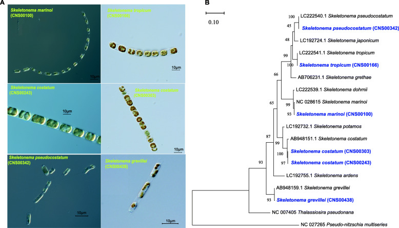
All candidate Skeletonema strains, which were readily identified by its cylindrical cells, usually formed long or short chains (Fig. 1A). S. pseudocostatum was observed with unicellular or two-three cells chains. Cell sizes of the candidate Skeletonema species varied substantially, which were not surprising because diatom cell size decreases as a result of the mechanism of cell division [31]. Thus, cell sizes alone could not be used as defining features for identifying Skeletonema species. Molecular information and more elaborate morphological characteristics are needed to facilitate species identification.
Fig. 1.
A Micrographs of six Skeletonema strains. B Phylogenetic tree of Skeletonema inferred from cox1 genes. The six Skeletonema strains in blue were obtained in this study, and their cox1 sequences have been uploaded to NCBI (MW438979-MW438984); the other sequences were downloaded from NCBI GenBank. Phylogenetic trees were generated using the Maximum Likelihood (ML) method with 1000 bootstrap replicates. Evolutionary model was Tamura-Nei model with gamma distribution (G = 0.187)
The cox1 gene sequences of all six Skeletonema strains (Fig. 1B) were used in the initial annotation of the Skeletonema strains. The cox1 gene sequence of the strain CNS00100 was identical to that of NC_028615.1 (percentage identity PID was 99.74%; Additional file 1), which was annotated as the species S. marinoi. Similarly, the strains CNS00166, CNS00243, CNS00303, CNS00342, and CNS00438 were annotated as S. tropicum (LC222541.1, PID = 100.00%), S. costatum (AB948151.1, PID = 99.20%), S. costatum (AB948151.1, PID = 99.20%), S. pseudocostatum (LC222540.1, PID = 100.00%) and S. grevillei (AB948159.1, PID = 99.90%; Additional file 1), respectively. The strains CNS00243 and CNS00303 were annotated to the same species, i.e. S. costatum.
Annotation using other molecular markers (18S rDNA, rbcL and 28S rDNA D1-D3) generally supported the cox1-based annotation of these six Skeletonema strains (Additional file 1; Additional file 2).
Construction and analysis of Skeletonema mtDNAs
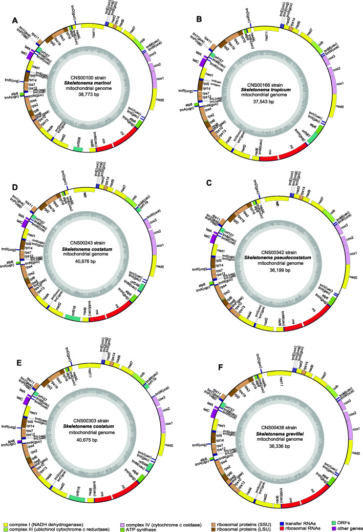
We constructed full-length mtDNAs for five Skeletonema species including S. marinoi, S. pseudocostatum, S. grevillei, S. costatum (with two strains), and S. tropicum. Notably, mtDNAs of S. pseudocostatum, S. grevillei, S. costatum, and S. tropicum were constructed in this project for the first time. Among the 36 reported mtDNAs in the Bacillariophyta (Additional file 3), 32 mtDNAs were full-length. The sizes of mtDNAs among the Bacillariophyta varied greatly (Additional file 4), ranging from 32,777 bp in Melosira undulata [46] to 103,605 bp in Halamphora calidilacuna [46]. The sizes of Skeletonema mtDNAs were relatively short, varying from 36,199 bp to 40,676 bp (Table 1). The AT contents were relatively similar (70.3–71.5%). All six Skeletonema mtDNAs contained 35 PCGs including atp6, 8, 9; cob; cox1–3; nad1–7, 4 L, 9, 11; rpl2, 5, 6, 14, 16; rps 2, 3, 4, 7, 8, 10, 11, 12, 13, 14, 19; and tatA and tatC), and two non-coding rRNA genes (rns and rnl; Table 1). Additionally, mtDNAs of S. tropicum, S. grevillei, S. pseudocostatum and S. marinoi contained 25 tRNAs, while that of S. costatum had 26 tRNAs and contain one more trnM. The start codon of atp8 gene in S. marinoi was GTG, while the start codon of atp8 was ATA in S. tropicum and S. pseudocostatum, and TTA in S. costatum and S. grevillei. No introns were found in any of these Skeletonema mtDNAs, although introns have been identified in many phytoplankton mtDNAs [46]. The intergenic regions of the mtDNAs of these five Skeletonema species were relatively small, with average sizes ranging from 58 bp in S. tropicum to 87 bp in S. marinoi, suggesting that the Skeletonema mtDNAs were all compact as other diatom mtDNAs [43].
Table 1.
The genome features and gene content of 12 mtDNAs in the class Mediophyceae belonging to the Phylum Bacillariophyta. The strains indicated in blue were obtained in this study
| Species | Strains | Habitat | Accession number | Size(bp) | A + T(%) | PCGs/rRNAs | rps2 | rrn5 | tRNA | Introns | Start codon of atp8 | Reference |
|---|---|---|---|---|---|---|---|---|---|---|---|---|
| Skeletonema marinoi | CNS00100 | Marine | MW438984 | 38,773 | 70.3 | 34/2 | + | – | 25 | 0 | GTG | This study |
| Skeletonema marinoi | JK029 | Marine | NC_028615 | 38,515 | 70.3 | 34/2 | + | – | 25 | 0 | GTG | [45] |
| Skeletonema tropicum | CNS00166 | Marine | MW438983 | 37,543 | 70.6 | 34/2 | + | – | 25 | 0 | ATA | This study |
| Skeletonema costatum | CNS00243 | Marine | MW438982 | 40,676 | 71.5 | 34/2 | + | – | 26 | 0 | TTA | This study |
| Skeletonema costatum | CNS00303 | Marine | MW438981 | 40,675 | 71.5 | 34/2 | + | – | 26 | 0 | TTA | This study |
| Skeletonema pseudocostatum | CNS00342 | Marine | MW438980 | 36,199 | 70.6 | 34/2 | + | – | 25 | 0 | ATA | This study |
| Skeletonema grevillei | CNS00438 | Marine | MW438979 | 36,336 | 70.8 | 34/2 | + | – | 25 | 0 | TTA | This study |
| Thalassiosira pseudonana | Marine | NC_007405 | 43,827 | 69.9 | 34/2 | + | – | 25 | 1 | TTA | [47] | |
| Thalassiosira profunda | CNS00050 | Marine | MW013551 | 40,470 | 69.0 | 34/2 | + | – | 25 | 0 | ATT | [48] |
| Odontella regia | CNS00380 | Marine | MW018491 | 37,617 | 73.4 | 34/2 | – | + | 24 | 0 | ATG | [43] |
| Lithodesmium undulatum | CNS00316 | Marine | MW023083 | 37,057 | 75.3 | 34/2 | – | + | 25 | 0 | ATT | [43] |
| Toxarium undulatum | ECT3802 | Marine | NC_037988 | 40,429 | 69.9 | 34/2 | + | – | 26 | 0 | ATG | [49] |
The core genes included 34 PCGs (atp6, 8, 9; cob; cox1, 2, 3; nad1–7, 4 L, 9, 11; rpl2, 5, 6, 14, 16; rps 3, 4, 7, 8, 10, 11, 12, 13, 14, 19; and tatA, tatC) and two rRNAs
Phylogenetic analysis of Skeletonema mtDNAs
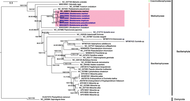
Among the 36 Bacillariophyta species with reported mtDNAs, the five Skeletonema species belonged to the class Mediophyceae (Additional file 3; Table 1; Fig. 2), including S. marinoi, which is the only Skeletonema species whose full-length mtDNA has been constructed. To explore the evolutionary relationship between Skeletonema species and other diatom species, we constructed a phylogenetic tree using 31 PCGs that were shared by mtDNAs of Bacillariophyta and Oomycota, which were used as outgroup taxa (Fig. 3). All species in Bacillariophyta fell into three clades corresponding to three classes including Mediophyceae, Bacillariophyceae, and Coscinodiscophyceae (Fig. 3). In the class Mediophyceae, Skeletonema species clustered closely with Thalassiosira pseudonana, which was consistent with previous studies [17, 45]. Both genera belong to the same Order Thalassiosirales [2]. Within Skeletonema, the S. marinoi mtDNA constructed in this study (MW438984) clustered closely with that of the other S. marinoi strain (JK029 strain from Korean seawater, NC_028615) as expected, so were the mtDNAs of two S. costatum strains (CNS00243 and CNS00303). S. tropicum clustered closely with S. pseudocostatum, consistent with previous report [3, 24]. The mtDNA of S. grevillei, which was isolated from the South China Sea, was independent from other clades.
Fig. 2.
Circular maps of the complete mtDNAs of six Skeletonema species. The PCGs, rRNAs and tRNAs are labeled outside the circles. The assignment of genes is indicated by colors according to the different functional groups. The ring of bar graphs inside the circle shows the GC content in dark gray
Fig. 3.
The phylogenetic tree based on concatenated amino acid sequences of 31 mitochondrial protein-coding genes (atp6, 8, 9; cob; cox1–3; nad1–7, 4 L, 9, 11; rpl2, 5, 6, 14, 16; rps 3, 4, 8, 10, 11, 13, 14, 19; and tatC) using Maximum likelihood (ML) methods. The branch values were SH-aLRT support (%) / aBayes support / ultrafast bootstrap support (%). The strains indicated in blue were obtained in this study. Phytophthora ramorum and Saprolegnia ferax were used as outgroups
Synteny conservation with divergent regions in mitochondrial genome
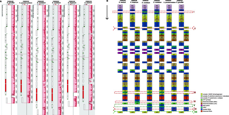
The mtDNA sizes of these Skeletonema species showed some variations with the difference between the longest mtDNA (S. costatum, CNS00243 strain, 40,676 bp) and the shortest mtDNA (S. pseudocostatum, 36,199 bp) being 4477 bp. However, comparative analysis found that there was nearly perfect synteny conservation among Skeletonema species (Fig. 4A). This is expected because previous study found high synteny conservation between the mtDNAs of S. marinoi and T. pseudonana [43], which is diatom species of a different genus in the order Thalassiosirales. At the single gene level, the genome organization was also well conserved among the five Skeletonema species with only minor differences (Fig. 4B). The numbers of open reading frames (orfs) and trnM genes were different among these Skeletonema mtDNAs, which were primarily responsible for the different lengths of mtDNAs. S. pseudocostatum and S. grevillei each had a single orf, S. marinoi and S. tropicum each had two orfs, while S. costatum each had three orfs. S. costatum each had an additional trnM. Compared with S. pseudocostatum, mtDNA of S. costatum had three more orfs (orf119, orf516 and orf248) and one more trnM, which in total accounted for 2730 bp. Another main reason for the big size difference between the two species was the length of intergenic regions. For example, the length of the combined intergenic regions in S. costatum (CNS00243 strain) was roughly 1745 bp longer than that of S. pseudocostatum.
Fig. 4.
A Synteny relationships among six Skeletonema mtDNAs based on Mauve analysis. B mtDNA gene arrangements of six Skeletonema species. Blocks with the same color represent the same type of genes
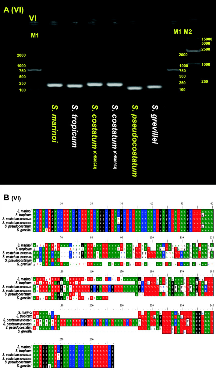
Interestingly, despite the deep conservation of these mtDNAs, comparative sequence analysis of these mtDNAs also revealed seven discrete regions with enhanced divergence, which were indicated using I, II, III, IV, V, VI and VII (Fig. 4B). Within these regions, DNA sequences were essentially unalignable (Additional file 5 and Additional file 6). Region I was located between trnW and trnM (Additional file 5A). The S. costatum mtDNAs have one big insertion (orf119) compared with other species, which were also validated by designed PCR primers (I-F and I-R in Additional file 7; Additional file 6). Insertions in mtDNAs of S. costatum (trnM and orf248) were also found in region V (located between atp6 and trnI; Additional file 5E and Additional file 6), which were validated by PCR primers (V-F and V-R in Additional file 7). In region III (located between nad2 and nad6; Additional file 5C and Additional file 6), the mtDNAs of S. marinoi (orf306), and S. costatum (orf516) had big insertions, respectively. In the region IV, only S. tropicum mtDNA had an insertion (orf387 between trnH and atp6) with 1786 bp length compared with other Skeletonema species with the lengths between 439 and 455 bp (Additional file 5D and Additional file 6). Divergent regions II, VI and VII, were located between nad11 and trnD (Additional file 5B and Additional file 6); between trnI and trnV (Fig. 5A-B; Additional file 5F); between trnV and nad5 (Additional file 5G), respectively. These divergent regions (Fig. 4B and Additional file 5) are generally located at the non-coding regions except the region V. The nature regarding the formation of these divergent regions remained unknown. However, these divergent regions may be used as potential molecular markers for distinguishing different Skeletonema species.
Fig. 5.
A Agarose gels image of PCR products for region VI among the six Skeletonema mtDNAs. The full-length gels are presented in Additional file 15. The brands sequences were S. marinoi, S. tropicum, S. costatum (CNS00243), S. costatum (CNS00303), S. pseudocostatum and S. grevillea, respectively. The brands of Marker M1 and M2 were on the right. B DNA alignment information of region VI for the six Skeletonema species
Among the seven regions, region VI could be used a molecular marker for distinguishing five Skeletonema species with both high resolution and high specificity. This region was different among five Skeletonema species, thus it could be used to distinguish different Skeletonema species (Fig. 5). To explore the specificity of the region VI as a molecular marker, primers (VI-F: ACTACTGACCTTACGCTTATC, VI-R: CGAAAAAGTTGGTGGTTCGATTCCA) were designed for PCR amplification of the region VI in 11 other phytoplankton species (i.e., Thalassiosira pseudonana, Chaetoceros muelleri, Heterosigma akashiwo, Aureococcus anophagefferens, Chattonella marina, Amphidinium carterae, Alexandrium tamarense, Karenia mikimotoi, Prorocentrum donghaiense, Isochrysis galbana, and Phaeocystis globosa). Agarose gel images of PCR products showed that only the PCR amplification was positive only for Chaetoceros muelleri with one bright band, whose length was much longer than that of Skeletonema species (Additional file 8), suggesting that the region VI can be used as a molecular marker to distinguish Skeletonema species with high specificity.
Divergence time estimation based on PCGs of mtDNAs
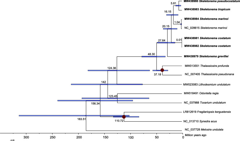
The time-calibrated phylogeny of the Mediophyceae and other species was constructed using DNA sequences of 34 PCGs from 15 mtDNAs. The divergence times of Mediophyceae species were estimated using the MCMC tree (Fig. 6). The divergence between the class Mediophyceae and the class Coscinodiscophyceae was inferred to have occurred at 183.51 Ma ago (Mya; 95% HPD (highest posterior density): 100.65–312.13 Mya). Furthermore, the Skeletonema species formed a monophyletic clade at approximately 48.30 Mya (95% HPD 31.52–76.79 Mya) apart from Thalassiosira species. Among the Skeletonema species: S. grevillei diverged from other Skeletonema species approximately 27.64 Mya (95% HPD 13.10–47.11 Mya). The sister clade of S. costatum with two strains diverged from the clades including S. marinoi, S. tropicum and S. pseudocostatum approximately 20.15 Mya (95% HPD 8.94–35.6). S. marinoi diverged from the sister clade between S. tropicum and S. pseudocostatum approximately 16.15 Mya (95% HPD 7.08–29.41 Mya).
Fig. 6.
Time-calibrated phylogeny of 15 mtDNAs based on 34 PCGs in the class Mediophyceae and outgroup (Melosira undulata). We added two Bacillariophyceae species (Synedra acus and Fragilariopsis kerguelensis) to establish appropriate fossil calibration points. The red dots represent calibration points and the 95% highest posterior density interval for node ages are shown with translucent blue bars
Discussion
Compact Skeletonema mtDNAs
The six Skeletonema mtDNAs constructed in this study had relatively small sizes (Additional file 4), with short intergenic regions, no introns, and no repeat regions. Introns have been found in many diatom mtDNA genes (Additional file 3), which greatly increased the length of mtDNA. For example, the largest mitochondrial genome ever discovered was Halamphora calidilacuna [46], reaching 103,605 bp. The cox1 genes of H. calidilacuna spanned 31,716 bp, with only 1506 bp (4.7% of the total length of cox1 gene) coding sequences. Some repeats were also found in some mtDNAs, which induced larger sizes of mtDNAs. For example, mtDNA of Phaeodactylum tricornutum contained a 35 kb-long repeat [50]. The intergenic regions also contribute significantly to the whole length of mtDNAs, and the larger size of intergenic regions is usually associated with larger size of mtDNAs. Thus another reason for the small size mtDNAs among Skeletonema species was relatively short intergenic regions compared to other diatom species (Additional file 4).
Synteny conservation of Skeletonema mtDNAs
Our analysis found that Skeletonema mtDNAs contained similar numbers of genes containing 35 common PCGs, 2 rRNA genes and 25 tRNA genes (except in the S. costatum, which contained an additional trnM gene), same as the two mtDNAs of Thalassiosira (T. pseudonana and T. profunda) [47, 48]. The Nitzschia mtDNAs also include 35 unique PCGs and 2 rRNA genes, but these mtDNAs only have 24 tRNA genes compared that in Thalassiosira with and Skeletonema genus [46, 49, 51, 52]. Interesting, the mtDNAs in Halamphora genus show difference: H. calidilacuna mtDNA includes 34 PCGs and 26 tRNA genes; while H. coffeaeformis mtDNA includes 35 PGCs and 24 tRNA genes [46]. The mtDNAs of the two Halamphora species, which were quite different from that of the above three genera, include 3 rRNA genes and introns. While for the genetic structure, the mtDNAs belonging to the same genus showed highly synteny although the number of genes and genome sizes within the genus varied greatly (Additional file 9 and Fig. 4A). We also found slight variation among the Thalassiosira genus with two inversions (Additional file 9), showing the interspecific variability in the Thalassiosira genus. In brief, the mtDNAs of five Skeletonema species showed high conservation, which was in agreement with other species belonging to the same genus according to the previously reported mtDNAs.
Phylogeny and evolution of Skeletonema species
Diatoms arose in the Triassic period, perhaps as early as 250 Mya according to the molecular clock estimate [53], and provided a main ecological role in carbon cycle about 100 Mya, when the CO2 level was five times higher than that nowadays. The timing coincided with the divergence of a second major lineage of diatoms, the bipolar and multipolar centrics, including Thalassiosirales clade [54]. Phylogenetic analysis of 31 core PCGs showed that Skeletonema was sister to Thalassiosira, both belonging to the order Thalassiosirales, consistent with the current classification [2, 55]. Our results (Fig. 6) show that Skeletonema species diverged from Thalassiosira species approximately 48.30 Mya (95% HPD: 31.52–76.79 Mya) during the Eocene, a period included a major burst of diversification affected by sea level, silica availability and interspecific competition with other planktonic groups [56]. This divergence time was estimated using 34 mtDNA genes, which was slightly earlier than that estimated using the two nuclear genes, seven plastid genes and two mitochondrial genes [55, 57], but was later than that estimated using two nuclear genes and two plastid genes [58], and using 18S rDNA [59].
Among the Skeletonema genus, the two HAB species S. marinoi and S. pseudocostatum diverged approximately 16.15 Mya (95% HPD 7.08–29.41 Mya), which was slight early than that results estimated previously using 18S rDNA [53]; and were later than that estimated using the two nuclear genes, seven plastid genes and two mitochondrial genes [55]. The divergence times estimated based on 34 PCGs of mtDNA among Skeletonema species in this study were generally consistent with previous results with minor fluctuations, which facilitating the understanding the evolution of Skeletonema species in the view of mitochondria with uniparental inheritance.
Conclusion
In this study, we reported mtDNAs of four Skeletonema species for the first time, and re-sequenced the S. marinoi mtDNA of a strain isolated in coastal waters in China. Through comparative analysis of the mtDNA structure, base composition and gene order, we found relatively conserved genetic arrangement among the Skeletonema species. We also reported discrete regions with high divergence among the mtDNAs of five Skeletonema species, which could potentially provide high resolution for the phylogenetic studies among the genus. We further reconstructed a phylogenetic tree, which was used to estimate the divergence times among Skeletonema species. However, more taxa and more complete mtDNAs data are needed to verify the evolutionary relationships and primers application.
The mtDNAs of five Skeletonema species showed similar sizes, conserved genetic structure and high syntenic relationships among the genus. While the five Skeletonema species showed different ecological geographical distribution (Additional file 10). For example, S. marinoi was easily found in seawaters of higher latitude with lower temperature (Additional file 10A); While S. tropicum was recorded in seawaters of lower latitude with higher temperature (Additional file 10B). We hypothesize that the nuclear genomes of the Skeletonema species may possess substantial differences, which facilitate the adaptation of different Skeletonema species. To further explore the specific ecological distribution mechanism, whole genomes of Skeletonema species could help to provide new ideas.
Methods
Strain isolation and whole genome sequencing
Skeletonema strains studied in this project were isolated from water samples collected during six expeditions in Chinese coastal waters (Additional file 11), using methods as described previously [43]. The seawater of culture was collected from Jiaozhou Bay, filtered through a 0.22 μm mixed-fiber membrane and sterilized at 121 °C for 30 min. The Skeletonema strains were maintained in L1 medium [60]. The culture salinity was 28–30 ‰. The culture temperature was 19 ± 1 °C, and the irradiance was from 72 μmol photons /(m2·s) with a 12:12 h light-dark cycle. The strains were maintained in our lab for 3 months to 13 months before sequencing.
The morphological features of the Skeletonema strains were observed using ZEISS Axio Imager 2 (ZEISS, Germany) at the 600× magnification. We selected reprehensive strains based on the preliminary morphological observation for Illumina sequencing. For DNA extraction, healthy cells at the exponential growth phase were harvested via centrifugation at 8000 rpm for 5 min. Total DNAs were extracted using DNAsecure Plant Kit (Tiangen Biotech, Beijing, China) using the manufacturers’ instructions. DNA samples were quantified using Qubit® DNA Assay Kit in Qubit® 3.0 Flurometer (Invitrogen, USA), 0.2 μg genomic DNA of each sample was fragmented by sonication (Covaris S220, Covaris, USA) to the length of 350 bp. DNA fragments were harvested, end polished, A-tailed, and ligated with adapters for Illumina sequencing, followed by PCR amplification. The PCR products were purified using AMPure XP system (Beckman Coulter, Beverly, USA). Qualified libraries were sequenced using NovaSeq PE150 (Illumina, San Diego, CA, USA) at Novogene (Beijing, China).
Genomic assessment, assembly and annotation
Clean data was obtained after trimming using Trimmomatic-0.39 [61]. From the K-mer analysis using Jellyfish [62] and GenomeScope [63], estimated nuclear genome sizes of six Skeletonema strains ranged from 51.72 M (S. grevillei) to 119.42 M (S. costatum, CNS00303 strain) (Additional file 11 and Additional file 12). The heterozygosity ranged from 0.11 to 2.90%. The repetitive rate ranged from 0.83 to 2.54%. Bacterial contamination was negligible (< 0.3%) in the DNA samples of S. marinoi, S. tropicum, S. costatum (CNS00243 strain) and S. grevillei strains, while the bacterial contaminations were relatively high in DNA samples of S. pseudocostatum (20.64%) and S. costatum (CNS00303 strain, 18.58%) strains. Common molecular markers, including full-length of 18S rDNA, cox1 and rbcL, 28S rDNA D1-D3 and complete mtDNAs were assembled using the GetOrganelle [64]. All of the assembled results were validated using BWA [65], SAMtools [66] and IGV [67].
MtDNAs were annotated using MFannot (https://github.com/BFL-lab/Mfannot), Annotation results were validated and corrected by the NCBI’s ORF Finder (https://www.ncbi.nlm.nih.gov/orffinder/) with the genetic code of 4 Mold, Protozoan, and Coelenterate Mitochondrial; Mycoplasma/Spiroplasma. For accurate comparison, we have also inspected and re-annotated mtDNAs downloaded from the NCBI (Additional file 3).
Phylogenetic analysis and divergence time estimations
The Phylogenetic trees of cox1 (Fig. 1), full-length 18S rDNA, rbcL and 28S rDNA D1-D3 (Additional file 1; Additional file 2) were generated using the Maximum Likelihood (ML) method with 1000 bootstrap replicates in MEGAX [68]. Appropriate evolutionary models were selected using Model Selection.
The phylogenetic tree of mitochondrial PCGs were constructed by extracting 31 PCGs, including atp6, 8, 9; cob; cox1, 2, 3; nad1–7, 4 L, 9, 11; rpl2, 5, 6, 14, 16; rps3, 4, 8, 10, 11, 13, 14, 19 and tatC, from the published Bacillariophyta mtDNAs and six Skeletonema mtDNAs in this study (Additional file 3) [43, 48]. The 31 genes amino acid sequences were individually aligned using MAFFT [69], trimmed using trimAl 1.2rev59 [70], and concatenated using Phyutility [71]. The evolutionary models were obtained using ModelFinder [72]. The phylogenetic tree was constructed using IQ-TREE [73] with SH-aLRT support (%) / aBayes support / ultrafast bootstrap support (%). Phytophthora ramorum and Saprolegnia ferax in Oomycota were used as outgroups (Fig. 3). FigTree 1.4.3 (http://tree.bio.ed.ac.uk/software/figtree/) was used to display the phylogenetic tree.
Phylogenetic analysis and molecular dating were analyzed using 34 mitochondrial PCGs DNA sequences shared by Mediophyceae species and outgroups Melosira undulata. The 34 PCGs included atp6, 8, 9; cob; cox1–3; nad1–7, 4 L, 9, 11; rpl2, 5, 6, 14, 16; rps 3, 4, 7, 8, 10, 11, 12, 13, 14, 19; and tatA and tatC, which were shared among the 15 species. These PCGs were aligned (MAFFT with Codon mode) and concatenated (Concatenate Sequence). The best-fit evolutionary model and partitioning scheme were selected by PartitionFinder2, which were shown in the Additional file 13. The phylogenetic trees (MrBayes) was constructed using the software PhyloSuite v1.2.2 [74]. Molecular dating was performed using the PAML package [75] in there three steps: (1) Rough estimation of substitution rate was analyzed using baseml; (2) Estimation of gradient and hessian of branch length; (3) Estimation of divergence times with the approximate likelihood method using the mcmctree. Two calibration points used in this analysis were shown in the Additional file 14. We added two Bacillariophyceae species (Synedra acus and Fragilariopsis kerguelensis) to establish appropriate fossil calibration points. The calibration point of Synedra and Fragilaria was based on fossil record [57]. The divergence times with fossil-calibrated deriving from ribosomal RNA and organelle genes, i.e. T. profunda and T. pseudonana [53, 58, 76]. The phylogenetic tree was displayed in the FigTree and visualized with 95% HPD interval for each node.
Synteny analysis and molecular marker development of six Skeletonema mtDNAs
Synteny analysis of six Skeletonema mtDNAs sequences was carried out with the program Mauve v2.3.1 [77]. The region with great sequence differences were aligned using MUSCLE algorithm in the MEGAX. Six primers (I-F/R to VI-F/R in Additional file 7) for PCR amplification of molecular markers (region I to VI in Fig. 4B) were developed using the Primer Premier 5.0 [78]. PCRs were performed in a final volume of 50 μL containing 2 μL diluted template DNA (about 50 ng), 1 μL forward primer, 1 μL reverse primer, 25 μL mix (Tiangen, China) and 21 μL ddH2O. The reaction was denatured at 94 °C for 4 min. Then, reactions were run for 32 cycles at 94 °C for 1 min, 52 °C for 1 min 50s, and 72 °C for 2 min and a final extension at 72 °C for 10 min. These PCR products were run on 1% agarose gels for checking amplicon lengths.
Supplementary Information
Additional file 1. The alignment results with highest PID for common molecular markers of six Skeletonema strains. PID means percentage identity.
Additional file 2. The phylogenetic analysis of Skeletonema species and outgroups using 18S rDNA (A), the D1-D3 region of 28S rDNA gene (B) and rbcL (C). Phylogenetic trees were generated using the Maximum Likelihood (ML) method with 1000 bootstrap replicates. Evolutionary models for 18S rDNA, the D1-D3 region of 28S rDNA gene, and rbcL were Tamura 3-parameter model with gamma distribution (G = 0.617), Kimura 2-parameter model with gamma distribution (G = 0.297), and Tamura 3-parameter with gamma distribution (G = 0.689).
Additional file 3. Genome features of 36 mtDNAs from the Phylum Bacillariophyta.
Additional file 4. The intergenic region sizes and mtDNAs sizes of completed mtDNAs in the Bacillariophyta.
Additional file 5. The alignment details of seven regions with great difference of six Skeletonema mtDNAs.
Additional file 6. The agarose gels image of PCR products of seven regions among the six Skeletonema mtDNAs. The full-length gels are presented in Additional file 15. For each region (I-VI), the brands sequences were S. marinoi, S. tropicum, S. costatum (CNS00243), S. costatum (CNS00303), S. pseudocostatum and S. grevillei, respectively. The brands of Marker M1 and M2 were on the right.
Additional file 7. The primes for the region I to region VI among the Skeletonema genus. F in the primer names meant forward primer; R in the primer names meant reverse primer. I to VI in the primer names were corresponding to region I to region VI.
Additional file 8. The agarose gels image of PCR products for cox1 and region VI among Skeletonema species and other eleven species (Thalassiosira pseudonana, Chaetoceros muelleri, Heterosigma akashiwo, Aureococcus anophagefferens, Chattonella marina, Amphidinium carterae, Alexandrium tamarense, Karenia mikimotoi, Prorocentrum donghaiense, Isochrysis galbana and Phaeocystis globosa). The primers of cox1 gene were, F: GGAACTTTATATTTAATYTTTGGWGC, R: AATACCAGAATTAGCAAGAACAAC [40]. The full-length gels are presented in Additional file 16.
Additional file 9. Synteny relationships among two Thalassiosira mtDNAs (A); eight Nitzschia mtDNAs (B); two Halamphora mtDNAs (C) based Mauve analysis.
Additional file 10. The distributions of five Skelentonema species from GBIF (https://www.gbif.org/).
Additional file 11. The sample collection and genomic assessment information of six Skeletonema strains.
Additional file 12. The genomic assessment of six Skeletonema strains.
Additional file 13. The best-fit evolutionary model and partitioning scheme for phylogenetic analysis and molecular dating.
Additional file 14. Calibration points used in the divergence time analysis by PAML.
Additional file 15. The full-length gels used in Fig. 5 and Additional file 7.
Additional file 16. The full-length gels used in Additional file 9.
Acknowledgements
We are grateful to colleagues from the Jiaozhou Bay Marine Ecosystem Research Station for the opportunity to participate in the investigation expeditions and for their help with field sampling. We are also thankful to the open research cruise (NORC2019-01, NORC2019-03-02 and NORC2019-07) supported by the NSFC shiptime sharing Project (41849901, U1606404, 41849007).
Abbreviations
- mtDNA
Mitochondrial genome
- HAB
Harmful algal bloom
- PCG
Protein-coding gene
- HPD
Highest posterior density
- orf
Open reading frame
Authors’ contributions
NC designed the experiments; SL collected, isolated, and characterized the species; SL, YW and MZ performed the experiments; SL, YW, QX and MZ analyzed the data; SL and NC wrote the paper. All authors contributed to the editing and gave final approval for publication.
Funding
This research was supported by the Strategic Priority Research Program of Chinese Academy of Sciences (XDB42000000), the Natural Science Foundation of China (41906118), the Chinese Academy of Sciences Pioneer Hundred Talents Program (to Nansheng Chen), the Taishan Scholar Project Special Fund (to Nansheng Chen), the Qingdao Innovation and Creation Plan (Talent Development Program - 5th Annual Pioneer and Innovator Leadership Award to Nansheng Chen, 19–3–2-16-zhc) and the National Key Research and Development Program of china (2017YFC1404300).
Availability of data and materials
The sequencing results (raw data) have been submitted to NCBI and the BioProject number is PRJN695365 (https://www.ncbi.nlm.nih.gov/sra/?term=PRJNA695365). The mtDNA sequences and annotation results of six Skeletonema species have been submitted to NCBI under specific accession numbers (MW438979-MW438984). The Skeletonema species are provided by the Key Laboratory of Marine Ecology and Environmental Science from the Institute of Oceanology, Chinese Academy of Sciences.
Declarations
Ethics approval and consent to participate
Not applicable.
Consent for publication
Not applicable.
Competing interests
The authors declare that they have no competing interests.
Footnotes
Publisher’s Note
Springer Nature remains neutral with regard to jurisdictional claims in published maps and institutional affiliations.
Shuya Liu and Yichao Wang contributed equally to this work.
References
- 1.Sarno D, Kooistra WHCF, Medlin LK, Percopo I, Zingone A. Diversity in the genus skeletonema (bacillariophyceae). II. An assessment of the taxonomy of s. costatum-like species with the description of four new species. J Phycol. 2005;41(1):151–176. doi: 10.1111/j.1529-8817.2005.04067.x. [DOI] [Google Scholar]
- 2.M.D. Guiry in Guiry MDG, G.M . National University of Ireland. Galway: World-wide electronic publication; 2021. AlgaeBase. [Google Scholar]
- 3.Kooistra W, Sarno D, Balzano S, Gu HF, Andersen RA, Zingone A. Global diversity and biogeography of Skeletonema species (Bacillariophyta) Protist. 2008;159(2):177–193. doi: 10.1016/j.protis.2007.09.004. [DOI] [PubMed] [Google Scholar]
- 4.Xu X, Yu ZM, Cheng FJ, He LY, Cao XH, Song XX. Molecular diversity and ecological characteristics of the eukaryotic phytoplankton community in the coastal waters of the Bohai Sea, China. Harmful Algae. 2017;61:13–22. doi: 10.1016/j.hal.2016.11.005. [DOI] [Google Scholar]
- 5.Liu S, Chen N. Advances in biodiversity analysis of phytoplankton and harmful algal bloom species in the Jiaozhou Bay (Chinese with English abstract). Mar Sci. 2021;45(4):170–88.
- 6.Cheng JF, Li Y, Liang JR, Gao YH, Wang P, Ho KC, et al. Morphological variability and genetic diversity in five species of Skeletonema (Bacillariophyta). Prog Nat Sci. 2008;18(11):1345–55. 10.1016/j.pnsc.2008.05.002.
- 7.Godhe A, Harnstrom K. Linking the planktonic and benthic habitat: genetic structure of the marine diatom Skeletonema marinoi. Mol Ecol. 2010;19(20):4478–4490. doi: 10.1111/j.1365-294X.2010.04841.x. [DOI] [PubMed] [Google Scholar]
- 8.Godhe A, Sjoqvist C, Sildever S, Sefbom J, Hardardottir S, Bertos-Fortis M, et al. Physical barriers and environmental gradients cause spatial and temporal genetic differentiation of an extensive algal bloom. J Biogeogr. 2016;43(6):1130–42. 10.1111/jbi.12722.
- 9.Pfannkuchen DM, Godrijan J, Tankovic MS, Baricevic A, Kuzat N, Djakovac T, et al. The ecology of one cosmopolitan, one newly introduced and one occasionally advected species from the genus Skeletonema in a highly structured ecosystem, the northern Adriatic. Microb Ecol. 2018;75(3):674–87. 10.1007/s00248-017-1069-9. [DOI] [PubMed]
- 10.Buzancic M, Arapov J, Bakrac A, Gladan ZN, Skejic S. Morphological characterization of skeletonema grevillei (bacillariophyta, thalasiosirales) in the eastern adriatic sea. Vie Et Milieu Life Environ. 2017;67(3–4):193–199. [Google Scholar]
- 11.Gu H, Zhang X, Sun J, Luo Z. Diversity and seasonal occurrence of Skeletonema (Bacillariophyta) species in Xiamen harbour and surrounding seas, China. Cryptogam Algol. 2012;33(3):245–263. doi: 10.7872/crya.v33.iss3.2012.245. [DOI] [Google Scholar]
- 12.Zingone A, Percopo I, Sims PA, Sarno D. Diversity in the genus Skeletonema (Bacillariophyceae). I. A reexamination of the type material of s. Costatum with the description of s. Grevillei sp Nov. J Phycol. 2005;41(1):140–50. 10.1111/j.1529-8817.2005.04066.x.
- 13.Liu D, Jiang J, Wang Y, Zhang Y, Di B. Large scale northward expansion of warm water species Skeletonema tropicum (Bacillariophyceae) in China seas. Chin J Oceanol Limnol. 2012;30(4):519–527. doi: 10.1007/s00343-012-1249-x. [DOI] [Google Scholar]
- 14.Castillo JA, Delcastillo MEM, Hernandezbecerril DU. Morphology and distribution of species of the diatom genus skeletonema in a tropical coastal lagoon. Eur J Phycol. 1995;30(2):107–115. doi: 10.1080/09670269500650871. [DOI] [Google Scholar]
- 15.Bergesch M, Garcia M, Odebrecht C. Diversity and morphology of skeletonema species in southern Brazil, southwestern Atlantic Ocean. J Phycol. 2009;45(6):1348–1352. doi: 10.1111/j.1529-8817.2009.00743.x. [DOI] [PubMed] [Google Scholar]
- 16.Hasle GR, Evensen DL. Brackish water and freshwater species of the diatom genus skeletonema. Ii. Skeletonema potamos comb. Nov. J Phycol. 1976;12(1):73–82. doi: 10.1111/j.1529-8817.1976.tb02829.x. [DOI] [Google Scholar]
- 17.Duleba M, Ector L, Horvath Z, Kiss KT, Molnar LF, Pohner Z, et al. Biogeography and phylogenetic position of a warm-stenotherm centric diatom, Skeletonema potamos (CI Weber) hasle and its long-term dynamics in the river Danube. Protist. 2014;165(5):715–29. 10.1016/j.protis.2014.08.001. [DOI] [PubMed]
- 18.Carvalho Torgan L, Becker V, Bahi dos Santos C. Skeletonema potamos (Bacillariophyta) en la Laguna dos Patos, sur del Brasil: Taxonomía y distribución. Rev Peru Biol. 2009;16(1):93–96. doi: 10.15381/rpb.v16i1.181. [DOI] [Google Scholar]
- 19.Mann DG. The species concept in diatoms. Phycologia. 1999;38(6):437–495. doi: 10.2216/i0031-8884-38-6-437.1. [DOI] [Google Scholar]
- 20.Ogura A, Akizuki Y, Imoda H, Mineta K, Gojobori T, Nagai S. Comparative genome and transcriptome analysis of diatom, Skeletonema costatum, reveals evolution of genes for harmful algal bloom. BMC Genomics. 2018;19(1):765. doi: 10.1186/s12864-018-5144-5. [DOI] [PMC free article] [PubMed] [Google Scholar]
- 21.Winder M, Sommer U. Phytoplankton response to a changing climate. Hydrobiologia. 2012;698(1):5–16. doi: 10.1007/s10750-012-1149-2. [DOI] [Google Scholar]
- 22.Anderson DM, Cembella AD, Hallegraeff GM. Progress in understanding harmful algal blooms: paradigm shifts and new technologies for research, monitoring, and management. In: Carlson CA, Giovannoni SJ, editors. Annual Review of Marine Science, Vol 4. 2012. p. 143–176. [DOI] [PMC free article] [PubMed]
- 23.Li C, Zhu B, Chen H, Liu Z, Cui B, Wu J, et al. The relationship between the Skeletonema costatum red tide and Enviromnental factors in Hongsha Bay of Sanya, South China Sea. J Coast Res. 2009;25(3):651–74. 10.2112/07-0967.1.
- 24.Chen G-F, Wang G-C, Zhang B-Y, Fan X-L. Morphological and phylogenetic analysis of Skeletonema costatum-like diatoms (Bacillariophyta) from the China Sea. Eur J Phycol. 2007;42(2):163–175. doi: 10.1080/09670260601149784. [DOI] [Google Scholar]
- 25.Li L, Lu S, Cen J. Spatio-temporal variations of harmful algal blooms along the coast of Guangdong, southern China during 1980-2016. Oceanol Limnol. 2019;37(2):535–51. 10.1007/s00343-019-8088-y.
- 26.Liu S, Gibson K, Cui Z, Chen Y, Sun X, Chen N. Metabarcoding analysis of harmful algal species in Jiaozhou Bay. Harmful Algae. 2020;92:101772. doi: 10.1016/j.hal.2020.101772. [DOI] [PubMed] [Google Scholar]
- 27.Mohamed ZA. Potentially harmful microalgae and algal blooms in the Red Sea: current knowledge and research needs. Mar Environ Res. 2018;140:234–242. doi: 10.1016/j.marenvres.2018.06.019. [DOI] [PubMed] [Google Scholar]
- 28.Hemaiswarya S, Raja R, Kumar RR, Ganesan V, Anbazhagan C. Microalgae: a sustainable feed source for aquaculture. World J Microbiol Biotechnol. 2011;27(8):1737–1746. doi: 10.1007/s11274-010-0632-z. [DOI] [Google Scholar]
- 29.Johansson ON, Töpel M, Pinder MIM, Kourtchenko O, Blomberg A, Godhe A, et al. Skeletonema marinoi as a new genetic model for marine chain-forming diatoms. Sci Rep. 2019;9(1):5391. 10.1038/s41598-019-41085-5. [DOI] [PMC free article] [PubMed]
- 30.Amato A, Sabatino V, Nylund GM, Bergkvist J, Basu S, Andersson MX, et al. Grazer-induced transcriptomic and metabolomic response of the chain-forming diatom Skeletonema marinoi. ISME J. 2018;12(6):1594–604. 10.1038/s41396-018-0094-0. [DOI] [PMC free article] [PubMed]
- 31.Ferrante MI, Entrambasaguas L, Johansson M, Topel M, Kremp A, Montresor M, et al. Exploring Molecular Signs of Sex in the Marine Diatom Skeletonema marinoi. Genes. 2019;10(7). 10.3390/genes10070494. [DOI] [PMC free article] [PubMed]
- 32.Gallo C, d'Ippolito G, Nuzzo G, Sardo A, Fontana A. Autoinhibitory sterol sulfates mediate programmed cell death in a bloom-forming marine diatom. Nat Commun. 2017;8(1). 10.1038/s41467-017-01300-1. [DOI] [PMC free article] [PubMed]
- 33.Fiori E, Pistocchi R. Skeletonema marinoi (Bacillariophyceae) sensitivity to herbicides and effects of temperature increase on cellular responses to terbuthylazine exposure. Aquat Toxicol. 2014;147:112–120. doi: 10.1016/j.aquatox.2013.12.014. [DOI] [PubMed] [Google Scholar]
- 34.Johansson ON, Topel M, Egardt J, Pinder MIM, Andersson MX, Godhe A, et al. Phenomics reveals a novel putative chloroplast fatty acid transporter in the marine diatom Skeletonema marinoi involved in temperature acclimation. Sci Rep. 2019;9(1). 10.1038/s41598-019-51683-y. [DOI] [PMC free article] [PubMed]
- 35.Haernstroem K, Ellegaard M, Andersen TJ, Godhe A. Hundred years of genetic structure in a sediment revived diatom population. Proc Natl Acad Sci U S A. 2011;108(10):4252–4257. doi: 10.1073/pnas.1013528108. [DOI] [PMC free article] [PubMed] [Google Scholar]
- 36.Stenow R, Olofsson M, Robertson EK, Kourtchenko O, Whitehouse MJ, Ploug H, et al. Resting stages of Skeletonema marinoi assimilate nitrogen from the ambient environment under dark, anoxic conditions. J Phycol. 2020;56(3):699–708. 10.1111/jpy.12975. [DOI] [PubMed]
- 37.Godhe A, McQuoid MR, Karunasagar I, Karunasagar I, Rehnstam-Holm AS. Comparison of three common molecular tools for distinguishing among geographically separated clones of the diatom Skeletonema marinoi Sarno et Zingone (bacillariophyceae) J Phycol. 2006;42(2):280–291. doi: 10.1111/j.1529-8817.2006.00197.x. [DOI] [Google Scholar]
- 38.Sarno D, Kooistra W, Balzano S, Hargraves PE, Zingone A. Diversity in the genus Skeletonema (Bacillariophyceae): III. Phylogenetic position and morphological variability of Skeletonema costatum and Skeletonema grevillei, with the description of Skeletonema ardens sp nov. J Phycol. 2007;43(1):156–170. doi: 10.1111/j.1529-8817.2006.00305.x. [DOI] [Google Scholar]
- 39.Hevia-Orube J, Orive E, David H, Laza-Martinez A, Seoane S. Skeletonema species in a temperate estuary: a morphological, molecular and physiological approach. Diatom Res. 2016;31(3):185–197. doi: 10.1080/0269249X.2016.1228548. [DOI] [Google Scholar]
- 40.Yamada M, Otsubo M, Tsutsumi Y, Mizota C, Nakamura Y, Takahashi K, et al. Utility of mitochondrial-encoded cytochrome c oxidase I gene for phylogenetic analysis and species identification of the planktonic diatom genus Skeletonema. Phycol Res. 2017;65(3):217–25. 10.1111/pre.12179.
- 41.Lv W, Jiang H, Bo J, Wang C, Yang L, He S. Comparative mitochondrial genome analysis of Neodontobutis hainanensis and Perccottus glenii reveals conserved genome organization and phylogeny. Genomics. 2020;112(6):3862–3870. doi: 10.1016/j.ygeno.2020.06.039. [DOI] [PubMed] [Google Scholar]
- 42.Zhang W, Sun Y, Liu J, Xu C, Zou X, Chen X, et al. DNA barcoding of Oryza: conventional, specific, and super barcodes. Plant Mol Biol. 2021;105(3):215–28. 10.1007/s11103-020-01054-3. [DOI] [PMC free article] [PubMed]
- 43.Wang Y, Chen Y, Wang J, Liu F, Chen N. Mitochondrial genome of the harmful algal bloom species Odontella regia (Mediophyceae, Bacillariophyta) J Appl Phycol. 2021;33(2):855–868. doi: 10.1007/s10811-020-02364-1. [DOI] [Google Scholar]
- 44.Smith DR, Arrigo KR, Alderkamp A-C, Allen AE. Massive difference in synonymous substitution rates among mitochondrial, plastid, and nuclear genes of Phaeocystis algae. Mol Phylogenet Evol. 2014;71:36–40. doi: 10.1016/j.ympev.2013.10.018. [DOI] [PubMed] [Google Scholar]
- 45.An SM, Kim SY, Noh JH, Yang EC. Complete mitochondrial genome of Skeletonema marinoi (Mediophyceae, Bacillariophyta), a clonal chain forming diatom in the west coast of Korea. Mitochondrial DNA. 2017;28(1–2):19–20. doi: 10.3109/19401736.2015.1106523. [DOI] [PubMed] [Google Scholar]
- 46.Pogoda CS, Keepers KG, Hamsher SE, Stepanek JG, Kane NC, Kociolek JP. Comparative analysis of the mitochondrial genomes of six newly sequenced diatoms reveals group II introns in the barcoding region of cox1. Mitochondrial DNA Part A. 2019;30(1):43–51. doi: 10.1080/24701394.2018.1450397. [DOI] [PubMed] [Google Scholar]
- 47.Armbrust EV, Berges JA, Bowler C, Green BR, Martinez D, Putnam NH, et al. The genome of the diatom Thalassiosira pseudonana: ecology, evolution, and metabolism. Science. 2004;306(5693):79–86. 10.1126/science.1101156. [DOI] [PubMed]
- 48.Liu K, Liu S, Chen Y, Liu F, Zhao Y, Chen N. Complete mitochondrial genome of Thalassiosira profunda (Mediophyceae, Bacillariophyta) Mitochondrial DNA Part B. 2021;6(4):1560–1562. doi: 10.1080/23802359.2021.1916409. [DOI] [PMC free article] [PubMed] [Google Scholar]
- 49.Guillory WX, Onyshchenko A, Ruck EC, Parks M, Nakov T, Wickett NJ, et al. Recurrent loss, horizontal transfer, and the obscure origins of mitochondrial introns in diatoms (Bacillariophyta). Genome Biol Evol. 2018;10(6):1504–15. 10.1093/gbe/evy103. [DOI] [PMC free article] [PubMed]
- 50.Secq M-PO-L, Green BR. Complex repeat structures and novel features in the mitochondrial genomes of the diatoms Phaeodactylum tricornutum and Thalassiosira pseudonana. Gene. 2011;476(1–2):20–26. doi: 10.1016/j.gene.2011.02.001. [DOI] [PubMed] [Google Scholar]
- 51.Crowell RM, Nienow JA, Cahoon AB. The complete chloroplast and mitochondrial genomes of the diatom Nitzschia palea (Bacillariophyceae) demonstrate high sequence similarity to the endosymbiont organelles of the dinotom Durinskia baltica. J Phycol. 2019;55(2):352–364. doi: 10.1111/jpy.12824. [DOI] [PubMed] [Google Scholar]
- 52.Kamikawa R, Azuma T, Ishii K-i, Matsuno Y, Miyashita H. Diversity of Organellar genomes in non-photosynthetic diatoms. Protist. 2018;169(3):351–361. doi: 10.1016/j.protis.2018.04.009. [DOI] [PubMed] [Google Scholar]
- 53.Sorhannus U. A nuclear-encoded small-subunit ribosomal RNA timescale for diatom evolution. Mar Micropaleontol. 2007;65(1–2):1–12. doi: 10.1016/j.marmicro.2007.05.002. [DOI] [Google Scholar]
- 54.Armbrust EV. The life of diatoms in the world's oceans. Nature. 2009;459(7244):185–192. doi: 10.1038/nature08057. [DOI] [PubMed] [Google Scholar]
- 55.Nakov T, Beaulieu JM, Alverson AJ. Insights into global planktonic diatom diversity: the importance of comparisons between phylogenetically equivalent units that account for time. ISME J. 2018;12(11):2807–2810. doi: 10.1038/s41396-018-0221-y. [DOI] [PMC free article] [PubMed] [Google Scholar]
- 56.Lewitus E, Bittner L, Malviya S, Bowler C, Morlon H. Clade-specific diversification dynamics of marine diatoms since the Jurassic. Nat Ecol Evol. 2018;2(11):1715–1723. doi: 10.1038/s41559-018-0691-3. [DOI] [PMC free article] [PubMed] [Google Scholar]
- 57.Nakov T, Beaulieu JM, Alverson AJ. Accelerated diversification is related to life history and locomotion in a hyperdiverse lineage of microbial eukaryotes (diatoms, Bacillariophyta) New Phytol. 2018;219(1):462–473. doi: 10.1111/nph.15137. [DOI] [PMC free article] [PubMed] [Google Scholar]
- 58.Alverson AJ. Timing marine-freshwater transitions in the diatom order Thalassiosirales. Paleobiology. 2014;40(1):91–101. doi: 10.1666/12055. [DOI] [Google Scholar]
- 59.Castro-Bugallo A, Rojas D, Rocha S, Cermeño P. Phylogenetic analyses reveal an increase in the speciation rate of raphid pennate diatoms in the Cretaceous. bioRxiv. 2017.
- 60.Guillard RRL, Hargraves PE. Stichochrysis immobilis is a diatom, not a chrysophyte. Phycologia. 1993;32(3):234–236. doi: 10.2216/i0031-8884-32-3-234.1. [DOI] [Google Scholar]
- 61.Bolger AM, Lohse M, Usadel B. Trimmomatic: a flexible trimmer for Illumina sequence data. Bioinformatics. 2014;30(15):2114–2120. doi: 10.1093/bioinformatics/btu170. [DOI] [PMC free article] [PubMed] [Google Scholar]
- 62.Marcais G, Kingsford C. A fast, lock-free approach for efficient parallel counting of occurrences of k-mers. Bioinformatics. 2011;27(6):764–770. doi: 10.1093/bioinformatics/btr011. [DOI] [PMC free article] [PubMed] [Google Scholar]
- 63.Vurture GW, Sedlazeck FJ, Nattestad M, Underwood CJ, Fang H, Gurtowski J, et al. GenomeScope: fast reference-free genome profiling from short reads. Bioinformatics. 2017;33(14):2202–4. 10.1093/bioinformatics/btx153. [DOI] [PMC free article] [PubMed]
- 64.Jin J-J, Yu W-B, Yang J-B, Song Y, dePamphilis CW, Yi T-S, et al. GetOrganelle: a fast and versatile toolkit for accurate de novo assembly of organelle genomes. Genome Biol. 2020;21(1). 10.1186/s13059-020-02154-5. [DOI] [PMC free article] [PubMed]
- 65.Li H, Durbin R. Fast and accurate short read alignment with burrows-wheeler transform. Bioinformatics. 2009;25(14):1754–1760. doi: 10.1093/bioinformatics/btp324. [DOI] [PMC free article] [PubMed] [Google Scholar]
- 66.Li H, Handsaker B, Wysoker A, Fennell T, Ruan J, Homer N, et al. Genome project data P: the sequence alignment/map format and SAMtools. Bioinformatics. 2009;25(16):2078–9. 10.1093/bioinformatics/btp352. [DOI] [PMC free article] [PubMed]
- 67.Thorvaldsdottir H, Robinson JT, Mesirov JP. Integrative genomics viewer (IGV): high-performance genomics data visualization and exploration. Brief Bioinform. 2013;14(2):178–192. doi: 10.1093/bib/bbs017. [DOI] [PMC free article] [PubMed] [Google Scholar]
- 68.Kumar S, Stecher G, Li M, Knyaz C, Tamura K. MEGA X: molecular evolutionary genetics analysis across computing platforms. Mol Biol Evol. 2018;35(6):1547–1549. doi: 10.1093/molbev/msy096. [DOI] [PMC free article] [PubMed] [Google Scholar]
- 69.Katoh K, Standley DM. MAFFT multiple sequence alignment software version 7: improvements in performance and usability. Mol Biol Evol. 2013;30(4):772–780. doi: 10.1093/molbev/mst010. [DOI] [PMC free article] [PubMed] [Google Scholar]
- 70.Capella-Gutiérrez S, Silla-Martínez JM, Gabaldón T. trimAl: a tool for automated alignment trimming in large-scale phylogenetic analyses. Bioinformatics. 2009;25(15):1972–1973. doi: 10.1093/bioinformatics/btp348. [DOI] [PMC free article] [PubMed] [Google Scholar]
- 71.Smith SA, Dunn CW. Phyutility: a phyloinformatics tool for trees, alignments and molecular data. Bioinformatics. 2008;24(5):715–716. doi: 10.1093/bioinformatics/btm619. [DOI] [PubMed] [Google Scholar]
- 72.Kalyaanamoorthy S, Minh BQ, Wong TKF, von Haeseler A, Jermiin LS. ModelFinder: fast model selection for accurate phylogenetic estimates. Nat Methods. 2017;14(6):587–589. doi: 10.1038/nmeth.4285. [DOI] [PMC free article] [PubMed] [Google Scholar]
- 73.Trifinopoulos J, Lam-Tung N, von Haeseler A, Minh BQ. W-IQ-TREE: a fast online phylogenetic tool for maximum likelihood analysis. Nucleic Acids Res. 2016;44(W1):W232–W235. doi: 10.1093/nar/gkw256. [DOI] [PMC free article] [PubMed] [Google Scholar]
- 74.Zhang D, Gao F, Jakovlic I, Zou H, Zhang J, Li WX, et al. PhyloSuite: An integrated and scalable desktop platform for streamlined molecular sequence data management and evolutionary phylogenetics studies. Mol Ecol Resour. 2020;20(1):348–55. 10.1111/1755-0998.13096. [DOI] [PubMed]
- 75.Yang ZH. PAML 4: phylogenetic analysis by maximum likelihood. Mol Biol Evol. 2007;24(8):1586–1591. doi: 10.1093/molbev/msm088. [DOI] [PubMed] [Google Scholar]
- 76.Whittaker KA, Rignanese DR, Olson RJ, Rynearson TA. Molecular subdivision of the marine diatom Thalassiosira rotula in relation to geographic distribution, genome size, and physiology. BMC Evol Biol. 2012;12(1):209. doi: 10.1186/1471-2148-12-209. [DOI] [PMC free article] [PubMed] [Google Scholar]
- 77.Darling AE, Mau B, Perna NT. ProgressiveMauve: Multiple Genome Alignment with Gene Gain, Loss and Rearrangement. PLoS One. 2010;5(6). 10.1371/journal.pone.0011147. [DOI] [PMC free article] [PubMed]
- 78.Singh VK, Mangalam AK, Dwivedi S, Naik S. Primer premier: program for design of degenerate primers from a protein sequence. Biotechniques. 1998;24(2):318–319. doi: 10.2144/98242pf02. [DOI] [PubMed] [Google Scholar]
Associated Data
This section collects any data citations, data availability statements, or supplementary materials included in this article.
Supplementary Materials
Additional file 1. The alignment results with highest PID for common molecular markers of six Skeletonema strains. PID means percentage identity.
Additional file 2. The phylogenetic analysis of Skeletonema species and outgroups using 18S rDNA (A), the D1-D3 region of 28S rDNA gene (B) and rbcL (C). Phylogenetic trees were generated using the Maximum Likelihood (ML) method with 1000 bootstrap replicates. Evolutionary models for 18S rDNA, the D1-D3 region of 28S rDNA gene, and rbcL were Tamura 3-parameter model with gamma distribution (G = 0.617), Kimura 2-parameter model with gamma distribution (G = 0.297), and Tamura 3-parameter with gamma distribution (G = 0.689).
Additional file 3. Genome features of 36 mtDNAs from the Phylum Bacillariophyta.
Additional file 4. The intergenic region sizes and mtDNAs sizes of completed mtDNAs in the Bacillariophyta.
Additional file 5. The alignment details of seven regions with great difference of six Skeletonema mtDNAs.
Additional file 6. The agarose gels image of PCR products of seven regions among the six Skeletonema mtDNAs. The full-length gels are presented in Additional file 15. For each region (I-VI), the brands sequences were S. marinoi, S. tropicum, S. costatum (CNS00243), S. costatum (CNS00303), S. pseudocostatum and S. grevillei, respectively. The brands of Marker M1 and M2 were on the right.
Additional file 7. The primes for the region I to region VI among the Skeletonema genus. F in the primer names meant forward primer; R in the primer names meant reverse primer. I to VI in the primer names were corresponding to region I to region VI.
Additional file 8. The agarose gels image of PCR products for cox1 and region VI among Skeletonema species and other eleven species (Thalassiosira pseudonana, Chaetoceros muelleri, Heterosigma akashiwo, Aureococcus anophagefferens, Chattonella marina, Amphidinium carterae, Alexandrium tamarense, Karenia mikimotoi, Prorocentrum donghaiense, Isochrysis galbana and Phaeocystis globosa). The primers of cox1 gene were, F: GGAACTTTATATTTAATYTTTGGWGC, R: AATACCAGAATTAGCAAGAACAAC [40]. The full-length gels are presented in Additional file 16.
Additional file 9. Synteny relationships among two Thalassiosira mtDNAs (A); eight Nitzschia mtDNAs (B); two Halamphora mtDNAs (C) based Mauve analysis.
Additional file 10. The distributions of five Skelentonema species from GBIF (https://www.gbif.org/).
Additional file 11. The sample collection and genomic assessment information of six Skeletonema strains.
Additional file 12. The genomic assessment of six Skeletonema strains.
Additional file 13. The best-fit evolutionary model and partitioning scheme for phylogenetic analysis and molecular dating.
Additional file 14. Calibration points used in the divergence time analysis by PAML.
Additional file 15. The full-length gels used in Fig. 5 and Additional file 7.
Additional file 16. The full-length gels used in Additional file 9.
Data Availability Statement
The sequencing results (raw data) have been submitted to NCBI and the BioProject number is PRJN695365 (https://www.ncbi.nlm.nih.gov/sra/?term=PRJNA695365). The mtDNA sequences and annotation results of six Skeletonema species have been submitted to NCBI under specific accession numbers (MW438979-MW438984). The Skeletonema species are provided by the Key Laboratory of Marine Ecology and Environmental Science from the Institute of Oceanology, Chinese Academy of Sciences.


