PLOS ONE logoLink to PLOS ONE
. 2022 Sep 7;17(9):e0273670. doi: 10.1371/journal.pone.0273670

Comparison of species-specific qPCR and metabarcoding methods to detect small pelagic fish distribution from open ocean environmental DNA

Zeshu Yu 1,2, Shin-ichi Ito 1,*, Marty Kwok-Shing Wong 1, Susumu Yoshizawa 1, Jun Inoue 1, Sachihiko Itoh 1, Ryuji Yukami 3, Kazuo Ishikawa 1,2, Chenying Guo 1,4, Minoru Ijichi 1,5, Susumu Hyodo 1
Editor: Arga Chandrashekar Anil6
PMCID: PMC9451083  PMID: 36070298

Abstract

Environmental DNA (eDNA) is increasingly used to noninvasively monitor aquatic animals in freshwater and coastal areas. However, the use of eDNA in the open ocean (hereafter referred to OceanDNA) is still limited because of the sparse distribution of eDNA in the open ocean. Small pelagic fish have a large biomass and are widely distributed in the open ocean. We tested the performance of two OceanDNA analysis methods—species-specific qPCR (quantitative polymerase chain reaction) and MiFish metabarcoding using universal primers—to determine the distribution of small pelagic fish in the open ocean. We focused on six small pelagic fish species (Sardinops melanostictus, Engraulis japonicus, Scomber japonicus, Scomber australasicus, Trachurus japonicus, and Cololabis saira) and selected the Kuroshio Extension area as a testbed, because distribution of the selected species is known to be influenced by the strong frontal structure. The results from OceanDNA methods were compared to those of net sampling to test for consistency. Then, we compared the detection performance in each target fish between the using of qPCR and MiFish methods. A positive correlation was evident between the qPCR and MiFish detection results. In the ranking of the species detection rates and spatial distribution estimations, comparable similarity was observed between results derived from the qPCR and MiFish methods. In contrast, the detection rate using the qPCR method was always higher than that of the MiFish method. Amplification bias on non-target DNA and low sample DNA quantity seemed to partially result in a lower detection rate for the MiFish method; the reason is still unclear. Considering the ability of MiFish to detect large numbers of species and the quantitative nature of qPCR, the combined usage of the two methods to monitor quantitative distribution of small pelagic fish species with information of fish community structures was recommended.

Introduction

Due to the vastness of the ocean and the difficulty of observation during stormy weather, many aspects of the migration and distribution patterns of marine fish species, including important economic species, remain unclear. Surveying fish distribution by net catching seems to be the most reliable method. However, the escaping ability of fish influences the catch efficiency, and this method requires an enormous amount of time in the open ocean, which makes it difficult to obtain high resolution data.

In recent years, a new technique for investigating fish distribution in water, environmental DNA method, has been developed. Environmental DNA (eDNA) is DNA that creatures shed into their living environment, including water, sediment, soil, and even air [14]. Surveys using eDNA have been performed in rivers, lakes, and coastal areas [59]. However, the eDNA method faces more challenges in the open ocean [10, 11]. The validity of ‘eDNA survey in the open ocean’ (hereafter OceanDNA) is unclear, given the lower density of creatures like fish and zooplankton in the open ocean [10, 11]. For example, zooplankton were detected in the coastal region, but not in the open waters, in 1.5 L water samples using COI metabarcoding [11]. The foregoing indicates that while OceanDNA has the potential to be a valuable fish survey method, careful feasibility testing and efficient performance protocols are necessary.

Monitoring aquatic species using eDNA always includes the following sequential steps: (1) collecting the environmental samples (water sampling and filtering in fish species surveys), (2) eDNA extraction, and (3) eDNA analysis [13]. eDNA analysis can obtain information that includes such as distribution patterns of specific species or species diversity in a habitat [12, 13]. The two primary eDNA analysis methods are: species-specific polymerase chain reaction (PCR) and eDNA metabarcoding [13]. Species-specific PCR includes conventional PCR (PCR), quantitative PCR (qPCR), and digital PCR (dPCR) [1416]. Species-specific PCR methods are simpler and quicker (several hours to perform PCR followed by analysis) and are economical when the focus is on a few species [14, 15]. In contrast, eDNA metabarcoding can detect many species in a single analysis, which is advantageous in biodiversity or multispecies resource surveys. eDNA metabarcoding uses primers designed for conserved regions to perform PCR, followed by high-throughput sequencing (HTS) and classification of the eDNA into taxonomic units by blasting with a DNA database [9, 1721]. For fish species, a new eDNA metabarcoding method called MiFish was developed in 2015. MiFish uses primers that target a hyper-variable region (~160–190 bp) of the fish 12S rRNA gene. This region contains sufficient information for identification to the species or genus levels [19]. MiFish has been widely applied over six continents and the Palmyra Atoll in the eastern equatorial Pacific [19, 21].

However, both qPCR and eDNA metabarcoding face observational biases due to the degradation of eDNA, advection by ocean currents, and the inhibition of DNA amplification by additional substances [2224]. In addition, amplification bias of high-throughput sequencing (HTS) can influence the sensitivity of eDNA metabarcoding [2528]. High abundance of eDNA may inhibit the amplification of low-abundance eDNA through the competition of binding with metabarcoding primers [2527]. Both issues can result in the loss of some species in eDNA metabarcoding analysis.

In a study that identified three invasive mosquito species (Aedes albopictus, Ae. japonicus and Ae. koreicus) from natural freshwater bodies, both eDNA analysis methods showed “reliable and congruent results” [29]. Another study involving the Mediterranean fanworm Sabella spallanzanii suggested that species-specific PCR (qPCR and ddPCR) had nearly double the probability of detection compared with eDNA metabarcoding [30]. Some other studies also used both species-specific PCR and eDNA metabarcoding [31, 32]. However, research comparison of the reliability and efficiency of species-specific PCR and eDNA metabarcoding in the open ocean has been insufficient. Thus, to help determine the proper OceanDNA method for marine fish surveys, we compared species-specific qPCR and eDNA metabarcoding for OceanDNA to determine whether their results are comparable and to develop a feasible and optimal protocol for a new open ocean fish survey.

A series of OceanDNA surveys on 247 seawater samples collected from the Kuroshio Extension was performed using the qPCR and eDNA metabarcoding analysis methods. For eDNA metabarcoding, we used the MiFish pipeline [19, 33]. We focused on six small pelagic fish: Japanese sardine (hereafter sardine, Sardinops melanostictus), Japanese anchovy (hereafter anchovy) Engraulis japonicus, chub mackerel (Scomber japonicus), blue mackerel (Scomber australasicus), Japanese jack mackerel (hereafter jack mackerel, Trachurus japonicus), and Pacific saury (hereafter saury, Cololabis saira). The selection of these species reflected their abundance in the surface layers, their importance as target fishery species, and the recent development of a real-time qPCR method for their detection (primers listed in S2 Table). We believe that the acquired data will aid in the establishment of OceanDNA, as a useful tool for evaluating fish distribution in the open ocean, where the concentration of fish eDNA is expected to be scarce, has been insufficient.

Materials and methods

Water sampling

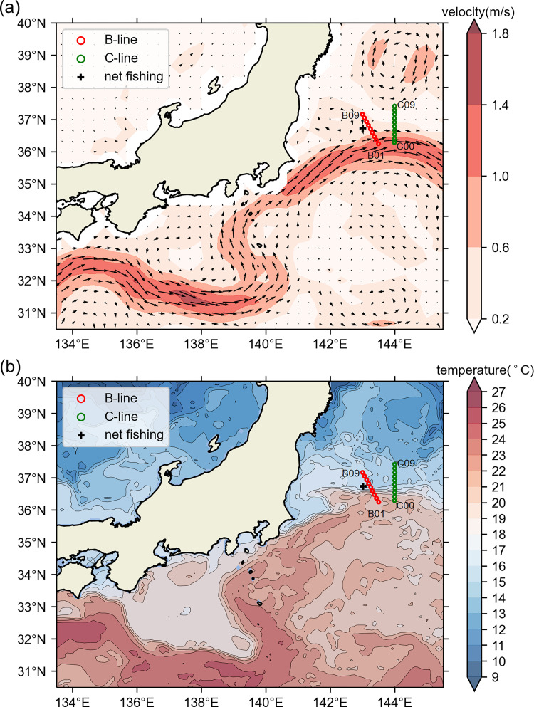
Sea water samples were collected during the KS-18-5 research cruise in the Kuroshio Extension area by the R/V Shinsei-Maru research vessel. OceanDNA seawater samples were collected from 19 water sampling stations. The stations were arranged on two lines (nine stations from B-line and ten stations from C-line; Fig 1 and S1 Table), and from 13 depths (0, 5, 10, 15, 25, 30, 50, 80, 100, 125, 150, 200, and 300 m). In total, 247 seawater samples were collected, including 117 B-line samples and 130 C-line samples. Compared with the sea surface velocity and temperature field, both lines extended from the Kuroshio Extension to the mixed water region in the north.

Fig 1. Positions of OceanDNA sampling stations in the Kuroshio Extension area.

Fig 1

The red circles represent stations B01–B09, the green circles represent stations C00–C09, and the plus mark represents the net sampling location. Maps were made in Spyder (Python 3.7) using Natural Earth data. Free vector and raster map data @ naturalearthdata.com. (a) Colors represent current speeds and arrows represent current velocity vectors on the sea surface (5-day mean from May 10 to 14, 2018) from Ocean Surface Current Analysis Real-time data [34] (https://podaac.jpl.nasa.gov/Data hosted and openly shared by the PO.DAAC, without restriction, in accordance with NASA’s Earth Science program Data and Information Policy). (b) The contours and colors represent sea surface temperature (˚C) distribution on May 10, 2018, from GHRSST Level 4 OSTIA Global Foundation Sea Surface Temperature Analysis data [35] (hosted and openly shared by the PO.DAAC, without restriction, in accordance with NASA’s Earth Science program Data and Information Policy).

Water samples were collected from water depths of 5 to 300 m using Niskin bottles combined with a conductivity temperature depth (CTD) system. This system can accurately determine depth. The surface (0 m) water samples were collected using a clean bucket. Each water sample was stored in a fresh plastic bag that had been co-washed three times (Rontainer, Sekisui Chemical Co., Ltd., Tokyo, Japan). Approximately 7 L was weighed and then immediately filtered using a Sterivex-GP pressure filter unit with a 0.22 μm pore size (Merck Millipore, Burlington, MA, USA). To perform filtering, the inlet end of the Sterivex-GP pressure filter unit was attached to the 1/4 inch HB to M Luer lock (XX3002564; Merck Biopharma Co., Ltd., Tokyo, Japan), which was assembled into one end of a peroxide-cured silicon pump tube (L/S25, 96400–25; Yamato Scientific Co., Ltd., Tokyo, Japan). The pump tube was then fixed by tube cartridge (07519–70; Yamato Scientific Co., Ltd.) to the multi-channel pump head (07519–06; Yamato Scientific Co., Ltd.). The pump head was assembled into a digital pump (07528–10; Yamato Scientific Co., Ltd.). Finally, through the peroxide treatment silicon pump tube, the digital pump (rotation speed set to 60 rpm) pushed the water sample through the Sterivex-GP pressure filter unit from the plastic bag. Before we collected water samples, all silicon tubes, tube connectors, and stoppers were treated with 1% bleach (sodium hypochlorite solution) and cleaned with Milli-Q water to prevent contamination before filtering. After filtering, the plastic bag with a residual water sample was weighed, and the weight difference before and after filtering was used as the filtered sea water mass. We connected a 50 mL disposable syringe to the Sterivex filter unit through a disposable connector (discarded every time after dealing with a sample) and then excluded the resident water in the Sterivex filter unit by air pressure applied manually. After excluding the sea water completely from the Sterivex filter unit, we used a 5 mL disposable syringe (discarded every time after dealing with a sample) to add 2.0 mL RNAlater Stabilization Solution (Thermo Fisher Scientific, Waltham, MA, USA) into each Sterivex filter unit to immerse the whole membrane, which was then kept for 12 h at 4 ˚C. Then, Sterivex filter units were immediately stored at -30 ˚C until DNA extraction.

The eDNA extraction and purification

The extraction and purification of eDNA from Sterivex were performed by Bioengineering Lab. Co., Ltd. (Kanagawa, Japan). The procedure was based on a previously described protocol [19]. Before starting the extraction, silicon tubes, tube connectors, and stoppers were treated with 1% bleach and cleaned with Milli-Q water to prevent contamination. Sterivex filter units were kept on ice until the RNAlater was thawed. DNA extraction was performed using Charge Switch Forensic DNA Purification Kit (Thermo Fisher Scientific). Briefly, after removing RNAlater, 2 mL lysis mix (containing Lysis Buffer and 20 μL Proteinase K) was added to each Sterivex filter unit. The filter units were incubated at 55 ˚C for 30 min. Supernatant from each filter unit was collected in a new microcentrifuge tube. ChargeSwitch® Magnetic Beads (Thermo Fisher Scientific) were added to bind to the DNA. After washing the magnetic beads with Wash Buffer, DNA combined with Magnetic Beads was eluted with 150 μL of Elution Buffer for each sample and then transferred to a new microcentrifuge tube. Finally, the eluted DNA solutions were purified by adding an equal volume of AMPure XP Reagent (Beckman Coulter Inc., Brea, CA, USA).

Metabarcoding of fish (MiFish)

MiFish primers were used to perform eDNA metabarcoding as previously described [19]. The primers and reaction settings are shown in the S1 Fig. The details of the MiFish method are provided in the S1 Text. The MiFish procedure was performed on all 247 samples. The library preparation of the MiFish method failed in 63 samples (49 B-line samples and 14 C-line samples). In the remaining 184 samples, the average number of raw and clean reads was 600,718 and 138,476, respectively. The details of the MiFish results are shown in the S3S5 Tables.

The qPCR assay

Real-time qPCR assays were performed on the sardine, anchovy, chub mackerel, blue mackerel, jack mackerel, and saury [36]. Six species-specific probes and qPCR primers were designed in D-loop region of mitochondrial DNA. The detailed primer sequence information is listed in S2 Table. Each 10 μL qPCR reaction system contained 5 μL of 2× TaKaRa Probe qPCR Mix (TaKaRa Bio Corporation, Kusatsu, Japan), 900 nM forward primers and 900 nM reverse primers, 250 nM fluorescent probe, and 2.5 μL of sample/standard eDNA. We performed qPCR using an ABI 7900HT real-time PCR system (Applied Biosystems, Foster City, CA, USA). The thermal cycling profile comprised 95 ˚C for 10 min and 50 cycles of 95 ˚C for 15 s, and 65 ˚C for 1 min. The qPCR standards were prepared by diluting the plasmids containing the specific sequences at 101–107 copies per 2.5 μL in each reaction. Each sample was assayed in duplicate, and the detailed qPCR results are shown in S6 Table.

Data analyses

Presence detection indicated that the target fish quantity was higher than 0 in any of the duplicate assays for the qPCR method, or the ratio of the target operational taxonomic units (OTUs) in the total OTU read was >1% in a sample using the MiFish method. The latter 1% criterion for the MiFish method was used to avoid false presence [37, 38]. In this study, species-specific qPCR was performed on the aforementioned six pelagic fish. However, because of the similarity in DNA sequences, OTU identification in MiFish could not clearly distinguish the DNA sequences of chub mackerel and blue mackerel. Thus, we used “chub/blue mackerel” to represent either chub mackerel or blue mackerel and treated “chub/blue mackerel” as the target fish. As a result, the comparison of eDNA metabarcoding and qPCR was performed on five targets: chub/blue mackerel (chub mackerel and blue mackerel), sardine, anchovy, jack mackerel, and saury. The MiFish procedure could not be applied to 63 OceanDNA samples in which the library preparation failed (S3 Table). Therefore, we excluded their data before comparing the performance of the qPCR and MiFish methods.

To examine the correspondence of the survey results of the species-specific qPCR and MiFish methods, Pearson’s Phi-coefficients were calculated based on presence/absence detection data to assess the correlation between qPCR (presence = 1/absence = 0) and MiFish (presence = 1/absence = 0). The value of the Pearson’s Phi-coefficient ranges from -1 to +1, where +1 (-1) indicates perfect agreement (disagreement) and 0 indicates no relationship. As the precise meaning of the coefficients (strengths of relationships) depends on the freedom of data, we also calculated the p-value of Fisher’s exact test.

To compare the presence data distributions between the qPCR and MiFish methods, spatial distribution figures were made using matplotlib with Spyder (Python 3.7) based on presence/absence data of the qPCR and MiFish methods, DNA quantity data of the qPCR method, space data (depth, longitude, latitude), and environmental data (temperature and chlorophyll-a concentration, showed in S9 Table). The data were recorded by the CTD system during the seawater sampling.

As shown in the Results section, the detection rate of the qPCR method was higher than that of the MiFish method, whereas the presence distributions of both methods were similar. To find the possible reasons for the different detection rate of the qPCR and MiFish methods, we tested two hypotheses. One hypothesis posited that the detection limit of the qPCR method is lower than that of the MiFish method. The other hypothesis posited that OceanDNA amplification of target species is inhibited by OceanDNA amplification of non-target species (amplification bias) in the MiFish method. To test whether the two methods have different DNA quantity detection limits, we divided the samples in which qPCR detected the target species into different groups according to the OceanDNA quantity of target species measured by qPCR and then compared the MiFish detection loss rate among all groups. The MiFish detection loss rate was defined as the percentage of samples in which MiFish failed to detect target species. To test the influence of the amplification bias in the MiFish method, we divided the samples in which qPCR detected the target species into the group in which target fish was not detected by MiFish (MiFish-0/qPCR-1 samples) and the group in which MiFish also detected the target fish (MiFish-1/qPCR-1 samples). We then compared the number of non-target OTUs and the chlorophyll-a concentration between these two groups using the Mann-Whitney U test to test the statistical significance. Non-target OTUs were OTUs that belonged to the non-target fish. Chlorophyll-a concentration was included for comparison because it represents the algae quantity, and algae DNA could not be amplified using the MiFish method.

Results

Comparison between results from OceanDNA and from net survey

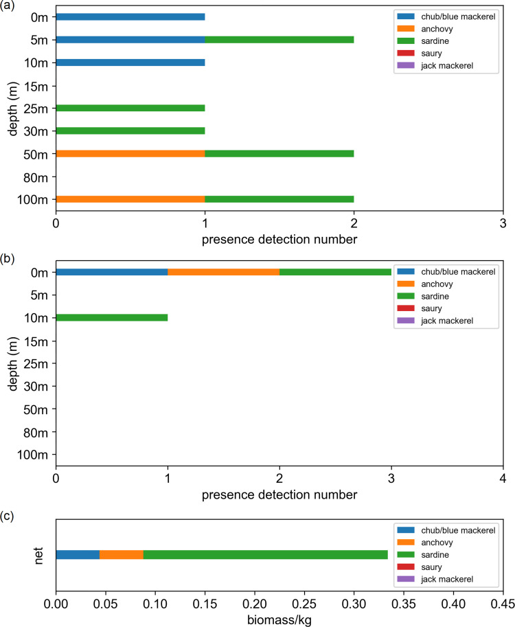
As our purpose of developing OceanDNA methods is to survey fish distribution in the open ocean, we firstly compared our OceanDNA data with direct net sampling survey results. The net sampling data used were obtained by a pelagic trawling covered depth of 0–25 m that were performed during a cruise by the R/V Soyo-Maru vessel of the Japan Fisheries Research and Education Agency (net sampling station is shown in Fig 1), on May 11, 2018. The target fish species caught in the net comprised 158 sardine (40.85–66.80 mm body length, 0.85–3.74 g body weight; 0.246 kg in total), 289 chub/blue mackerel (16.99–64.08 mm fork length. 0.04–2.75 g in body weight; 0.044 kg in total), and 40 anchovies (34.99–68.68 mm body length, 0.35–3.24 g in body weight; 0.044 kg in total). The chub/blue mackerel were small in size and it was difficult to distinguish the two species morphologically. The abundance was highest in sardine, followed by chub/blue mackerel and anchovy. We chose the OceanDNA data from station B06 (sampled on May 11, 2018) for comparison because it is the closest to the net sampling point (Fig 1). Both the qPCR and MiFish methods detected the presence of sardine, chub/blue mackerel, and anchovy at B06, with sardine being most prevalent (Fig 2). The result of OceanDNA method mirrored that of the net sampling.

Fig 2. OceanDNA detection results of five target fish at station B06 and fish caught in net sampling.

Fig 2

Detection results using (a) qPCR method and (b) MiFish method, and (c) fish biomass data from the net sampling (pelagic trawling, at depth of 0–25 m).

Detection rate comparison of target species between MiFish and qPCR

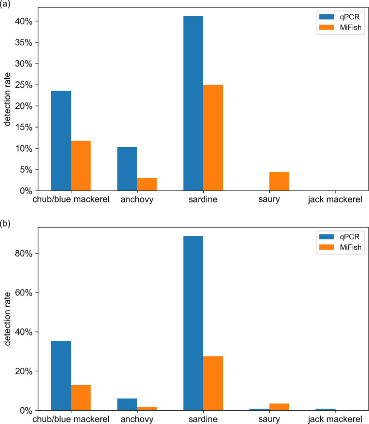
The detection results (presence/absence) from both qPCR and MiFish methods showed that sardine and chub/blue mackerel were the most common among the five target fish in both B-line and C-line (“all detection rate” always >20%; S7 Table). For these two common fish, as well as for anchovy, the detection rate using the qPCR method appeared to always be much higher (more than twice) than that of the MiFish method, in both B-line and C-line samples (Fig 3). In the case of saury, the detection rate using the MiFish method was higher than that of the qPCR method (the detection rates were all <4% using both methods and on both lines). Jack mackerel was almost undetectable in both the B- and C-lines, by qPCR or MiFish methods.

Fig 3. Detection rate ranking of five target fish based on qPCR data and MiFish data.

Fig 3

Data based on samples collected on the B-line (a) and C-line (b). The ‘detection rate’ is calculated as: detection number / whole sample number of B-line (or C-line). chub/blue mackerel = Scomber japonicus and Scomber australasicus, sardine = Sardinops melanostictus, anchovy = Engraulis japonicus, jack mackerel = Trachurus japonicus, saury = Cololabis saira.

On the contrary, the detection rate ranking of the five target fish was the same on the B-line and C-line in each method (Fig 3). For qPCR data, the detection rate ranking for both lines was sardine > chub/blue mackerel > anchovy > saury > jack mackerel. The detection rate ranking of MiFish data on both lines was sardine > chub/blue mackerel > saury > anchovy > jack mackerel. Although the qPCR and MiFish methods have different detection rates for each target fish, the detection rate rankings from the two methods were comparable.

Correlation between MiFish and qPCR on target species

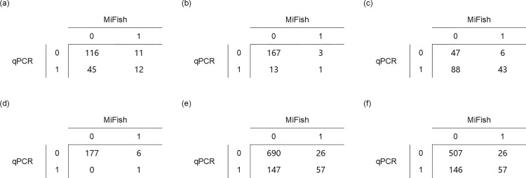
Since similarities and differences were observed between the results of the MiFish and qPCR methods, we examined the Phi-coefficients between the detection results from the two methods. Since the relative abundance ranking among the five target fish was the same in the B-line and C-line samples and the two lines were not far from each other (Fig 1), we summed the data of these two lines in the Phi-coefficient analyses. Phi-coefficient examination was performed on presence/absence data (0 = absence, 1 = presence) of each target fish (except jack mackerel, as it was only detected in one sample) from all 184 OceanDNA samples in which the MiFish library preparation was successful (Fig 4 and Table 1), and a positive Phi-correlation was detected in every examination (Table 1). Statistically significant positive correlations were found for chub/blue mackerel, sardine, and saury (chub/blue mackerel: Phi ≈ 0.17, p ≈ 0.0199; sardine: Phi ≈ 0.22, p ≈ 0.0017; saury: Phi ≈ 0.37, p ≈ 0.0380; Fisher’s exact test). In addition, statistically significant positive correlations were observed (when not including jack mackerel: Phi ≈ 0.33, p ≈ 6.6e-19; when including jack mackerel: Phi ≈ 0.35, p ≈ 1.0e-21) from the pooled data (Table 1). The Phi- coefficient examination also revealed the similarity of detection results between the MiFish and qPCR methods.

Fig 4. Comparison tables for Phi-coefficients analysis of detection performance among the five target fish.

Fig 4

(a) chub/blue mackerel (Scomber japonicus and Sc. australasicus), (b) anchovy (Engraulis japonicus), (c) sardine (Sardinops melanostictus), (d) saury (Cololabis saira), (e) pooling (sum data of all five target fish, chub/blue mackerel, sardine, anchovy, saury and jack mackerel), (f) pooling-4 (sum data of (a) to (d)). 0 = absence, 1 = presence.

Table 1. Coefficients (Phi-coefficient value and Fisher’s exact test) of detection performance among the five target fish.

chub/blue mackerel anchovy sardine saury pooling-4a poolingb
Phi-coefficient 0.17 0.10 0.22 0.37 0.33 0.35
p <0.05 >0.05 <0.05 <0.05 <0.05 <0.05

a pooling-4 is the sum of chub/blue mackerel, anchovy, sardine, and saury

b pooling refers to the sum of chub/blue mackerel, sardine, anchovy, saury, and jack mackerel.

Spatial distribution comparison of target species between MiFish and qPCR

The spatial distribution was analyzed in each target fish using detection data from the qPCR and MiFish methods, as well as the position information of each OceanDNA sample. Because the number of samples detected in anchovy, saury, and jack mackerel were very low (presence detection sample numbers were always <10), only the spatial distribution results of the high-detection-rate chub/blue mackerel and sardine were compared (Figs 5 and 6).

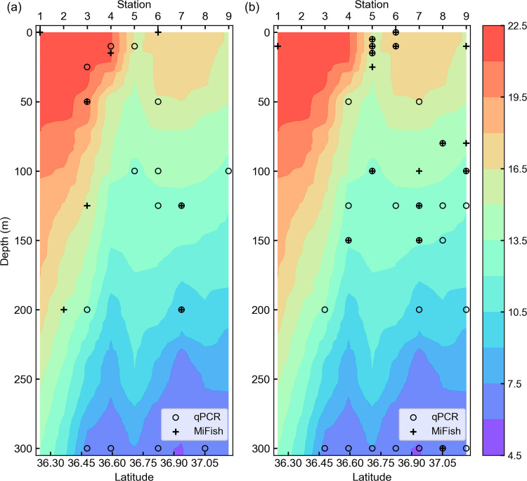
Fig 5. Comparison of spatial distribution pattern detected by qPCR and MiFish methods in the B-line vertical section.

Fig 5

(a) Spatial distribution of chub/blue mackerel (Scomber japonicus and Sc. australasicus). (b) Spatial distribution of sardine (Sardinops melanostictus). Plus marks represent the presence detection samples found by the MiFish method and open circles represent the presence detection samples found by the qPCR method. Background colors represent water temperature (˚C).

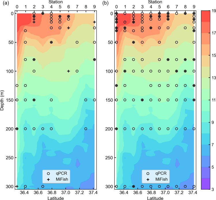
Fig 6. Comparison of spatial distribution pattern detected by qPCR and MiFish methods in the C-line vertical section.

Fig 6

(a) Spatial distribution of chub/blue mackerel (Scomber japonicus and Sc. australasicus). (b) Spatial distribution of sardine (Sardinops melanostictus). Plus marks represent the presence detection samples found by the MiFish method and open circles represent the presence detection samples found by the qPCR method. Background colors represent water temperature (˚C).

The spatial distribution area of the MiFish presence detection samples was narrower than that from the qPCR method, as expected based on the lower detection rate of the MiFish method (Figs 5 and 6). However, similar distribution patterns were found of the MiFish and qPCR methods for both sardine and chub/blue mackerel on the two lines, especially for B-line sardine and C-line chub/blue mackerel. In detail, for B-line collected sardines, the qPCR and MiFish results showed a distribution pattern comprising no sardine distribution at stations 1 and 2, distribution depth of sardine shifted to shallower from stations 4 to 5, and the distribution depth of sardine shifted to deeper from stations 6 to 7 and 8. For C-line chub/blue mackerel, both the qPCR and MiFish results showed similar patterns, with the distribution of chub/blue mackerel around stations 4–6 showing shallower distribution than the others.

Influences of detection limit and amplification bias on detection rate

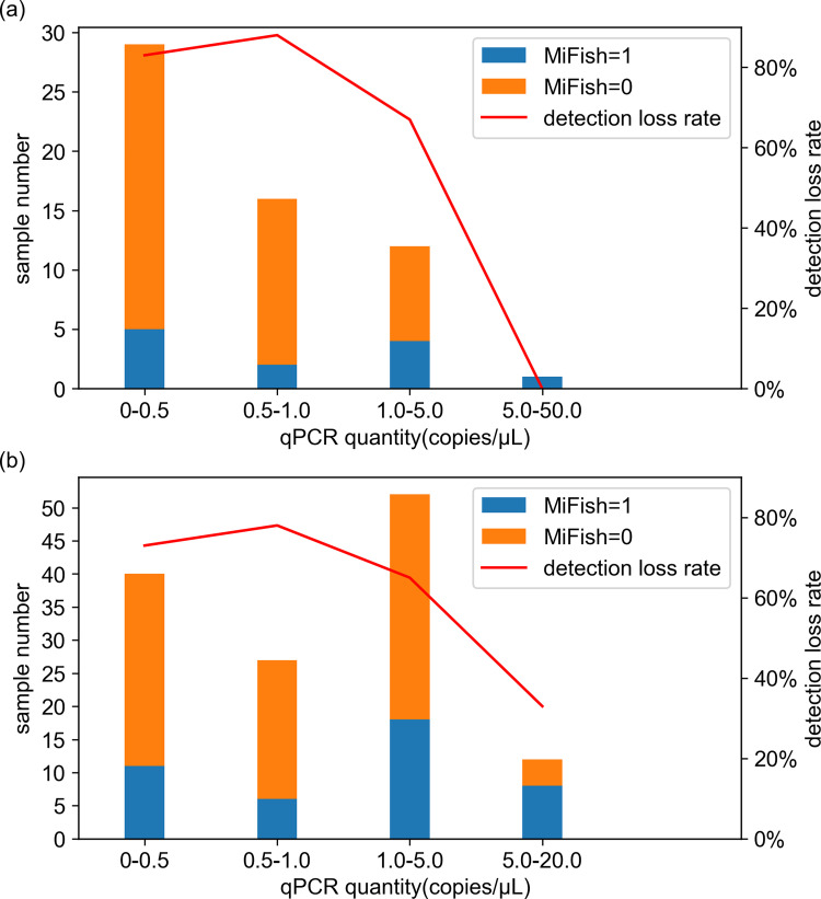
As possible sources to generate the detection rate difference between MiFish and qPCR found in this study (Fig 3 and S7 Table), we tested the influence of the detection limit and amplification bias in MiFish. For the detection limit effect, the dependency of MiFish detection loss rate on OceanDNA quantity of target species measured by qPCR was investigated. The histograms of the MiFish presence/absence detection based on the qPCR determined OceanDNA quantity showed that the highest MiFish detection loss rate (percentage of the samples in which MiFish failed to detect the target species) of chub/blue mackerel and sardine appeared in the bins of 0–0.5 copies/μL and 0.5–1.0 copies/μL OceanDNA quantity (Fig 7). The MiFish detection loss rate decreased markedly for samples in which OceanDNA quantity exceeded 5.0 copies/μL (Fig 7). However, the MiFish detection loss rate was still high (66.7% for chub/blue mackerel and 65.4% for sardine) in the bin of 1.0–5.0 copies/μL OceanDNA quantity (Fig 7).

Fig 7. Comparison of MiFish presence/absence detection samples based on OceanDNA quantity measured by qPCR.

Fig 7

(a) Result of chub/blue mackerel (Scomber japonicus and Sc. australasicus) and (b) result of sardine (Sardinops melanostictus). The figures were prepared from pooling data of B-line and C-line. Samples in which qPCR did not detect OceanDNA are not included in this figure. Numbers in x axis represent the OceanDNA quantity (copies/μL) measured by qPCR.

For amplification bias in MiFish, we investigated the influence of non-target OTUs and chlorophyll-a. Non-target OTUs were those that did not belong to the target fish; these represented fish DNA that was amplified in the library preparation but did not belong to the target fish. In the MiFish-0/qPCR-1 samples, the number of non-target OTUs was higher than that in the MiFish-1/qPCR-1 samples (Table 2). The increase was approximately 35% higher in chub/blue mackerel, 153% in sardine, and 115% in anchovy. All increases were statistically significant (p < 0.001, Mann-Whitney U test). Conversely, the chlorophyll-a concentration was lower in the MiFish-0/qPCR-1 samples than in the MiFish-1/qPCR-1 samples for anchovy and sardine (Table 2).

Table 2. Difference of non-target OTU number (average per sample) and chlorophyll-a concentration (average per sample) between MiFish-0/qPCR-1 and MiFish-1/qPCR-1 samples.

Non-target OTU number Chlorophyll-a concentration (mg/m3)
chub/blue mackerel anchovy sardine chub/blue mackerel anchovy sardine
MiFish-0/qPCR-1 18.29 16.92 29.16 0.65 0.57 0.59
MiFish-1/qPCR-1 13.58 8.00 11.53 0.59 1.35 0.90
P <0.001 <0.001 <0.001 <0.001 <0.001 <0.001

The data were based on samples after excluding library failure samples. MiFish-0/qPCR-1 samples represent samples in which the qPCR method detected the target fish, whereas the MiFish method did not. MiFish-1/qPCR-1 samples represent samples in which both the qPCR and MiFish methods detected the target fish.

Discussion

eDNA has received much attention as a means of investigating the distribution of open ocean fish. Metabarcoding and species-specific PCR are the two main eDNA analysis methods. The MiFish method is a metabarcoding method developed for fish that focuses on a part of the fish 12S rRNA gene. Although it is a relatively new method, the MiFish method has been widely used by researchers for several years to obtain fish species information from eDNA. As shown in Fig 2, the present findings also partially confirmed the reliability of MiFish through the consistency between net sampling data and MiFish fish detection data. On the contrary, considering the simplicity of the procedure and its lower cost, the species-specific PCR method was also used for eDNA when only one or several species were analyzed. Our qPCR data were consistent with the net sampling data. Therefore, as the main focus of this study, we compared the characteristics of qPCR and MiFish methods.

Quality control of data

Before discussing about the comparison between the characteristics of qPCR and MiFish methods, we must discuss about data quality of this study. Our seawater sampling protocol was relatively old and it was designed based on previous eDNA studies on the marine microorganisms, in which negative controls were not common [39, 40]. Thus, the KS-18-5 samples did not contain the negative controls. However, the later studies have shown the importance of negative controls to monitor possible contamination during the sampling [41, 42]. Therefore, in the cruises after KS-18-5, negative controls by filtering the Milli-Q water during water sampling for OceanDNA were performed. From the negative controls of the seven cruises after KS-18-5, no target fish DNA have been detected by the qPCR method and no reads have been detected by the MiFish methods (in the form of “library preparation failed as no PCR products obtained”). Although this cannot justify the lack of negative control in KS-18-5, the use of the same seawater sampling method suggested that there could be no contamination in the KS-18-5 sampling.

Similarity between the detection performance of the qPCR and MiFish methods

In this study, we compared the presence/absence detection results, which represent the existence/absence of the target fish. The results demonstrated the similarity between the detection performance of the qPCR and MiFish methods. First, similar relative abundance rankings among the different target fish were shown using the two eDNA analysis methods. Second, correlation analyses indicated that presence/absence estimations obtained from the qPCR data and the MiFish data always exhibited positive correspondence with each other (Table 1). Based on the combined data of the B- and C- lines, pairwise Phi-coefficients showed positive correlations in four of the five target fish as well as in multiple target pooling data. These results suggested that both qPCR and MiFish methods can detect small pelagic fish. Third, a similar spatial distribution pattern was evident between the MiFish and qPCR detection results, especially for B-line sardine and C-line chub/blue mackerel (Figs 5 and 6). The findings showed comparable similarity and positive correlation between the detection performance of the qPCR and MiFish methods.

Higher detection rate in the qPCR method than in the MiFish method

Despite the similarity and positive correlation shown above, the difference between the detection performance of the qPCR and MiFish methods was also clearly shown in this study. According to the detection results (S7 Table), among the five target fish, the qPCR method showed a much higher detection rate than that of the MiFish method for the three targets, including the most and second most common fish. Combining B-line and C-line data together, the detection rate was 2.48 times for chub/blue mackerel, 2.67 times for sardine, and 3.50 times for anchovy in the qPCR method compared with in the MiFish method. In addition, only one detection of jack mackerel was detected by qPCR.

Previous studies have also shown that species-specific qPCR has a higher sensitivity than eDNA metabarcoding. For example, in a study in Blakney Creek, New South Wales, metabarcoding failed to detect redfin perch (Perca fluviatilis) DNA in six of eight samples where qPCR showed a positive result [43]. In two studies in the same survey area, TaqMan qPCR detected catfish (Silurus glanis) at two sampling sites and European eel (Anguilla anguilla) at one sampling site, whereas eDNA metabarcoding failed to detect these two fish [44, 45]. In addition, previous eDNA survey studies on other aquatic animals also showed a higher detection ability of qPCR than of the metabarcoding method, such as in great crested newt (Triturus cristatus) [15] and Mediterranean fanworm Sabella spallanzanii [30]. Our study and the aforementioned studies indicate there is a difference in detection sensitivity between the qPCR method and metabarcoding method in the survey of aquatic creatures using eDNA.

Relationship between OceanDNA quantity and difference in the detection rate between MiFish and qPCR

Including MiFish, eDNA metabarcoding methods have been widely used in wildlife surveys, especially in species diversity surveys [6, 4648]. The ability to detect a large number of species in a single performance is a great advantage of metabarcoding compared with species-specific PCR. However, as shown above, the lower sensitivity of the eDNA metabarcoding evident in many studies is a problem that researchers need to consider.

To help overcome this problem, we tested some possible reasons for the low detection rate of the MiFish method compared to the qPCR method. The first hypothesis is that the detection limit of the MiFish method is higher than that of the qPCR method. In this case, when the OceanDNA quantity of the target fish is very low, it would still be detectable by qPCR, but very difficult to detect using the MiFish method. Given the wide distribution of these fish in the survey area, we focused on the sardine and chub/blue mackerel test results. Samples with the highest OceanDNA quantity had the lowest MiFish detection loss rate, as expected (Fig 7). However, the MiFish detection loss rate did not change appreciably between samples with 1.0–5.0 copies/μL OceanDNA quantity and lower OceanDNA quantity (Fig 7). Therefore, the presently-observed lower detection rate of the MiFish method compared with that of the qPCR method can only be partially attributed to the lower detection limit of the MiFish method with low eDNA quantity in the open ocean.

Other possible reasons for difference in detection rate between MiFish and qPCR

Several previous studies have found that amplification bias is a reason for missing some species in eDNA metabarcoding [43, 4951]. Normally, amplification bias is thought to be the preferential amplification on some DNA, especially high relative abundance DNA [5255]. Including the MiFish used in this study, the primers used in eDNA metabarcoding focus on universal sequences to cover a wide range of species. Therefore, preferential amplification revealed the existence of species that released less eDNA and were masked by species that left more DNA in the sample [23, 24]. In the present study, in MiFish-0/qPCR-1 samples, the number of non-target OTUs tended to be higher than that in the MiFish-1/qPCR-1 samples (Table 2). However, the chlorophyll-a concentrations did not show such a tendency (Table 2). The chlorophyll-a concentration represents the amount of algae; algae DNA is not the target of MiFish primers, and it should not be amplified (while chlorophyll-a itself inhibits PCR reactions [56]). Therefore, our results also seem to support amplification bias on non-target-species DNA to be a reason for the detection rate difference between the MiFish method and the qPCR method, while inhibition from chlorophyll-a seemed to not be the reason. However, amplification bias cannot completely explain this difference.

Library preparation failures in MiFish

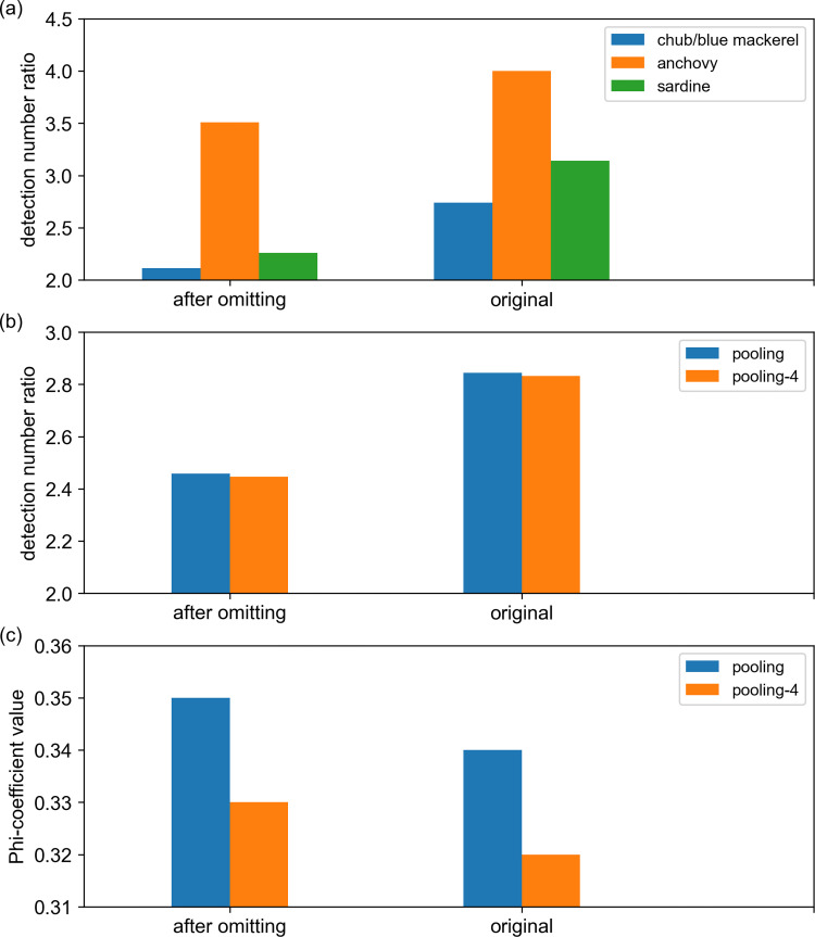
As mentioned before, library preparation using the MiFish method failed in 63 OceanDNA samples (S3 Table). These samples were not included in the above analyses. If these samples were included, the comparison more obviously revealed the difference in the detection rates between the two methods (Fig 8, S8 Table). The difference in the detection rates between the qPCR and MiFish methods for chub/blue mackerel, anchovy, and sardine increased (Fig 8A), and the detection rate differences also increased from 2.46 to 2.84 for the pooled data (Fig 8B). On the contrary, the Phi-coefficient value between qPCR and MiFish presence/absence data decreased for the pooling data (Fig 8C).

Fig 8. Differences between the detection results of the qPCR and MiFish methods before and after exclusion of samples in which the library preparation failed.

Fig 8

(a) The detection number ratios (qPCR detection number / MiFish detection number) of chub/blue mackerel (Scomber japonicus and Sc. australasicus), anchovy (Engraulis japonicus) and sardine (Sardinops melanostictus). (b) The detection number ratios of pooling and pooling-4 (pooling-4 = sum of chub/blue mackerel, anchovy, sardine and saury, pooling = chub/blue mackerel, sardine, anchovy, saury, and jack mackerel). (c) Phi-coefficient values of pooling and pooling-4. ‘original’ means the data came from all 247 OceanDNA samples, ‘after omitting’ means those samples, in which the library preparation failed, were excluded.

One critical step in the MiFish library preparation is the first-round PCR that the universal PCR primers amplify the target gene fragments across the target taxa [21]. Therefore, the library preparation failure is likely to happen in the samples with virtually no DNA of target taxa. Indeed, in the negative controls we collected during the later cruises, the library preparation also failed, as mentioned earlier. The lack of target taxa DNA is also possible in seawater samples for the high proportion of microbial DNA and the sparse distribution of fish eDNA [10]. Although MiFish primers were reported to be very effective [57, 58], there are still possible problems like PCR dropout which can lead to amplification failures of existing fish DNA in samples [21]. As shown in our study, practical library preparation of OceanDNA could be challenging in the MiFish method.

Perspectives for future development of OceanDNA analysis methods in marine fish distribution surveys

The present comparison between OceanDNA results and real direct net survey results (Figs 1 and 2; although the net sampling data was limited to one station) confirmed the ability of the qPCR and MiFish methods, when used on OceanDNA, to be useful tools for small pelagic fish distribution surveys. We believe it is valuable to further develop these two OceanDNA analysis methods. The MiFish method has been proven to be a sensitive and effective tool in eDNA studies for fish. However, the present results and those of previous studies also showed that metabarcoding methods, such as MiFish, sometimes suffer a lower detection rate than the qPCR method. In addition, primer efficiency can influence the detection by the eDNA metabarcoding method. For example, in one study, three metabarcoding primers gave different primer efficiencies in silico tests, and 16S rRNA or CytB primers sometimes displayed an efficiency close to 0 [59].Another study also showed that in the same water samples, the metabarcoding method targeting the COI gene primer had a much higher detection rate compared to the 18S rRNA [30]. On the contrary, the results from eDNA metabarcoding cannot be replaced by those from qPCR. The present findings highlight that there are always some unique detection results found only by the MiFish method for each of the five target fish (S7 Table), especially for the detection of saury. Moreover, when conducting research aimed at large numbers of species, such as biodiversity studies, the use of the eDNA metabarcoding method is essential.

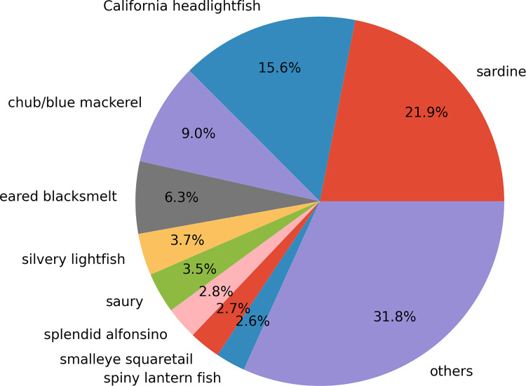
Given the information above, we propose combining the qPCR and MiFish methods in OceanDNA analysis to monitor quantitative distribution of small pelagic fish species with information of fish community structures. The qPCR method provides quantitative estimates and special distribution of target fish species, as shown in Figs 5 and 6. On the other hand, using MiFish data of the water samples in which the qPCR method detected the target species (we called them selected samples), fish community coexisting with the target fish can be inferred. For example, we estimated the fish community coexisting with sardine and chub/blue mackerel (Fig 9). This calculation assumed that DNA quantity is proportional to the MiFish read number over the sum of the samples. At this stage, the MiFish data do not have quantitative information. Therefore, the ratios in Fig 9 do not represent relative abundance. To overcome this issue, quantitative sequencing (qSeq) technique, which adds a random sequence tag to the target sequence before PCR in the library preparation to estimate the DNA quantity from read number found in the metabarcoding [60, 61], is required. However, we could not find any previous study on fish with qSeq in the open ocean. Besides, even with qSeq, there is still a risk that the ‘read number-DNA quantity’ relationship varies among different species, which can be examined by qPCR data of several target fish. In addition, the added random sequence tag for qSeq consumed some reads in the sequencing, which may reduce the read number we can obtain from the target fish DNA. In our preliminary experiments (data has not been submitted), adding the sequence tag increased the number of non-detected species in MiFish, which suggested the possible difficulty of qSeq in OceanDNA. Thus, we suggest the combination of the qPCR and MiFish methods as a possible way to detect a large number of species that coexist with the target fish. The combined methods will permit a comprehensive understanding of the quantitative distribution of small pelagic fish within fish communities in the open waters.

Fig 9. Pie chart based on read percentages of fish species detected by the MiFish method.

Fig 9

Here, the pie chart was made based on MiFish data from C-line samples that at least one of sardine, chub/blue mackerel and anchovy was detected by the qPCR method, where sardine = Sardinops melanostictus, California headlightfish = Diaphus theta, chub/blue mackerel = Scomber japonicus + Sc. australasicus, eared blacksmelt = Lipolagus ochotensis, silvery lightfish = Maurolicus muelleri, saury = Cololabis saira, splendid alfonsino = Beryx splendens, smalleye squaretail = Tetragonurus cuvieri, spiny lantern fish = Myctophum spinosum.

Conclusions

The eDNA analysis methods of species-specific PCR and eDNA metabarcoding are widely used in eDNA survey research [10, 62, 63]. eDNA metabarcoding methods, such as MiFish, can detect a large number of species in one working series, and so have been actively used in research that aims to investigate multiple species [10, 63, 64]. For studies that focus on a single target species, species-specific PCR can also be a good choice because of its simplicity, high sensitivity, and low cost [14]. For OceanDNA research, which is still being developed, the selection of eDNA analysis methods is an important decision for researchers. To provide some clarity for this decision, we conducted an eDNA survey in the open ocean area of the Kuroshio Extension using both the species-specific qPCR and MiFish methods. Five target small pelagic fish (chub/blue mackerel, sardine, anchovy, jack mackerel, and saury) were selected to compare the detection performance of the two eDNA analysis methods. The similar existence/absence detection performance and spatial distribution pattern estimation were evidence of the consistency between the detection results of qPCR and MiFish. At the same time, the obvious detection rate difference between the qPCR method and the MiFish method showed that one method cannot simply replace the other. Our study results indicate that the influence of non-target species DNA on the amplification in MiFish and the lower sensitivity of the MiFish method on lower DNA quantity can partially explain the detection rate difference between the qPCR and MiFish methods. However, more studies are needed to determine the difference between the survey results of the two methods. Thus, we suggest the value of these two OceanDNA analysis methods as well as the necessity for further developing.

Supporting information

S1 Text. Detailed procedure for the MiFish method.

(PDF)

S1 Fig. The library procedure, thermal cycle settings, and primer sequences of the MiFish method in this study.

(PDF)

S2 Fig. Comparison between the spatial distribution of presence detection samples found by MiFish and the eDNA quantity obtained from the qPCR results.

(PDF)

S1 Table. Longitude, latitude, and CTD starting time of each sampling station.

(XLSX)

S2 Table. Species-specific qPCR primers and probes in this study.

(XLSX)

S3 Table. Library preparation results for each sample.

(XLSX)

S4 Table. Number of OTU reads in B-line OceanDNA samples.

(XLSX)

S5 Table. Number of OTU reads in C-line OceanDNA samples.

(XLSX)

S6 Table. qPCR quantity (copies/μL) for all OceanDNA samples.

(XLSX)

S7 Table. Detection results of target fish by the qPCR and MiFish methods.

(XLSX)

S8 Table. Comparison tables for Phi-coefficients analysis of detection performance.

(PDF)

S9 Table. Temperature and chlorophyll-a concentration data recorded in the sampling area.

(XLSX)

Acknowledgments

The OceanDNA survey was conducted by the R/V Shinsei-Maru. We thank the captain and all members of cruise KS-18-5. We also appreciate the assistance of Dr. Megumi Enomoto for sea water sampling. We express special thanks to Mr. Shinsuke Toyoda of Marine Work Japan for his operation of the CTD water sampling device. MiFish method analysis was performed with the help of Bioengineering Lab. Co., Ltd.

Data Availability

All relevant data are within the manuscript and its Supporting Information files.

Funding Statement

SI, JP21H04735, The Japan Society for the Promotion of Science (JSPS) KAKENHI, https://www.jsps.go.jp/english/. The funders had no role in study design, data collection and analysis, decision to publish, or preparation of the manuscript. SI & SH, The OceanDNA project, The University of Tokyo Future Society Initiative, https://www.u-tokyo.ac.jp/adm/fsi/ja/projects/sdgs/projects_00103.html. The funders had no role in study design, data collection and analysis, decision to publish, or preparation of the manuscript.

References

  • 1.Thomsen Philip F., Willerslev Eske. Environmental DNA–An emerging tool in conservation for monitoring past and present biodiversity. Biol Conserv. 2015. March. doi: 10.1016/j.biocon.2014.11.019 [DOI] [Google Scholar]
  • 2.Barnes Matthew A., Turner Cameron R. The ecology of environmental DNA and implications for conservation genetics. Conserv Genet. 2015. September 08. doi: 10.1007/s10592-015-0775-4 [DOI] [Google Scholar]
  • 3.Sassoubre Lauren M., Yamahara Kevan M., Gardner Luke D., Block Barbara A., Boehm Alexandria B. Quantification of environmental DNA (eDNA) shedding and decay rates for three marine fish. Environ Sci Technol. 2016. August 31. doi: 10.1021/acs.est.6b03114 [DOI] [PubMed] [Google Scholar]
  • 4.Nevers Meredith B., Kasia Przybyla-Kelly Dawn Shively, Morris Charles C., Dickey Joshua, Byappanahalli Murulee N. Influence of sediment and stream transport on detecting a source of environmental DNA. PLOS ONE. 2020. December 28. doi: 10.1371/journal.pone.0244086 [DOI] [PMC free article] [PubMed] [Google Scholar]
  • 5.Irene Adrian-Kalchhauser Patricia Burkhardt-Holm. An eDNA assay to monitor a globally invasive fish species from flowing freshwater. PLOS ONE. 2016. January 27. doi: 10.1371/journal.pone.0147558 [DOI] [PMC free article] [PubMed] [Google Scholar]
  • 6.Stat Michael, John Jeffrey, D. DiBattista, Stephen J. Newman, Michael Bunce, Euan S. Harvey. Combined use of eDNA metabarcoding and video surveillance for the assessment of fish biodiversity. Conserv Biol. 2018. July 13. doi: 10.1111/cobi.13183 [DOI] [PMC free article] [PubMed] [Google Scholar]
  • 7.Plough Louis V., Ogburn Matthew B., Fitzgerald Catherine L., Geranio Rose, Marafino Gabriella A., Richie Kimberly D. Environmental DNA analysis of river herring in Chesapeake Bay: A powerful tool for monitoring threatened keystone species. PLOS ONE. 2018. November 1. doi: 10.1371/journal.pone.0205578 [DOI] [PMC free article] [PubMed] [Google Scholar]
  • 8.Minegishi Yuki, Marty Kwok-Shing Wong Takashi Kanbe, Araki Hitoshi, Kashiwabara Tomomi, Ijichi Minoru, et al. Spatiotemporal distribution of juvenile chum salmon in Otsuchi Bay, Iwate, Japan, inferred from environmental DNA. PLOS ONE. 2019. September 4. doi: 10.1371/journal.pone.0222052 [DOI] [PMC free article] [PubMed] [Google Scholar]
  • 9.Gold Zachary, Sprague Joshua, Kushner David J., Erick Zerecero Marin Paul H. Barber. eDNA metabarcoding as a biomonitoring tool for marine protected areas. PLOS ONE. 2021. February 24. doi: 10.1371/journal.pone.0238557 [DOI] [PMC free article] [PubMed] [Google Scholar]
  • 10.Stat Michael, Huggett Megan J., Bernasconi Rachele, Joseph D. DiBattista, Tina E. Berry, Stephen J. Newman, et al. Ecosystem biomonitoring with eDNA: Metabarcoding across the tree of life in a tropical marine environment. Sci Rep. 2017. Sepetember 25. doi: 10.1038/s41598-017-12501-5 [DOI] [PMC free article] [PubMed] [Google Scholar]
  • 11.10.1002/edn3.32 Eva Garcia-Vazquez, Oriane Georges, Sara Fernandez, Alba Ardura. eDNA metabarcoding of small plankton samples to detect fish larvae and their preys from Atlantic and Pacific waters. Sci Rep. 2021. March 31. [DOI] [PMC free article] [PubMed] [Google Scholar]
  • 12.Takahara Teruhiko, Minamoto Toshifumi, Yamanaka Hiroki, Doi Hideyuki, Zen’ichiro Kawabata. Estimation of fish biomass using environmental DNA. PLOS ONE. 2012. April 26. doi: 10.1371/journal.pone.0035868 [DOI] [PMC free article] [PubMed] [Google Scholar]
  • 13.Rees Helen C., Maddison Ben C., Middleditch David J., Patmore James R. M., Gough Kevin C. REVIEW: The detection of aquatic animal species using environmental DNA–A review of eDNA as a survey tool in ecology. J Appl Ecol. 2014. June 20. doi: 10.1111/1365-2664.12306 [DOI] [Google Scholar]
  • 14.Tsuji Satsuki, Takahara Teruhiko, Doi Hideyuki, Shibata Naoki, Yamanaka Hiroki. The detection of aquatic macroorganisms using environmental DNA analysis—A review of methods for collection, extraction, and detection. Environ DNA. 2019. June 20. doi: 10.1002/edn3.21 [DOI] [Google Scholar]
  • 15.Harper Lynsey R., Lori Lawson Handley Christoph Hahn, Boonham Neil, Rees Helen C., Gough Kevin C., et al. Needle in a haystack? A comparison of eDNA metabarcoding and targeted qPCR for detection of the great crested newt (Triturus cristatus). Ecol Evol. 2018. May 29. doi: 10.1002/ece3.4013 [DOI] [PMC free article] [PubMed] [Google Scholar]
  • 16.Goldberg Caren S., Turner Cameron R., Deiner Kristy, Klymus Katy E., Philip Francis Thomsen Melanie A. Murphy, et al. Critical considerations for the application of environmental DNA methods to detect aquatic species. Methods Ecol Evol. 2016. May 26. doi: 10.1111/2041-210X.12595 [DOI] [Google Scholar]
  • 17.Shaw Jennifer L. A., Weyrich Laura, Cooper Alan. Using environmental (e)DNA sequencing for aquatic biodiversity surveys: A beginner’s guide. Mar Freshw Res. 2016. March 1. doi: 10.1071/MF15361 [DOI] [Google Scholar]
  • 18.Deiner Kristy, Bik Holly M., Elvira Mächler Mathew Seymour, Anaïs Lacoursière-Roussel Florian Altermatt, et al. Environmental DNA metabarcoding: Transforming how we survey animal and plant communities. Mol Ecol. 2017. September 16. doi: 10.1111/mec.14350 [DOI] [PubMed] [Google Scholar]
  • 19.Miya Masaki, Sato Yukuto, Fukunaga Tsukasa, Sado Tetsuya, Sato Keiichi, et al. MiFish, a set of universal PCR primers for metabarcoding environmental DNA from fishes: Detection of more than 230 subtropical marine species. R Soc Open Sci. 2015. July 01. doi: 10.1098/rsos.150088 [DOI] [PMC free article] [PubMed] [Google Scholar]
  • 20.Valentini Alice, Taberlet Pierre, Miaud Claude, Civade Raphaël, Herder Jelger, Thomsen Philip Francis, et al. Next-generation monitoring of aquatic biodiversity using environmental DNA metabarcoding. Mol Ecol. 2015. October 19. doi: 10.1111/mec.13428 [DOI] [PubMed] [Google Scholar]
  • 21.Miya Masaki, Gotoh Ryo O., Sado Tetsuya. MiFish metabarcoding: A high-throughput approach for simultaneous detection of multiple fish species from environmental DNA and other samples. Fish Sci 86. 2020. September 15. doi: 10.1007/s12562-020-01461-x [DOI] [Google Scholar]
  • 22.Collins Rupert A., Wangensteen Owen S., Eoin J. O’Gorman, Stefano Mariani, David W. Sims, Martin J. Genner. Persistence of environmental DNA in marine systems. Commun Biol. 2018. November 5. doi: 10.1038/s42003-018-0192-6 [DOI] [PMC free article] [PubMed] [Google Scholar]
  • 23.Andruszkiewicz Elizabeth A., Koseff Jeffrey R., Fringer Oliver B., Ouellette Nicholas T., Lowe Anna B., Edwards Christopher A., et al. Modeling environmental DNA transport in the coastal ocean using Lagrangian particle tracking. Front Mar Sci. 2019. August 07. doi: 10.3389/fmars.2019.00477 [DOI] [Google Scholar]
  • 24.Scott Baker C., Debbie Steel, Sharon Nieukirk, Holger Klinck. Environmental DNA (eDNA) from the wake of the whales: Droplet digital PCR for detection and species identification. Front Mar Sci. 2018. April 19. doi: 10.3389/fmars.2018.00133 [DOI] [Google Scholar]
  • 25.Aird Daniel, Ross Michael G., Chen Wei-Sheng, Danielsson Maxwell, Fennell Timothy, Russ Carsten, et al. Analyzing and minimizing PCR amplification bias in Illumina sequencing libraries. Genome Biology. 2011. February 21. doi: 10.1186/gb-2011-12-2-r18 [DOI] [PMC free article] [PubMed] [Google Scholar]
  • 26.Guo-Jie Brandon-Mong Han Ming Gan, Sing K. W., Lee P. S., Lim P. E., Wilson J. J. DNA metabarcoding of insects and allies: An evaluation of primers and pipelines. Bull Entomol Res. 2015. September 07. doi: 10.1017/S0007485315000681 [DOI] [PubMed] [Google Scholar]
  • 27.Evans Nathan T., Olds Brett P., Renshaw Mark A., Turner Cameron R., Li Yiyuan, Jerde Christopher L., et al. Quantification of mesocosm fish and amphibian species diversity via environmental DNA metabarcoding. Mol Ecol Resour. 2015. May 28. doi: 10.1111/1755-0998.12433 [DOI] [PMC free article] [PubMed] [Google Scholar]
  • 28.Kelly Ryan P., Port Jesse A., Yamahara Kevan M., Crowder Larry B. Using environmental DNA to census marine fishes in a large mesocosm. PLOS ONE. 2014. January 15. doi: 10.1371/journal.pone.0086175 [DOI] [PMC free article] [PubMed] [Google Scholar]
  • 29.Schneider Judith, Valentini Alice, Dejean Tony, Montarsi Fabrizio, Taberlet Pierre, Glaizot Olivier, et al. Detection of invasive mosquito vectors using environmental DNA (eDNA) from water samples. PLOS ONE. 2016. September 14. doi: 10.1371/journal.pone.0162493 [DOI] [PMC free article] [PubMed] [Google Scholar]
  • 30.Wood Susanna A., Pochon Xavier, Laroche Olivier, Ulla von Ammon Janet Adamson, Zaiko Anastasija. A comparison of droplet digital polymerase chain reaction (PCR), quantitative PCR and metabarcoding for species-specific detection in environmental DNA. Mol Ecol Resour. 2019. July 10. doi: 10.1111/1755-0998.13055 [DOI] [PubMed] [Google Scholar]
  • 31.Stoeckle Mark Y., Mithun Das Mishu Zachary Charlop-Powers. GoFish: A versatile nested PCR strategy for environmental DNA assays for marine vertebrates. PLOS ONE. 2018. December 11. doi: 10.1371/journal.pone.0198717 [DOI] [PMC free article] [PubMed] [Google Scholar]
  • 32.Andruszkiewicz Elizabeth A., Sassoubre Lauren M., Boehm Alexandria B. Persistence of marine fish environmental DNA and the influence of sunlight. PLOS ONE. 2017. September 15. doi: 10.1371/journal.pone.0185043 [DOI] [PMC free article] [PubMed] [Google Scholar]
  • 33.Sato Yukuto, Miya Masaki, Fukunaga Tsukasa, Sado Tetsuya, Iwasaki Wataru. MitoFish and MiFish pipeline: A mitochondrial genome database of fish with an analysis pipeline for environmental DNA metabarcoding. Mol Biol Evol. 2018. April 14. doi: 10.1093/molbev/msy074 [DOI] [PMC free article] [PubMed] [Google Scholar]
  • 34.ESR. 2009. OSCAR third deg. Ver. 1. PO.DAAC, CA, USA. Dataset accessed [2021-07-08] at 10.5067/OSCAR-03D01. [DOI]
  • 35.UK Met Office. 2005. OSTIA L4 SST Analysis. Ver. 1.0. PO.DAAC, CA, USA. Dataset accessed [2021-07-06] at 10.5067/GHOST-4FK01. [DOI]
  • 36.Marty Kwok-Shing Wong Shigenori Nobata, Ito Shin-ichi, Hyodo Susumu. Development of species-specific multiplex real-time PCR assays for tracing the small pelagic fishes of North Pacific with environmental DNA. Environ DNA. 2022. January 14. doi: 10.1002/edn3.275 [DOI] [Google Scholar]
  • 37.Bernd Hänfling Lori Lawson Handley, Read Daniel S., Hahn Christoph, Li Jianlong, Nichols Paul, et al. Environmental DNA metabarcoding of lake fish communities reflects long-term data from established survey methods. Mol Ecol. 2016. April 20. doi: 10.1111/mec.13660 [DOI] [PubMed] [Google Scholar]
  • 38.Li Jianlong, Tristan W. Hatton-Ellis, Lori-Jayne Lawson Handley, Helen S. Kimbell, Marco Benucci, Graeme Peirson, et al. Ground-truthing of a fish-based environmental DNA metabarcoding method for assessing the quality of lakes. J Appl Ecol. 2019. February 03. doi: 10.1111/1365-2664.13352 [DOI] [Google Scholar]
  • 39.Stoeck Thorsten, Bass David, Nebel Markus, Christen Richard, Jones Meredith D. M., Breiner Hans-werner, et al. Multiple marker parallel tag environmental DNA sequencing reveals a highly complex eukaryotic community in marine anoxic water. Mol Ecol. 2010. February 10. doi: 10.1111/j.1365-294X.2009.04480.x [DOI] [PubMed] [Google Scholar]
  • 40.Behnke Anke, Bunge John, Barger Kathryn, Breiner Hans-Werner, Alla Victoria, Stoeck Thorsten. Microeukaryote community patterns along an O2/H2S gradient in a supersulfidic anoxic fjord (Framvaren, Norway). Appl Environ Microbiol. 2006. May. doi: [DOI] [PMC free article] [PubMed] [Google Scholar]
  • 41.Yamamoto Satoshi, Masuda Reiji, Sato Yukuto, Sado Tetsuya, Araki Hitoshi, Kondoh Michio, et al. Environmental DNA metabarcoding reveals local fish communities in a species-rich coastal sea. Sci Rep. 2017. January 12. doi: 10.1038/srep40368 [DOI] [PMC free article] [PubMed] [Google Scholar]
  • 42.Sato Masaaki, Inoue Nariaki, Nambu Ryogen, Furuichi Naoki, Imaizumi Tomohito, Ushio Masayuki. Quantitative assessment of multiple fish species around artificial reefs combining environmental DNA metabarcoding and acoustic survey. Sci Rep. 2021. September 30. doi: 10.1038/s41598-021-98926-5 [DOI] [PMC free article] [PubMed] [Google Scholar]
  • 43.Bylemans Jonas, Gleeson Dianne M., Duncan Richard P., Hardy Christopher M., Furlan Elise M. A performance evaluation of targeted eDNA and eDNA metabarcoding analyses for freshwater fishes. Environ DNA. 2019. October 16. doi: 10.1002/edn3.41 [DOI] [Google Scholar]
  • 44.Schenekar Tamara, Schletterer Martin, Lecaudey Laurène A., Weiss Steven J. Reference databases, primer choice, and assay sensitivity for environmental metabarcoding: Lessons learnt from a re-evaluation of an eDNA fish assessment in the Volga headwaters. River Res Appl. 2020. March 01. doi: 10.1002/rra.3610 [DOI] [Google Scholar]
  • 45.Lecaudey Laurène A., Schletterer Martin, Kuzovlev Vyacheslav V., Hahn Christoph, Weiss Steven J. Fish diversity assessment in the headwaters of the Volga River using environmental DNA metabarcoding. Aquat Conserv. 2019. August 01. doi: 10.1002/aqc.3163 [DOI] [Google Scholar]
  • 46.Nakagawa Hikaru, Yamamoto Satoshi, Sato Yukuto, Sado Tetsuya, Minamoto Toshifumi, Miya Masaki. Comparing local- and regional-scale estimations of the diversity of stream fish using eDNA metabarcoding and conventional observation methods. Freshw Biol. 2018. February 27. doi: 10.1111/fwb.13094 [DOI] [Google Scholar]
  • 47.West Katrina M., Stat Michael, Harvey Euan S., Skepper Craig L., Joseph D. DiBattista, Zoe T. Richards, et al. eDNA metabarcoding survey reveals fine-scale coral reef community variation across a remote, tropical island ecosystem. Mol Ecol. 2020. February 11. doi: 10.1111/mec.15382 [DOI] [PubMed] [Google Scholar]
  • 48.Naiara Guimarães Sales Owen Simon Wangensteen, Daniel Cardoso Carvalho Kristy Deiner, KimPræbel IlariaCoscia, et al. Space-time dynamics in monitoring Neotropical fish communities using eDNA metabarcoding. Sci Total Environ. 2021. February 1. doi: 10.1016/j.scitotenv.2020.142096 [DOI] [PubMed] [Google Scholar]
  • 49.Tremblay Julien, Singh Kanwar, Fern Alison, Kirton Edward S., He Shaomei, Woyke Tanja, et al. Primer and platform effects on 16S rRNA tag sequencing. Front Microbiol. 2015. August 04. doi: 10.3389/fmicb.2015.00771 [DOI] [PMC free article] [PubMed] [Google Scholar]
  • 50.Elbrecht Vasco, Leese Florian. Can DNA-based ecosystem assessments quantify species abundance? Testing primer bias and biomass—Sequence relationships with an innovative metabarcoding protocol. PLOS ONE. 2015. July 8. doi: 10.1371/journal.pone.0130324 [DOI] [PMC free article] [PubMed] [Google Scholar]
  • 51.Kelly Ryan P., Andrew Olaf Shelton Ramón Gallego. Understanding PCR processes to draw meaningful conclusions from environmental DNA studies. Sci Rep. 2019. August 20. doi: 10.1038/s41598-019-48546-x [DOI] [PMC free article] [PubMed] [Google Scholar]
  • 52.Alberdi Antton, Aizpurua Ostaizka, Thomas Marcus, Gilbert Pius, Bohmann Kristine. Scrutinizing key steps for reliable metabarcoding of environmental samples. Methods Ecol Evol. 2017. July 01. doi: 10.1111/2041-210X.12849 [DOI] [Google Scholar]
  • 53.Nichols Ruth V., Vollmers Christopher, Newsom Lee A., Wang Yue, Heintzman Peter D., Leighton McKenna, et al. Minimizing polymerase biases in metabarcoding. Mol Ecol Resour. 2018. May 24. doi: 10.1111/1755-0998.12895 [DOI] [PubMed] [Google Scholar]
  • 54.Josep Piñol Miquel A. Senar, Symondson William O. C. The choice of universal primers and the characteristics of the species mixture determine when DNA metabarcoding can be quantitative. Mol Ecol. 2018. June 25. doi: 10.1111/mec.14776 [DOI] [PubMed] [Google Scholar]
  • 55.Gargan Laura M., Brooks Paul R., Vye Siobhan R., Ironside Joseph E., Jenkins Stuart R., Crowe Tasman P., et al. The use of environmental DNA metabarcoding and quantitative PCR for molecular detection of marine invasive non-native species associated with artificial structures. Biol Invasions. 2021. November 09. doi: 10.1007/s10530-021-02672-8 [DOI] [Google Scholar]
  • 56.Kang Tae-Jin, Yang Moon-Sik. Rapid and reliable extraction of genomic DNA from various wild-type and transgenic plants. BMC Biotechnol. 2004. September 02. doi: 10.1186/1472-6750-4-20 [DOI] [PMC free article] [PubMed] [Google Scholar]
  • 57.Bylemans J, Gleeson DM, Hardy CM, Furlan E. Toward an ecoregion scale evaluation of eDNA metabarcoding primers: a case study for the freshwater fish biodiversity of the Murray-Darling Basin (Australia). Ecol Evol. 2018. August 05. doi: 10.1002/ece3.4387 [DOI] [PMC free article] [PubMed] [Google Scholar]
  • 58.Collins RA, Bakker J, Wangensteen OS, Soto AZ, Corrigan L, Sims DW, et al. Non-specific amplification compromises environmental DNA metabarcoding with COI. Methods Ecol Evol. 2019. August 02. doi: 10.1111/2041-210x.13276 [DOI] [Google Scholar]
  • 59.Burgener M., Hübner P. Mitochondrial DNA enrichment for species identification and evolutionary analysis. Z Lebensm Unters Forsch A Food Res Technol. 1998. October. doi: 10.1007/s002170050329 [DOI] [Google Scholar]
  • 60.Hoshino Tatsuhiko, Inagaki Fumio. Application of stochastic labeling with random sequence barcodes for simultaneous quantification and sequencing of environmental 16S rRNA genes. PLOS ONE. 2017. January 4. doi: 10.1371/journal.pone.0169431 [DOI] [PMC free article] [PubMed] [Google Scholar]
  • 61.Hoshino Tatsuhiko, Nakao Ryohei, Doi Hideyuki, Minamoto Toshifumi. Simultaneous absolute quantification and sequencing of fish environmental DNA in a mesocosm by quantitative sequencing technique. Sci Rep. 2021. February 23. doi: 10.1038/s41598-021-83318-6 [DOI] [PMC free article] [PubMed] [Google Scholar]
  • 62.Doi Hideyuki, Uchii Kimiko, Takahara Teruhiko, Matsuhashi Saeko, Yamanaka Hiroki, Minamoto Toshifumi. Use of droplet digital PCR for estimation of fish abundance and biomass in environmental DNA surveys. PLOS ONE. 2015. March 23. doi: 10.1371/journal.pone.0122763 [DOI] [PMC free article] [PubMed] [Google Scholar]
  • 63.Bessey Cindy, Jarman Simon N., Berry Oliver, Olsen Ylva S., Bunce Michael, Simpson Tiffany, et al. Maximizing fish detection with eDNA metabarcoding. Environ DNA. 2020. February 20. doi: 10.1002/edn3.74 [DOI] [Google Scholar]
  • 64.Ruppert Krista M., Richard J., Klinea Md Saydur Rahman. Past, present, and future perspectives of environmental DNA (eDNA) metabarcoding: A systematic review in methods, monitoring, and applications of global eDNA. Glob Ecol Conserv. 2019. January. doi: 10.1016/j.gecco.2019.e00547 [DOI] [Google Scholar]

Decision Letter 0

Arga Chandrashekar Anil

15 Feb 2022

PONE-D-21-36080Comparison of species-specific qPCR and metabarcoding methods to detect small pelagic fish distribution from open ocean environmental DNAPLOS ONE

Dear Dr. Ito,

Thank you for submitting your manuscript to PLOS ONE. After careful consideration, we feel that it has merit but does not fully meet PLOS ONE’s publication criteria as it currently stands. Therefore, we invite you to submit a revised version of the manuscript that addresses the points raised during the review process.

 I have one review which points several corrections and urge the authors to carefully consider them and revise suitably for further consideration

Please submit your revised manuscript by Apr 01 2022 11:59PM. If you will need more time than this to complete your revisions, please reply to this message or contact the journal office at plosone@plos.org. When you're ready to submit your revision, log on to https://www.editorialmanager.com/pone/ and select the 'Submissions Needing Revision' folder to locate your manuscript file.

Please include the following items when submitting your revised manuscript:

  • A rebuttal letter that responds to each point raised by the academic editor and reviewer(s). You should upload this letter as a separate file labeled 'Response to Reviewers'.

  • A marked-up copy of your manuscript that highlights changes made to the original version. You should upload this as a separate file labeled 'Revised Manuscript with Track Changes'.

  • An unmarked version of your revised paper without tracked changes. You should upload this as a separate file labeled 'Manuscript'.

If you would like to make changes to your financial disclosure, please include your updated statement in your cover letter. Guidelines for resubmitting your figure files are available below the reviewer comments at the end of this letter.

If applicable, we recommend that you deposit your laboratory protocols in protocols.io to enhance the reproducibility of your results. Protocols.io assigns your protocol its own identifier (DOI) so that it can be cited independently in the future. For instructions see: https://journals.plos.org/plosone/s/submission-guidelines#loc-laboratory-protocols. Additionally, PLOS ONE offers an option for publishing peer-reviewed Lab Protocol articles, which describe protocols hosted on protocols.io. Read more information on sharing protocols at https://plos.org/protocols?utm_medium=editorial-email&utm_source=authorletters&utm_campaign=protocols.

We look forward to receiving your revised manuscript.

Kind regards,

Arga Chandrashekar Anil, Ph. D., D. Agr.,

Academic Editor

PLOS ONE

Journal Requirements:

When submitting your revision, we need you to address these additional requirements.

1. Please ensure that your manuscript meets PLOS ONE's style requirements, including those for file naming. The PLOS ONE style templates can be found at

https://journals.plos.org/plosone/s/file?id=wjVg/PLOSOne_formatting_sample_main_body.pdf  and

https://journals.plos.org/plosone/s/file?id=ba62/PLOSOne_formatting_sample_title_authors_affiliations.pdf

2. Thank you for stating the following financial disclosure:

“SI, JP21H04735, The Japan Society for the Promotion of Science (JSPS) KAKENHI, https://www.jsps.go.jp/english/, NO

SI & SH, The OceanDNA project, The University of Tokyo Future Society Initiative, https://www.u-tokyo.ac.jp/adm/fsi/ja/projects/sdgs/projects_00103.html, NO”

Please state what role the funders took in the study.  If the funders had no role, please state: "The funders had no role in study design, data collection and analysis, decision to publish, or preparation of the manuscript."

If this statement is not correct you must amend it as needed.

Please include this amended Role of Funder statement in your cover letter; we will change the online submission form on your behalf.

3. Thank you for stating the following in the Acknowledgments Section of your manuscript:

“The contribution of SI was supported by the Japan Society for the Promotion of Science (JSPS) KAKENHI (Grant Number JP21H04735) and the OceanDNA project under the University of Tokyo Future Society Initiative. The OceanDNA survey was conducted by the R/V Shinsei-Maru. We thank the captain and all members of cruise KS-18-5. We also appreciate the assistance of Dr. Megumi Enomoto for sea water sampling.”

We note that you have provided additional information within the Acknowledgements Section that is not currently declared in your Funding Statement. Please note that funding information should not appear in the Acknowledgments section or other areas of your manuscript. We will only publish funding information present in the Funding Statement section of the online submission form.

Please remove any funding-related text from the manuscript and let us know how you would like to update your Funding Statement. Currently, your Funding Statement reads as follows:

“SI, JP21H04735, The Japan Society for the Promotion of Science (JSPS) KAKENHI, https://www.jsps.go.jp/english/, NO

SI & SH, The OceanDNA project, The University of Tokyo Future Society Initiative, https://www.u-tokyo.ac.jp/adm/fsi/ja/projects/sdgs/projects_00103.html, NO”

Please include your amended statements within your cover letter; we will change the online submission form on your behalf.

4. We note that figure 1 in your submission contain [map/satellite] images which may be copyrighted. All PLOS content is published under the Creative Commons Attribution License (CC BY 4.0), which means that the manuscript, images, and Supporting Information files will be freely available online, and any third party is permitted to access, download, copy, distribute, and use these materials in any way, even commercially, with proper attribution. For these reasons, we cannot publish previously copyrighted maps or satellite images created using proprietary data, such as Google software (Google Maps, Street View, and Earth). For more information, see our copyright guidelines: http://journals.plos.org/plosone/s/licenses-and-copyright.

 We require you to either (1) present written permission from the copyright holder to publish these figures specifically under the CC BY 4.0 license, or (2) remove the figures from your submission:

   1. You may seek permission from the original copyright holder of figure 1 to publish the content specifically under the CC BY 4.0 license.  

We recommend that you contact the original copyright holder with the Content Permission Form (http://journals.plos.org/plosone/s/file?id=7c09/content-permission-form.pdf) and the following text:

“I request permission for the open-access journal PLOS ONE to publish XXX under the Creative Commons Attribution License (CCAL) CC BY 4.0 (http://creativecommons.org/licenses/by/4.0/).  Please be aware that this license allows unrestricted use and distribution, even commercially, by third parties. Please reply and provide explicit written permission to publish XXX under a CC BY license and complete the attached form.”

Please upload the completed Content Permission Form or other proof of granted permissions as an "Other" file with your submission.

 In the figure caption of the copyrighted figure, please include the following text: “Reprinted from [ref] under a CC BY license, with permission from [name of publisher], original copyright [original copyright year].”

   2. If you are unable to obtain permission from the original copyright holder to publish these figures under the CC BY 4.0 license or if the copyright holder’s requirements are incompatible with the CC BY 4.0 license, please either i) remove the figure or ii) supply a replacement figure that complies with the CC BY 4.0 license. Please check copyright information on all replacement figures and update the figure caption with source information. If applicable, please specify in the figure caption text when a figure is similar but not identical to the original image and is therefore for illustrative purposes only.

The following resources for replacing copyrighted map figures may be helpful:

 USGS National Map Viewer (public domain): http://viewer.nationalmap.gov/viewer/

The Gateway to Astronaut Photography of Earth (public domain): http://eol.jsc.nasa.gov/sseop/clickmap/

Maps at the CIA (public domain): https://www.cia.gov/library/publications/the-world- factbook/index.html and https://www.cia.gov/library/publications/cia-maps-publications/index.html

NASA Earth Observatory (public domain): http://earthobservatory.nasa.gov/

Landsat: http://landsat.visibleearth.nasa.gov/

USGS EROS (Earth Resources Observatory and Science (EROS) Center) (public domain): http://eros.usgs.gov/#

Natural Earth (public domain): http://www.naturalearthdata.com/

[Note: HTML markup is below. Please do not edit.]

Reviewers' comments:

Reviewer's Responses to Questions

Comments to the Author

1. Is the manuscript technically sound, and do the data support the conclusions?

The manuscript must describe a technically sound piece of scientific research with data that supports the conclusions. Experiments must have been conducted rigorously, with appropriate controls, replication, and sample sizes. The conclusions must be drawn appropriately based on the data presented.

Reviewer #1: Partly

Reviewer #2: Yes

**********

2. Has the statistical analysis been performed appropriately and rigorously?

Reviewer #1: Yes

Reviewer #2: Yes

**********

3. Have the authors made all data underlying the findings in their manuscript fully available?

The PLOS Data policy requires authors to make all data underlying the findings described in their manuscript fully available without restriction, with rare exception (please refer to the Data Availability Statement in the manuscript PDF file). The data should be provided as part of the manuscript or its supporting information, or deposited to a public repository. For example, in addition to summary statistics, the data points behind means, medians and variance measures should be available. If there are restrictions on publicly sharing data—e.g. participant privacy or use of data from a third party—those must be specified.

Reviewer #1: Yes

Reviewer #2: Yes

**********

4. Is the manuscript presented in an intelligible fashion and written in standard English?

PLOS ONE does not copyedit accepted manuscripts, so the language in submitted articles must be clear, correct, and unambiguous. Any typographical or grammatical errors should be corrected at revision, so please note any specific errors here.

Reviewer #1: No

Reviewer #2: Yes

**********

5. Review Comments to the Author

Please use the space provided to explain your answers to the questions above. You may also include additional comments for the author, including concerns about dual publication, research ethics, or publication ethics. (Please upload your review as an attachment if it exceeds 20,000 characters)

Reviewer #1: General comments to authors

Authors collected large number of eDNA samples from open ocean environment and performed eDNA analysis using both species-specific qPCR and MiFish metabarcoding. The results indicated both methods have good agreement, while the qPCR provide better detection rates for the target species than the metabarcoding. I consider this study provide important information and knowledge for ocean ecology. However, the one analysis to estimate “corrected” read number is bit confusing (L492-516). In addition, some explanation is insufficient and there are several typos in this MS. I added detailed comments below.

Detailed comments to authors

L26: correct “invasively” to “noninvasively”.

L52-54: Correct to “~the most reliable method. However, the ability of fish influences the catch efficiency and this method requires enormous amount of time in the open ocean, these factors making difficult to obtain high resolution data.”

L61-62: Is this due to open ocean? Reading this paper [11], it seems to be rather due to difficulty in detecting eDNA from Cetaceans. Other studies (e.g., Garcia-Vazquez et al. 2021) on open oceans are more suitable to refer to here.

eDNA metabarcoding of small plankton samples to detect fish larvae and their preys from Atlantic and Pacific waters Eva Garcia-Vazquez, Oriane Georges, Sara Fernandez & Alba Ardura. Scientific Reports volume 11, Article number: 7224 (2021)

L72: “require only a small volume of sample (<10μL). I think this is a minimum volume rather than standard for species specific PCR. You should describe a standard volume of qPCR samples and it is smaller than metabarcoding.

L86-88: Why does this sentence appear in the paragraph of metabarcoding? This does not fit here.

L92: Typo. Correct ”species□specific PCR” to “species-specific PCR.”

L94: Typo. Correct to “However, research comparison….”

L138: Typo. Correct to “An approximately…”

L138: How did you filter 10L of water using a Sterivex? Using aspirator? You should describe the details of procedure.

L155: You should briefly explain the eDNA extraction procedure.

L167: Correct to “the average number of raw and clean reads was 600,718 and 138,476, respectively.”

L172: Don’t refer to an unpublished material.

L191: Add the interpretation of value of Pearon’s Phi-coefficient (e.g. 0.5 or higher means strong positive relationship, 0.3-0.5 means …, 0.1-0.3 mean… ).

L196:Typo. Delete “We”.

L218-220: Add the information regarding body size of the target species base on net sampling.

Fig.3 is not necessary because this information is described in Table1.

L274-: You mentioned that Mifish metabarcoding failed for 63 samples. Even so, did you use the results of all the samples for this analysis?

L274-279: As above mentioned, I don’t know how large the value of Phi-coefficient means a strong relationship between two variables. Please explain this in the statistical analysis section.

L299-322 and Fig.5: It is difficult to distinguish the results from MiFish and qPCR in Fig 5 because both marks are small and overlayed. Enlarge the mark of each mark and add a graph legend for them.

L386-400: Although this analysis is important and valuable for discussion, this paragraph describes the analysis procedure and its results, which should be moved to “Materials and method” and “Results”.

L419: Typo: “hada”?

L416-427 and 444-452: These sentences should be also moved to “Materials and method” and “Results”.

L484: Typo. Correct to ”In the present study”.

L491: Is it really difficult to apply qSeq to Ocean DNA study? Have you ever confirmed? Are there any references?

L496: Fig 9 should be corrected to Fig 8?

L519-520: Grammar error. Correct this sentence.

L487-489: Quantitative MiSeq sequencing (qMiSeq)has been also utilized in marine environments to monitor eDNA concentrations of multiple species simultaneously (Ushio et al. 2018 Metabarcoding and Metagenomics; Ushio 2019 Methods in Ecology and Evolution; Sato et al. 2021 Scientific reports).

L492-516: I do not understand this calculation from your sentences. Why the relationship in Fig. 8 can be true for other samples? MiFish read percentages in this figure may also have amplification bias. In that case, you cannot use this relationship to estimate the correct fish composition. At least, you should use the equation to explain this calculation for easier understanding to readers. In addition, these sentences also include “Materials and method” and “Results” as above mentioned. Distinguish them from “Discussion”.

Reviewer #2: "Environmental DNA (eDNA) is increasingly used to invasively monitor aquatic animals in

27 freshwater and coastal areas."

The word "invasively" in the first line of the abstract is not correct. eDNA is a non-invasive technique compare to the traditional invasive technique. So application of this world here may cause misunderstanding. I recommend to replace this (invasively) with more relative and meaningful world.

**********

6. PLOS authors have the option to publish the peer review history of their article (what does this mean?). If published, this will include your full peer review and any attached files.

If you choose “no”, your identity will remain anonymous but your review may still be made public.

Do you want your identity to be public for this peer review? For information about this choice, including consent withdrawal, please see our Privacy Policy.

Reviewer #1: No

Reviewer #2: Yes: Dr. Rose S.F Afzali

[NOTE: If reviewer comments were submitted as an attachment file, they will be attached to this email and accessible via the submission site. Please log into your account, locate the manuscript record, and check for the action link "View Attachments". If this link does not appear, there are no attachment files.]

While revising your submission, please upload your figure files to the Preflight Analysis and Conversion Engine (PACE) digital diagnostic tool, https://pacev2.apexcovantage.com/. PACE helps ensure that figures meet PLOS requirements. To use PACE, you must first register as a user. Registration is free. Then, login and navigate to the UPLOAD tab, where you will find detailed instructions on how to use the tool. If you encounter any issues or have any questions when using PACE, please email PLOS at figures@plos.org. Please note that Supporting Information files do not need this step.

PLoS One. 2022 Sep 7;17(9):e0273670. doi: 10.1371/journal.pone.0273670.r002

Author response to Decision Letter 0


29 Apr 2022

Response to academic editor and reviewers

We would like to thank the academic editor and reviewers for their encouragement and their devoted efforts to review the manuscript. We have done our best to address the concerns below. We much appreciate the reviewers for your thorough and constructive reviews, which we think have greatly improved the manuscript. Our responses are in blue and red (red color marked what we modified in the manuscript), while the original comments are in black and italicized.

Detailed response to academic editor

1. Please ensure that your manuscript meets PLOS ONE's style

requirements, including those for file naming. The PLOS ONE style

templates can be found at

https://journals.plos.org/plosone/s/file?id=wjVg/PLOSOne_formatting_sample_main_body.pdf

<https://journals.plos.org/plosone/s/file?id=wjVg/PLOSOne_formatting_sample_main_body.pdf>

and

https://journals.plos.org/plosone/s/file?id=ba62/PLOSOne_formatting_sample_title_authors_affiliations.pdf

<https://journals.plos.org/plosone/s/file?id=ba62/PLOSOne_formatting_sample_title_authors_affiliations.pdf>

> Thank you very much for your reminding. This time we carefully checked to make the manuscript meets PLOS ONE's style requirements.

2. Thank you for stating the following financial disclosure:

“SI, JP21H04735, The Japan Society for the Promotion of Science (JSPS)

KAKENHI, https://www.jsps.go.jp/english/, NO

SI & SH, The OceanDNA project, The University of Tokyo Future Society

Initiative, https://www.u-tokyo.ac.jp/adm/fsi/ja/projects/sdgs/projects_00103.html, NO”

Please state what role the funders took in the study. If the funders had no role, please state: "The funders had no role in study design, data collection and analysis, decision to publish, or preparation of the manuscript."

If this statement is not correct you must amend it as needed.

Please include this amended Role of Funder statement in your cover letter; we will change the online submission form on your behalf.

> Thank you for your reminding. Yes, as you said, the funders had no role in the study, this time we included the statement in the Financial Disclosure section as:

“SI, JP21H04735, The Japan Society for the Promotion of Science (JSPS) KAKENHI, https://www.jsps.go.jp/english/. The funders had no role in study design, data collection and analysis, decision to publish, or preparation of the manuscript.

SI & SH, The OceanDNA project, The University of Tokyo Future Society Initiative,

https://www.u-tokyo.ac.jp/adm/fsi/ja/projects/sdgs/projects_00103.html. The funders had no role in study design, data collection and analysis, decision to publish, or preparation of the manuscript.”

3. Thank you for stating the following in the Acknowledgments Section of

your manuscript:

“The contribution of SI was supported by the Japan Society for the Promotion of Science (JSPS) KAKENHI (Grant Number JP21H04735) and the OceanDNA project under the University of Tokyo Future Society Initiative. The OceanDNA survey was conducted by the R/V Shinsei-Maru.

We thank the captain and all members of cruise KS-18-5. We also appreciate the assistance of Dr. Megumi Enomoto for sea water sampling.”

We note that you have provided additional information within the Acknowledgements Section that is not currently declared in your Funding Statement. Please note that funding information should not appear in the Acknowledgments section or other areas of your manuscript. We will only

publish funding information present in the Funding Statement section of

the online submission form.

Please remove any funding-related text from the manuscript and let us know how you would like to update your Funding Statement. Currently, your Funding Statement reads as follows:

“SI, JP21H04735, The Japan Society for the Promotion of Science (JSPS)

KAKENHI, https://www.jsps.go.jp/english/, NO

SI & SH, The OceanDNA project, The University of Tokyo Future Society

Initiative, https://www.u-tokyo.ac.jp/adm/fsi/ja/projects/sdgs/projects_00103.html, NO”

Please include your amended statements within your cover letter; we will

change the online submission form on your behalf.

> We are sorry for this mistake, this time we deleted the funding-related text in the manuscript and stated the Funding Statement again in the Financial Disclosure section.

4. We note that figure 1 in your submission contain [map/satellite] images which may be copyrighted. All PLOS content is published under the Creative Commons Attribution License (CC BY 4.0), which means that the manuscript, images, and Supporting Information files will be freely available online, and any third party is permitted to access, download, copy, distribute, and use these materials in any way, even commercially, with proper attribution. For these reasons, we cannot publish previously

copyrighted maps or satellite images created using proprietary data, such as Google software (Google Maps, Street View, and Earth). For more information, see our copyright guidelines:

http://journals.plos.org/plosone/s/licenses-and-copyright

<http://journals.plos.org/plosone/s/licenses-and-copyright>.

We require you to either (1) present written permission from the

copyright holder to publish these figures specifically under the CC BY

4.0 license, or (2) remove the figures from your submission:

1. You may seek permission from the original copyright holder of figure 1 to publish the content specifically under the CC BY 4.0 license.

We recommend that you contact the original copyright holder with the Content Permission Form

(http://journals.plos.org/plosone/s/file?id=7c09/content-permission-form.pdf

<http://journals.plos.org/plosone/s/file?id=7c09/content-permission-form.pdf>)

and the following text:

“I request permission for the open-access journal PLOS ONE to publish XXX under the Creative Commons Attribution License (CCAL) CC BY 4.0

(http://creativecommons.org/licenses/by/4.0/

<http://creativecommons.org/licenses/by/4.0/>). Please be aware that this license allows unrestricted use and distribution, even commercially, by third parties. Please reply and provide explicit

written permission to publish XXX under a CC BY license and complete the attached form.”

Please upload the completed Content Permission Form or other proof of granted permissions as an "Other" file with your submission.

In the figure caption of the copyrighted figure, please include the following text: “Reprinted from [ref] under a CC BY license, with permission from [name of publisher], original copyright [original

copyright year].”

2. If you are unable to obtain permission from the original copyright holder to publish these figures under the CC BY 4.0 license or if the copyright holder’s requirements are incompatible with the CC BY 4.0 license, please either i) remove the figure or ii) supply a replacement figure that complies with the CC BY 4.0 license. Please check copyright information on all replacement figures and update the figure caption with source information. If applicable, please specify in the figure caption text when a figure is similar but not identical to the original image and is therefore for illustrative purposes only.

The following resources for replacing copyrighted map figures may be

helpful:

USGS National Map Viewer (public domain):

http://viewer.nationalmap.gov/viewer/

<http://viewer.nationalmap.gov/viewer/>

The Gateway to Astronaut Photography of Earth (public domain):

http://eol.jsc.nasa.gov/sseop/clickmap/

<http://eol.jsc.nasa.gov/sseop/clickmap/>

Maps at the CIA (public domain):

https://www.cia.gov/library/publications/the-world- factbook/index.html

<https://www.cia.gov/library/publications/the-world-%20factbook/index.html>

and

https://www.cia.gov/library/publications/cia-maps-publications/index.html <https://www.cia.gov/library/publications/cia-maps-publications/index.html>

NASA Earth Observatory (public domain):

http://earthobservatory.nasa.gov/ <http://earthobservatory.nasa.gov/>

Landsat: http://landsat.visibleearth.nasa.gov/

<http://landsat.visibleearth.nasa.gov/>

USGS EROS (Earth Resources Observatory and Science (EROS) Center)

(public domain): http://eros.usgs.gov/# <http://eros.usgs.gov/>

Natural Earth (public domain): http://www.naturalearthdata.com/

<http://www.naturalearthdata.com/>

[Note: HTML markup is below. Please do not edit.]

> Thank you very much for your statement about the copyright concerns. When drawing the map in Fig 1, we used the data of Natural Earth. But as stated on the website of Natural Earth (https://www.naturalearthdata.com/about/terms-of-use/) as follows, they don’t need users to ask them for permission.

“All versions of Natural Earth raster + vector map data found on this website are in the public domain. You may use the maps in any manner, including modifying the content and design, electronic dissemination, and offset printing. The primary authors, Tom Patterson and Nathaniel Vaughn Kelso, and all other contributors renounce all financial claim to the maps and invites you to use them for personal, educational, and commercial purposes.

No permission is needed to use Natural Earth. Crediting the authors is unnecessary.

However, if you wish to cite the map data, simply use one of the following.

Short text:

Made with Natural Earth.

Long text:

Made with Natural Earth. Free vector and raster map data @ naturalearthdata.com.”

Thus, we added the statement as “Maps were made in Spyder (Python 3.7) using Natural Earth data. Free vector and raster map data @ naturalearthdata.com.” in the caption of Fig 1.

When drawing Fig 1, we also used the current velocity data (Ocean Surface Current Analysis Real-time) and sea surface temperature data (GHRSST Level 4 OSTIA Global Foundation Sea Surface Temperature Analysis) downloaded from the PO.DAAC. But as stated on the website of PO.DAAC (https://podaac.jpl.nasa.gov/CitingPODAAC) as follows, they also don’t need users to ask them for permission.

“Data hosted by the PO.DAAC is openly shared, without restriction, in accordance with NASA's Earth Science program Data and Information Policy.

Bibliographic citations should be included in the References section of publications and other media to acknowledge those who have created the data, services, and tools provided by the PO.DAAC. Proper citations include the authors, title, publisher, and Digital Object Identifier (DOI) and will allow the products to be discovered and re-used by others. The proper citation for each PO.DAAC product is provided on its landing page and user guide.

Example Data Citation

…”

Thus, we modified the statement as “(a) Colors represent current speeds and arrows represent current velocity vectors on the sea surface (5-day mean from May 10 to 14, 2018) from Ocean Surface Current Analysis Real-time data [34] (hosted and openly shared by the PO.DAAC, without restriction, in accordance with NASA's Earth Science program Data and Information Policy). (b) The contours and colors represent sea surface temperature (℃) distribution on May 10, 2018, from GHRSST Level 4 OSTIA Global Foundation Sea Surface Temperature Analysis data [35] (hosted and openly shared by the PO.DAAC, without restriction, in accordance with NASA's Earth Science program Data and Information Policy)” in the caption of Fig 1. And we also added the Citation in the “References” as the follows, like the PO.DAAC website asked for.

“34. ESR. 2009. OSCAR third deg. Ver. 1. PO.DAAC, CA, USA. Dataset accessed [2021-07-08] at https://doi.org/10.5067/OSCAR-03D01.

35. UK Met Office. 2005. OSTIA L4 SST Analysis. Ver. 1.0. PO.DAAC, CA, USA. Dataset accessed [2021-07-06] at https://doi.org/10.5067/GHOST-4FK01.”

And finally, we wish to keep the original Fig 1 in the manuscript with the added citation information showed above.

Detailed response to reviewers

Reviewer #1:

General comments to authors

Authors collected large number of eDNA samples from open ocean environment and performed eDNA analysis using both species-specific qPCR and MiFish metabarcoding. The results indicated both methods have good agreement, while the qPCR provide better detection rates for the target species than the metabarcoding. I consider this study provide important information and knowledge for ocean ecology. However, the one analysis to estimate “corrected” read number is bit confusing (L492-516). In addition, some explanation is insufficient and there are several typos in this MS. I added detailed comments below.

> Thank you very much for your comment and suggestion. We are sorry that we didn’t make it clear of explaining the MiFish read number adjustment and some other points. This time we added explanation about how we did the “quantifying adjustment of MiFish reads result with qPCR data” in the Materials and methods. We also added some other explanations (such as the explanation of water filtering method and the explanation of eDNA purification method) according to your suggestion. We deeply apologize for the typos, this time we made it more carefully in checking the MS to clean up the typographical errors.

Detailed comments

L26: correct “invasively” to “noninvasively”.

>We deeply apologize for the typo, we have changed the “invasively” to “noninvasively” (L26, line number is based on 'Revised Manuscript with Track Changes' file at "Hidden revisions" status for here and below).

L52-54: Correct to “~the most reliable method. However, the ability of fish influences the catch efficiency and this method requires enormous amount of time in the open ocean, these factors making difficult to obtain high resolution data.”

> Thank you very much for your advice. Wave changed the sentence based on your advice: “~the most reliable method. However, the escaping ability of fish influences the catch efficiency, and this method requires an enormous amount of time in the open ocean, which makes it difficult to obtain high resolution data.” (L50-53).

L61-62: Is this due to open ocean? Reading this paper [11], it seems to be rather due to difficulty in detecting eDNA from Cetaceans. Other studies (e.g., Garcia-Vazquez et al. 2021) on open oceans are more suitable to refer to here.

> Thank you for the advice. As you mentioned it seems that the failure of detecting killer whale here may be due to the difficulty in detecting eDNA from Cetaceans. Therefore, we have changed the Róisín Pinfield et al. 2019 to the Garcia-Vazquez et al. 2021 which showed the difficulty of for detecting planktons in open ocean compared with coastal area for the reference [11]. We also changed the “fish and marine mammals” to “fish and zooplankton” (L59) and changed “in one study, although water samples were collected very near offshore killer whales (Orcinus orca), killer whale eDNA was not detected” to “For example, zooplankton were detected in the coastal region, but not in the open waters, in 1.5 L water samples using COI metabarcoding” (L60-61).

(Garcia-Vazquez et al. 2021, doi: 10.1038/s41598-021-86731-z.)

L72: “require only a small volume of sample (<10μL). I think this is a minimum volume rather than standard for species specific PCR. You should describe a standard volume of qPCR samples and it is smaller than metabarcoding.

> Thank you for your suggestion. Standard volume required for one qPCR reaction is about 2-2.5 μL (Minegishi et al. 2019, Takahara et al. 2013) and standard volume required for one reaction in metabarcoding varies among different studies (2μL in Markus et al. 2018, 10μL in Nathan et al. 2015). As sometimes required volume of metabarcoding is as small as qPCR, we decided to delete the sentence “require only a small volume of sample (<10μL)” when explaining advantages of qPCR (L71).

(Minegishi et al. 2019, doi: 10.1371/journal.pone.0222052.

Takahara et al. 2013, doi: 10.1371/journal.pone.0056584.

Markus et al. 2018, doi: 10.1038/s41598-018-23052-8.

Nathan et al. 2015, doi: 10.1111/1755-0998.12433.)

L86-88: Why does this sentence appear in the paragraph of metabarcoding?

This does not fit here.

> Sorry for causing the misunderstanding here. In this part, we wanted to introduce the disadvantage of both qPCR and metabarcoding here. To make it clear, we moved the introduction of MiFish (“For fish species, a new eDNA metabarcoding … in the eastern equatorial Pacific [19, 21].”) to the previous paragraph that introduced the qPCR and eDNA metabarcoding and their advantages. Then, we changed the remaining part in this paragraph to “However, both qPCR and eDNA metabarcoding face observational biases due to the degradation of eDNA, advection by ocean currents, and the inhibition of DNA amplification by additional substances [22-24]. In addition, amplification bias of high-throughput sequencing (HTS) can influence the sensitivity of eDNA metabarcoding [25-28]. High abundance of eDNA may inhibit the amplification of low-abundance eDNA through the competition of binding with metabarcoding primers [25-27]. Both issues can result in the loss of some species in eDNA metabarcoding analysis.” to introduce the disadvantage of both qPCR and metabarcoding here (L81-86).

L92: Typo. Correct ”species□specific PCR” to “species-specific PCR.”.

>Sorry for the special character, we have corrected it (L90-91).

L94: Typo. Correct to “However, research comparison….”

>Sorry for our mistake, we have corrected it (L92).

L138: Typo. Correct to “An approximately…”

>Sorry for our mistake, we have corrected it (L136). And we are very sorry that we made another mistake here. In fact, we filter approximately 7L water for each sample in this study, so we finally corrected here as “Approximately 7 L…” (L136).

L138: How did you filter 10L of water using a Sterivex? Using aspirator?

You should describe the details of procedure.

> Thank you for your suggestion. Now we added the details to show the procedure of water filtering here (L137-145).

“To perform filtering, inlet end of the Sterivex-GP pressure filter unit was attached to the 1/4 inch HB to M Luer lock (XX3002564; Merck Biopharma Co., Ltd., Tokyo, Japan), which was assembled into one end of a peroxide-cured silicon pump tube (L/S25, 96400-25; Yamato Scientific Co., Ltd., Tokyo, Japan). The pump tube was then fixed by tube cartridge (07519-70; Yamato Scientific Co., Ltd.) to the multi-channel pump head (07519-06; Yamato Scientific Co., Ltd.). The pump head was assembled into a digital pump (07528-10; Yamato Scientific Co., Ltd.). Finally, through the peroxide treatment silicon pump tube, the digital pump (rotation speed set to 60 rpm) pushed the water sample through the Sterivex-GP pressure filter unit from the plastic bag.”

L155: You should briefly explain the eDNA extraction procedure.

> Thank you for your suggestion. Now we added some explanation of eDNA extraction and purification here (L163-170).

“DNA extraction was performed using Charge Switch Forensic DNA Purification Kit (Thermo Fisher Scientific). Briefly, after removing RNAlater, 2 mL lysis mix (containing Lysis Buffer and 20 µL Proteinase K) was added to each Sterivex filter unit. The filter units were incubated at 55℃ for 30 min. Supernatant from each filter unit was collected in a new microcentrifuge tube. ChargeSwitch® Magnetic Beads (Thermo Fisher Scientific) were added to bind to the DNA. After washing the magnetic beads with Wash Buffer, DNA combined with Magnetic Beads was eluted with 150 µL of Elution Buffer for each sample and then transferred to a new microcentrifuge tube.”

L167: Correct to “the average number of raw and clean reads was 600,718 and 138,476, respectively.”

>Sorry for our mistake, we have corrected it (L177-178).

L172: Don’t refer to an unpublished material.

>As this article is already published now, we included it in the references list as [36] this time (L181).

36. Marty Kwok-Shing Wong, Shigenori Nobata, Shin-ichi Ito, Susumu Hyodo. Development of species-specific multiplex real-time PCR assays for tracing the small pelagic fishes of North Pacific with environmental DNA. Environ DNA. 2022 January 14. doi: 10.1002/edn3.275.

L191: Add the interpretation of value of Pearon’s Phi-coefficient (e.g. 0.5 or higher means strong positive relationship, 0.3-0.5 means …, 0.1-0.3 mean… )..

>Thank you for your suggestion. We added some interpretation of Phi-coefficient value. However, as precise meaning of coefficient value depends on the freedom of data, we more depended on the p-value (Fisher's exact test) here rather than using “crude estimates” coefficients meaning list. (L211-214)

“The value of the Pearson’s Phi-coefficient ranges from -1 to +1, where +1 (-1) indicates perfect agreement (disagreement) and 0 indicates no relationship. As the precise meaning of the coefficients (strengths of relationships) depends on the freedom of data, we also calculated the p-value of Fisher's exact test.”

L196:Typo. Delete “We”.

>Sorry for the typo, we have corrected it (L197).

L218-220: Add the information regarding body size of the target species base on net sampling.

>Thank you for the suggestion. We added the information of the body size of the target species collected by the net sampling here (L265-268).

“The target fish species caught in the net comprised 158 sardine (40.85-66.80 mm body length, 0.85-3.74 g body weight; 0.246 kg in total), 289 chub/blue mackerel (16.99-64.08 mm fork length. 0.04-2.75 g in body weight; 0.044 kg in total), and 40 anchovies (34.99-68.68 mm body length, 0.35-3.24 g in body weight; 0.044 kg in total).”

Fig.3 is not necessary because this information is described in Table1.

>Thank you for your advice. As you said, the information of Fig 3 is also included in the Table 1. However, when comparing the detection rate from qPCR and from MiFish among five target fish, we think the figure version will be clearer to understand. So, here we kept the Fig 3 (L300) and to prevent the information repetition, we moved the original Table 1 to the Supporting information part as S7 Table.

L274-: You mentioned that Mifish metabarcoding failed for 63 samples. Even so, did you use the results of all the samples for this analysis?

>Thank you for your question. By your question, we realized that we made a mistake. We did not include the 63 samples that failed in this Phi-coefficient examination, thus we corrected “247 OceanDNA samples” to “184 OceanDNA samples” here (L312).

L274-279: As above mentioned, I don’t know how large the value of Phi-coefficient means a strong relationship between two variables. Please explain this in the statistical analysis section..

>As our answers for your question to original L191, we have added some interpretation of Phi-coefficient value, but we more depended on the p-value (Fisher's exact test) here. From the p-value we can see statistically significant positive correlations were found for chub/blue mackerel, sardine, and saury as well as for pooled data (L316-320).

L299-322 and Fig.5: It is difficult to distinguish the results from MiFish and qPCR in Fig 5 because both marks are small and overlayed. Enlarge the mark of each mark and add a graph legend for them.

>Thank you for suggestion. We divided the original Fig 5 into two figures, Figs 5 and 6, for PLOS limited the max size of one single figure. Now the figures and the marks are larger, and we also added the legend of qPCR and MiFish. We hope the figures now are enough for reading the detection results from MiFish and qPCR (L342-354).

L386-400: Although this analysis is important and valuable for discussion, this paragraph describes the analysis procedure and its results, which should be moved to “Materials and method” and “Results”.

>Thank you for suggestion. We put the detailed procedure in the “Materials and method” (L221-230).

“To find the possible reasons for the different detection rate of the qPCR and MiFish methods, we tested two hypotheses. One hypothesis posited that the detection limit of the qPCR method is lower than that of the MiFish method. The other hypothesis posited that OceanDNA amplification of target species is inhibited by OceanDNA amplification of non-target species (amplification bias) in the MiFish method. To test whether the two methods have different DNA quantity detection limits, we divided the samples in which qPCR detected the target species into different groups according to the OceanDNA quantity of target species measured by qPCR and then compared the MiFish detection loss rate among all groups. The MiFish detection loss rate was defined as the percentage of samples in which MiFish failed to detect target species.”

And then, as you suggested, we built a part as “Influences of detection limit and amplification bias on detection rate” inside “Results” part to include the analyzing results (L370-387). Besides, we did some slight modification in the “Discussion” (L465-471).

L419: Typo: “hada”?

>Sorry for our mistake, we have corrected “hada tended” to “tended to be” (L479).

L416-427 and 444-452: These sentences should be also moved to “Materials and method” and “Results”.

>Thank you for your comment. We moved the explanation of methods in the “Materials and method” (L230-237).

“~To test the influence of the amplification bias in the MiFish method, we divided the samples in which qPCR detected the target species into the group in which target fish was not detected by MiFish (MiFish-0/qPCR-1 samples) and the group in which MiFish also detected the target fish (MiFish-1/qPCR-1 samples). We then compared the number of non-target OTUs and the chlorophyll-a concentration between these two groups using the Mann-Whitney U test to test the statistical significance. Non-target OTUs were OTUs that belonged to the non-target fish. Chlorophyll-a concentration was included for comparison because it represents the algae quantity, and algae DNA could not be amplified using the MiFish method.”

As for the analysis results in original L416-427, as we explained in the responses to the L386-400 question, we built a part inside “Results” to include them (L387-400). We also did some slight modification in the “Discussion” (such as in L480-486).

As for original L444-452, like we introduced in the “Materials and method” (S1 Text) the library preparation is the first step of the MiFish procedure. So, the MiFish method cannot be applied to samples in which the library preparation method failed. Till now, the reason of library preparation failure is undermined. Therefore, we thought it is better not to include it in “Results” part. However, library preparation failure did influence the detection ability of MiFish. Then, we included this part in “Discussion” to remind other researchers of this unsolved problem in MiFish method.

L484: Typo. Correct to ”In the present study”.

>Sorry for the typo. We have modified this sentence. (L532-535)

L491: Is it really difficult to apply qSeq to Ocean DNA study? Have you ever confirmed? Are there any references?

>Thank you for the question. When searching for the previous researches on fish metabarcoding, we didn’t find the record of using qSeq in open ocean eDNA samples. As you mentioned latter, several researchers succeeded performed qSeq for marine fish eDNA, but what they have in common is their samples were collected in coastal area, such as in the bay. For example, in Ushio et al. 2018, their marine eDNA samples were collected in coastal marine ecosystem in Maizuru Bay. We could not find any qSeq studies on open ocean fish. Therefore, we considered that it may be difficult to apply qSeq to OceanDNA when studying on fish (which was collected far away from the land, just as the Kuroshio Extension where we collected our samples). However, there is one research that performed qSeq in open ocean successfully for microorganisms (Lin et al. 2019), so we agreed that our original statement here is not proper enough.

To make the explanation proper and clear, we modified it as follows (L535-540).

“qSeq has also been used in eDNA studies on marine fish, but these studies were performed in environments close to the coast or in the bay [54]. For samples collected in open ocean far away from the land, such as the samples in this study, which were collected in the Kuroshio Extension, qSeq application may be difficult for fish study. Indeed, we could not find any previous study on fish with qSeq in the open ocean, although there has been a successful attempt with the microbiome community [55].”

(Ushio et al. 2018, doi: 10.3897/mbmg.2.23297.

Lin et al. 2019, doi: 10.1128/AEM.02634-18.)

L496: Fig 9 should be corrected to Fig 8?

>Thank you for the reminding. Sorry that we made a mistake in citing figure here. And because of the manuscript modification, now Fig 9 here is correct (L545).

L519-520: Grammar error. Correct this sentence.

>Thank you for your comment. Now we made it clear as follows (L567-569).

“MiFish read and qPCR quantity in this figure used the data of chub/blue mackerel (Scomber japonicus and Sc. australasicus) in the B-line, chub/blue mackerel in the C-line, anchovy (Engraulis japonicus) in the B-line, anchovy in the C-line, sardine (Sardinops melanostictus) in the B-line, and sardine in the C-line.”

L487-489: Quantitative MiSeq sequencing (qMiSeq)has been also utilized in marine environments to monitor eDNA concentrations of multiple species simultaneously (Ushio et al. 2018 Metabarcoding and Metagenomics; Ushio 2019 Methods in Ecology and Evolution; Sato et al. 2021 Scientific reports).

> Thank you very much for your supplementary introduction. As you mentioned, there are successful usage of Quantitative MiSeq sequencing (qMiSeq) in marine eDNA study. However, as mentioned above, rather than in the open ocean area away from the land, their sampling works are all performed in the areas close to the land, like in the coastal marine ecosystem (Ushio et al. 2018 and 2019) or around artificial reefs in Tateyama Bay (Sato et al. 2021). In fact, we also agree with the worth of trying qMiSeq in open ocean, but it may be difficult. So, we showed another possible way, to combine the use of qPCR and MiFish methods to achieve quantitative sequencing. To make it clear, we modified the text (L536-541).

L492-516: I do not understand this calculation from your sentences. Why the relationship in Fig. 8 can be true for other samples? MiFish read percentages in this figure may also have amplification bias. In that case, you cannot use this relationship to estimate the correct fish composition. At least, you should use the equation to explain this calculation for easier understanding to readers. In addition, these sentences also include “Materials and method” and “Results” as above mentioned. Distinguish them from “Discussion”.

>Thank you for the question and advice. To make the statement clearer, we added the explanation how we did the quantifying adjustment of MiFish reads result with qPCR data as follows in the “Materials and method” (L238-256).

“Considering the ability of MiFish to detect large numbers of species and the quantitative nature of qPCR, we attempted to combine the two methods to simultaneously determine the quantitative distribution of small pelagic fish species within fish community structures. We wanted to convert the OceanDNA read percentages detected by MiFish to OceanDNA quantities as an average view of the observation vertical sections. First, we investigated whether the MiFish read percentage (y) of the target species and qPCR quantity (x) showed a positive correlation. We used the data of sardine, anchovy, and chub/blue mackerel in the B- and C-line, since their detection rates were high (see Results). The MiFish read percentage indicates the selected species read percentage to the total OTU reads of all B-or C-line samples. Linear regressions were performed using statsmodels.regression.linear_model.OLS of Python. If a significant positive linear correlation between the MiFish read percentage (y) and qPCR quantity (x) was found, the adjusted DNA quantity of species n of all selected samples, Σ(qn), was calculated as:

Σ〖(q〗_n)=Σ[〖 y〗_n*(q_sardine+q_anchovy+q_mackerel)/(y_sardine+y_anchovy+y_mackerel)],

where qsardine, qanchovy, and qmackerel are the qPCR quantities for sardine, anchovy, and chub/ blue mackerel, respectively; and ysardine, yanchovy, and ymackerel are the MiFish read percentages of sardine, anchovy, and chub/ blue mackerel, respectively. The selected sample was one in which at least one of the sardine, anchovy and chub/blue mackerel was detected by both the MiFish and qPCR methods. The fish species composition was calculated using Σqn and compared with the original composition.”

In our samples, we found a significant positive linear correlation between the MiFish read percentage and the qPCR DNA quantity (L548). Using the method above, we calculated the adjusted DNA quantity from MiFish read number of each fish species. We apologize for causing the misunderstanding, but the method here was not for solving amplification bias problem. What we tried to do is estimating the DNA quantity from MiFish read number.

As you pointed out, the linear MiFish-qPCR correlation we got here was based on data in this study, so may not apply to other eDNA studies. In fact, we did not intend that we discovered a universal quantitative relationship between qPCR and MiFish data. What we wanted to indicate is a possible way of combining usage of MiFish and qPCR. Other researchers can simultaneously quantify and sequence multiple fish species if they discover the MiFish-qPCR quantitative relationship in their own OceanDNA samples.

In this study, we studied about the validity of each eDNA analysis methods (represented by MiFish and qPCR) to help developing eDNA method for fish distribution survey in open ocean. After a series of study, we confirmed the validity of both methods but also recognized the lower detection rate of MiFish method. Although we also tested some possible reasons for lower detection rate of MiFish, our results were still not enough for overcoming this problem. On the other hand, the MiFish and other metabarcoding methods have the irreplaceable capability in multi-species analysis. Thus, it is difficult to choose one method as the “best” eDNA analysis method. So, in the final part of “Discussion”, we wanted to introduce this new idea, combining the usage of qPCR and MiFish, to show a new possible way for open ocean fish survey study in the future. Thus, we wished to keep this part in the position like in the original manuscript. But, to make our purpose clearer, we also did some modification (L530-532, 562-564).

Reviewer #2: "Environmental DNA (eDNA) is increasingly used to invasively monitor aquatic animals in 27 freshwater and coastal areas." The word "invasively" in the first line of the abstract is not correct.

eDNA is a non-invasive technique compare to the traditional invasive technique. So application of this world here may cause misunderstanding. I recommend to replace this (invasively) with more relative and meaningful world.

>Thank you for the comment. We are sorry for the typo here. We have replaced it with “noninvasively” (L26).

Attachment

Submitted filename: Response_to_Reviewers_YUzeshu_20220429.docx

Decision Letter 1

Arga Chandrashekar Anil

20 May 2022

PONE-D-21-36080R1Comparison of species-specific qPCR and metabarcoding methods to detect small pelagic fish distribution from open ocean environmental DNAPLOS ONE

Dear Dr. Ito,

Thank you for submitting your manuscript to PLOS ONE. After careful consideration, we feel that it has merit but does not fully meet PLOS ONE’s publication criteria as it currently stands. Therefore, we invite you to submit a revised version of the manuscript that addresses the points raised during the review process. One of the reviewer has raised several questions related to methodology and deserve careful attention in the revision .

Please submit your revised manuscript by Jul 04 2022 11:59PM. If you will need more time than this to complete your revisions, please reply to this message or contact the journal office at plosone@plos.org. When you're ready to submit your revision, log on to https://www.editorialmanager.com/pone/ and select the 'Submissions Needing Revision' folder to locate your manuscript file.

Please include the following items when submitting your revised manuscript:

  • A rebuttal letter that responds to each point raised by the academic editor and reviewer(s). You should upload this letter as a separate file labeled 'Response to Reviewers'.

  • A marked-up copy of your manuscript that highlights changes made to the original version. You should upload this as a separate file labeled 'Revised Manuscript with Track Changes'.

  • An unmarked version of your revised paper without tracked changes. You should upload this as a separate file labeled 'Manuscript'.

If you would like to make changes to your financial disclosure, please include your updated statement in your cover letter. Guidelines for resubmitting your figure files are available below the reviewer comments at the end of this letter.

If applicable, we recommend that you deposit your laboratory protocols in protocols.io to enhance the reproducibility of your results. Protocols.io assigns your protocol its own identifier (DOI) so that it can be cited independently in the future. For instructions see: https://journals.plos.org/plosone/s/submission-guidelines#loc-laboratory-protocols. Additionally, PLOS ONE offers an option for publishing peer-reviewed Lab Protocol articles, which describe protocols hosted on protocols.io. Read more information on sharing protocols at https://plos.org/protocols?utm_medium=editorial-email&utm_source=authorletters&utm_campaign=protocols.

We look forward to receiving your revised manuscript.

Kind regards,

Arga Chandrashekar Anil, Ph. D., D. Agr.,

Academic Editor

PLOS ONE

[Note: HTML markup is below. Please do not edit.]

Reviewers' comments:

Reviewer's Responses to Questions

Comments to the Author

1. If the authors have adequately addressed your comments raised in a previous round of review and you feel that this manuscript is now acceptable for publication, you may indicate that here to bypass the “Comments to the Author” section, enter your conflict of interest statement in the “Confidential to Editor” section, and submit your "Accept" recommendation.

Reviewer #1: (No Response)

Reviewer #2: All comments have been addressed

**********

2. Is the manuscript technically sound, and do the data support the conclusions?

The manuscript must describe a technically sound piece of scientific research with data that supports the conclusions. Experiments must have been conducted rigorously, with appropriate controls, replication, and sample sizes. The conclusions must be drawn appropriately based on the data presented.

Reviewer #1: Yes

Reviewer #2: Yes

**********

3. Has the statistical analysis been performed appropriately and rigorously?

Reviewer #1: Yes

Reviewer #2: Yes

**********

4. Have the authors made all data underlying the findings in their manuscript fully available?

The PLOS Data policy requires authors to make all data underlying the findings described in their manuscript fully available without restriction, with rare exception (please refer to the Data Availability Statement in the manuscript PDF file). The data should be provided as part of the manuscript or its supporting information, or deposited to a public repository. For example, in addition to summary statistics, the data points behind means, medians and variance measures should be available. If there are restrictions on publicly sharing data—e.g. participant privacy or use of data from a third party—those must be specified.

Reviewer #1: Yes

Reviewer #2: Yes

**********

5. Is the manuscript presented in an intelligible fashion and written in standard English?

PLOS ONE does not copyedit accepted manuscripts, so the language in submitted articles must be clear, correct, and unambiguous. Any typographical or grammatical errors should be corrected at revision, so please note any specific errors here.

Reviewer #1: Yes

Reviewer #2: Yes

**********

6. Review Comments to the Author

Please use the space provided to explain your answers to the questions above. You may also include additional comments for the author, including concerns about dual publication, research ethics, or publication ethics. (Please upload your review as an attachment if it exceeds 20,000 characters)

Reviewer #1: General comments to authors

Authors addressed the reviewer’s comments and revised the manuscript, which got largely improved. Meanwhile, I noticed that this paper does not contain information about negative controls of eDNA samples (i.e., filtering of distilled water or Milli-Q). Because sampling negative controls is important to check possible contamination during field and laboratory processes, authors should add sentences about it. Besides, I do not consider that the combining method of metabarcoding and qPCR results of the target species can provide reliable estimates of eDNA quantity of the non-target species (L238-256, L529-564 and 603-605). Therefore, I recommend authors to delete the related sentences and figures about this method. I provided detailed comments about these points, as well as minor comments below.

Detailed comments to authors

L62 The sentence of “The foregoing indicates that~” should be included in the previous paragraph.

L108 Please add information of “scarce eDNA in open ocean” as “~ in the open ocean, where the concentration of fish eDNAs are expected to be scarce, has been insufficient.“

This will increase a necessity for this study

L151 Correct to “~were collected from water depths of 5 to 300 m”.

L131-177 I noticed that this paper does not mention about negative controls of eDNA samples (i.e., filtering of distilled water or Milli-Q). Sampling negative controls is necessary to check possible contamination during field and laboratory processes. Please add sentences about it.

L229-232 You should decide this detection threshold of metabarcoding based on the result of negative controls (Yamamoto et al. 2016; Sato et al. 2021). At least, you should show read number of negative controls.

L238-256 I understand your calculation for the adjusted DNA quantity of species n of all selected samples. However, I do not consider this calculation reliably estimate DNA quantity of the non-target species of qPCR because MiFish read percentage can vary with read number of other species as shown around 20 copies/μl of qPCR quantity in figure 9. MiFish read percentage can increase or decrease without change of its DNA quantity or read number if read numbers of other species change low or high, respectively. I consider such variability of MiFish read percentage can prevent from estimating reliable DNA quantity of non-target species. Therefore, I recommend authors delete the sentences about this method and Figure 9-11.

L262 Please add information about a depth range or depth layer (bottom or middle?) of net sampling in the main body and caption of Fig.2.

L325 and Fig.4 Checking figure 4, explanation in the caption seems to be incorrect. Pooling-4 and pooling should correspond to (f) and (e), respectively.

L488 In the discussion, you should infer possible reasons for preparation failures in MiFish libraries.

L492-494 How could you still calculate the detection number ratio with samples failed library preparation? Even if you failed library preparations, could you still get these results?

L529-564 and 603-605. As mentioned above, I am not convinced that combining method of qPCR and MiFish can reliably estimate the adjusted quantity of DNA in the non-target species of qPCR because MiFish read percentage can vary with other species reads. I recommend authors to delete these sentences and Figure 9-11.

Reviewer #2: (No Response)

**********

7. PLOS authors have the option to publish the peer review history of their article (what does this mean?). If published, this will include your full peer review and any attached files.

If you choose “no”, your identity will remain anonymous but your review may still be made public.

Do you want your identity to be public for this peer review? For information about this choice, including consent withdrawal, please see our Privacy Policy.

Reviewer #1: No

Reviewer #2: Yes: Rose Afzali

[NOTE: If reviewer comments were submitted as an attachment file, they will be attached to this email and accessible via the submission site. Please log into your account, locate the manuscript record, and check for the action link "View Attachments". If this link does not appear, there are no attachment files.]

While revising your submission, please upload your figure files to the Preflight Analysis and Conversion Engine (PACE) digital diagnostic tool, https://pacev2.apexcovantage.com/. PACE helps ensure that figures meet PLOS requirements. To use PACE, you must first register as a user. Registration is free. Then, login and navigate to the UPLOAD tab, where you will find detailed instructions on how to use the tool. If you encounter any issues or have any questions when using PACE, please email PLOS at figures@plos.org. Please note that Supporting Information files do not need this step.

PLoS One. 2022 Sep 7;17(9):e0273670. doi: 10.1371/journal.pone.0273670.r004

Author response to Decision Letter 1


2 Jul 2022

We would like to thank the academic editor and reviewers again for their encouragement and efforts to review the manuscript. We believed we have done our best to address the comments below, including the methodology question raised by reviewer. We much appreciate the reviewers for your thorough and constructive reviews, which we think have greatly improved the manuscript. Our responses are in blue and red (red color marked what we modified in the manuscript), while the original comments from editor and reviewers are in black and italicized, in the "Response_to_Reviewers" file.

Please see details in the "Response_to_Reviewers" file.

Sincerely yours, Shin-ichi Ito

Response to reviewers

Reviewer #1:

General comments to authors

Authors addressed the reviewer’s comments and revised the manuscript, which got largely improved. Meanwhile, I noticed that this paper does not contain information about negative controls of eDNA samples (i.e., filtering of distilled water or Milli-Q). Because sampling negative controls is important to check possible contamination during field and laboratory processes, authors should add sentences about it. Besides, I do not consider that the combining method of metabarcoding and qPCR results of the target species can provide reliable estimates of eDNA quantity of the non-target species (L238-256, L529-564 and 603-605). Therefore, I recommend authors to delete the related sentences and

figures about this method. I provided detailed comments about these points, as well as minor comments below.

>Thank you very much for your comments. We are very sorry for our omission of negative control sample here. In the later cruises we collected the negative controls, and the MiFish/qPCR results showed no sign of contamination. We gave the detailed explanation about the problem of negative control below. As for the combining method of metabarcoding and qPCR, we agreed that it is difficult to be confirmed based on the present data, so we deleted the sentences and figures about it (we keep only one figure to illustrate our ideas for the future usage of both methods). And we also addressed other points you raised as showed below.

Detailed comments

L62 The sentence of “The foregoing indicates that~” should be included in the previous paragraph.

> Sorry for our mistake, we have corrected it (L61).

L108 Please add information of “scarce eDNA in open ocean” as “~ in the open ocean, where the concentration of fish eDNAs are expected to be scarce, has been insufficient.”

This will increase a necessity for this study.

> Thank you very much for your advice, so now we wrote it as “~ in the open ocean, where the concentration of fish eDNA is expected to be scarce, has been insufficient.” (L107-108).

L131 Correct to “~were collected from water depths of 5 to 300 m”.

> Sorry for our mistake, we have corrected it (L132).

L131-177 I noticed that this paper does not mention about negative controls of eDNA samples (i.e., filtering of distilled water or Milli-Q). Sampling negative controls is necessary to check possible contamination during field and laboratory processes. Please add sentences about it.

> Thank you very much for pointing out the issue. We developed our water sampling protocols by learning from the previous eDNA studies on marine microorganisms. In those studies, the negative control is not common (like in Stoeck et al. 2010; Behnke et al. 2006). So, our sampling in this study also did not include negative control. However, as you pointed out, we also recognized the importance of sampling negative control in eDNA studies from the later studies. And we performed the negative control sampling in the later cruises (the data from which has not been submitted yet). We performed both qPCR and MiFish methods to the negative controls and we found no sign of contamination. As we used the same sea water sampling protocol in the later cruises, we also speculated that there is also no contamination in the KS-18-5 sampling.

Stoeck et al. 2010, https://doi.org/10.1111/j.1365-294X.2009.04480.x

Behnke et al. 2006, https://doi.org/10.1128/AEM.72.5.3626-3636.2006

We have added the explanation about the negative control problem in Discussion as follows (L402-413).

“Before discussing about the comparison between the characteristics of qPCR and MiFish methods, we must discuss about data quality of this study. Our seawater sampling protocol was relatively old and it was designed based on previous eDNA studies on the marine microorganisms, in which negative controls were not common [39, 40]. Thus, the KS-18-5 samples did not contain the negative controls. However, the later studies have shown the importance of negative controls to monitor possible contamination during the sampling [41, 42]. Therefore, in the cruises after KS-18-5, negative controls by filtering the Milli-Q water during water sampling for OceanDNA were performed. From the negative controls of the seven cruises after KS-18-15, no target fish DNA have been detected by the qPCR method and no reads have been detected by the MiFish methods (in the form of “library preparation failed as no PCR products obtained”). Although this cannot justify the lack of negative control in KS-18-15, the use of the same seawater sampling method suggested that there could be no contamination in the KS-18-5 sampling.”

L229-232 You should decide this detection threshold of metabarcoding based on the result of negative controls (Yamamoto et al. 2016; Sato et al. 2021). At least, you should show read number of negative controls.

> As we explained above, although we did not have negative control for KS-18-5, in the negative control we collected in later cruises, no read number or qPCR quantity was detected so we speculated there was also no contamination in KS-18-5. Even when contamination was not found, a threshold to avoid false presence is still needed for MiFish (just like in Sato et al. 2021). In our study, we set the threshold as 1% total read in each sample, which is similar but higher than in some previous studies (Li et al. 2019; Hänfling et al. 2016). In our study, the number (sum of all samples) of target OTUs meeting the 1% threshold is 149 and if we halve the threshold (to 0.5%), the number of target OTUs meeting threshold is 158, which only increased slightly. So, we speculated the 1% threshold is not very likely to cause serious change in our results.

Li et al. 2019, https://doi.org/10.1111/1365-2664.13352

Hänfling et al. 2016, https://doi.org/10.1111/mec.13660

(We also added these two articles into References to support our threshold setting. L200)

L238-256 I understand your calculation for the adjusted DNA quantity of species n of all selected samples. However, I do not consider this calculation reliably estimate DNA quantity of the non-target species of qPCR because MiFish read percentage can vary with read number of other species as shown around 20 copies/μl of qPCR quantity in figure 9. MiFish read percentage can increase or decrease without change of its DNA quantity or read number if read numbers of other species change low or high, respectively. I consider such variability of MiFish read percentage can prevent from estimating reliable DNA quantity of non-target species. Therefore, I recommend authors delete the sentences

about this method and Figure 9-11.

>Thank you very much for your advice. We analyzed and thought it again and as we felt it difficult to confirm the method confidently, so we deleted the sentences about this method and Figure 9, Figure 11. We added a new Fig 9 (modified from original Fig 10a) to illustrate our ideas about future usage of both methods.

L262 Please add information about a depth range or depth layer (bottom or middle?) of net sampling in the main body and caption of Fig.2.

> Thank you very much for your advice. Now we modified the description of net sampling as “The net sampling data used were obtained by a pelagic trawling covered depth of 0-25 m that were performed during a cruise by the R/V Soyo-Maru vessel of the Japan Fisheries Research and Education Agency (net sampling station is shown in Fig 1), on May 11, 2018.”(L245-246), and we also added the information in caption of Fig.2 as “~net sampling (pelagic trawling, at depth of 0-25 m)” (L261). Although the net sampling only covered a narrow depth range, small pelagic fish are known for their diel vertical migration behavior, as showed in (Yasuda et al. 2018; Matsuo et al. 1997; Ohshimo 1996; Stenevik et al. 2007;), so we thought it is better to compare the net sampling data here with OceanDNA sampling data in a wider depth rage.

Yasuda et al. 2018, https://doi.org/10.3354/meps12636

Matsuo et al. 1997, https://agris.fao.org/agris-search/search.do?recordID=JP1997005975

Ohshimo 1996, https://doi.org/10.2331/fishsci.62.344

Stenevik et al. 2007, https://doi.org/10.2989/AJMS.2007.29.1.12.77

L325 and Fig.4 Checking figure 4, explanation in the caption seems to be incorrect. Pooling-4 and pooling should correspond to (f) and (e), respectively.

>We are very sorry for this mistake. As you said, the pooling-4 and pooling correspond to (f) and (e), respectively. And we have corrected it in the caption of Fig 4 (L308-310).

L488 In the discussion, you should infer possible reasons for preparation failures in MiFish libraries.

> Thank you very much for your advice, now we have added the discussion about the possible reasons for library preparation failures in MiFish as follows (L496-504).

“One critical step in the MiFish library preparation is the first-round PCR that the universal PCR primers amplify the target gene fragments across the target taxa [57]. Therefore, the library preparation failure is likely to happen in the samples with virtually no DNA of target taxa. Indeed, in the negative controls we collected during the later cruises, the library preparation also failed, as mentioned earlier. The lack of target taxa DNA is also possible in seawater samples for the high proportion of microbial DNA and the sparse distribution of fish eDNA [10]. Although MiFish primers were reported to be very effective [58, 59], there are still possible problems like PCR dropout which can lead to amplification failures of existed fish DNA in samples [57]. As shown in our study, practical library preparation of OceanDNA could be challenging in the MiFish method.”

L492-494 How could you still calculate the detection number ratio with samples failed library preparation? Even if you failed library preparations, could you still get these results?

> Sorry for causing the misunderstanding. As you said, if we failed the library preparation in a sample, the MiFish procedure cannot work (like we introduced in the Materials and methods), so target fish detection cannot be found in it. Here we introduced the detection number ratio when included the samples with failed library preparation because we wish to highlight its negative impact to MiFish and call attention from other researchers to deal with the problem of library preparation failure.

L529-564 and 603-605. As mentioned above, I am not convinced that combining method of qPCR and MiFish can reliably estimate the adjusted quantity of DNA in the non-target species of qPCR because MiFish read percentage can vary with other species reads. I recommend authors to delete these sentences and Figure 9-11.

> Thank you very much for your advice. As mentioned above, we deleted the sentences about the combining method and Figure 9, Figure 11. And we made a little modification of original L 509-528 and used it as the end part of discussion as “Perspectives for future development of OceanDNA analysis methods in marine fish distribution surveys” (L516-559), in which we emphasized the value of both qPCR and MiFish and suggested the further development as follows. Although we agreed the combined method is not reliable at the present, we added a new Fig 9, (modified from original Fig 10a) to illustrate our ideas that combining usage of both methods could be feasible and useful in the future.

“Given the information above, we propose combining the qPCR and MiFish methods in OceanDNA analysis to monitor quantitative distribution of small pelagic fish species with information of fish community structures. The qPCR method provides quantitative estimates and special distribution of target fish species as shown in Figs 5 and 6. On the other hand, using MiFish data of the water samples in which the qPCR method detected the target species (we called them selected samples), fish community coexisting with the target fish can be inferred. For example, we estimated the fish community coexisting with sardine and chub/blue mackerel (Fig 9). This calculation assumed that DNA quantity is proportional to the MiFish read number over the sum of the samples. At this stage, the MiFish data does not have quantitative information. Therefore, the ratios in Fig 9 do not represent relative abundance. To overcome this issue, quantitative sequencing (qSeq) technique, which adds a random sequence tag to the target sequence before PCR in the library preparation to estimate the DNA quantity from read number found in the metabarcoding [61, 62], is required. qSeq has been used in eDNA studies on marine fish, but these studies were performed in environments close to the coast or in the bay [63]. We could not find any previous study on fish with qSeq in the open ocean, although there has been a successful attempt with the microbiome community [64]. Even with qSeq, there is still a risk that the ‘read number-DNA quantity’ relationship varies among different species, which can be examined by qPCR data of several target fish. In addition, the added random sequence tag for qSeq consumed some reads in the sequencing, which may reduce the read number we can obtain from the target fish DNA. In our preliminary experiments (data has not been submitted), adding the sequence tag increased number of non-detected species in MiFish, which suggested the possible difficulty of qSeq in OceanDNA. Thus, we suggest the combination of the qPCR and MiFish methods as a possible way to detect a large number of species that coexist with the target fish. The combined methods will permit a comprehensive understanding of the quantitative distribution of small pelagic fish within fish community in the open waters.”

We also made a little modification in the Abstract and Conclusions and (L44-46, L587-588).

Attachment

Submitted filename: Response_to_Reviewers_YUzeshu _ 20220702.docx

Decision Letter 2

Arga Chandrashekar Anil

2 Aug 2022

PONE-D-21-36080R2Comparison of species-specific qPCR and metabarcoding methods to detect small pelagic fish distribution from open ocean environmental DNAPLOS ONE

Dear Dr. Ito,

Thank you for submitting your manuscript to PLOS ONE. After careful consideration, we feel that it has merit but does not fully meet PLOS ONE’s publication criteria as it currently stands. Therefore, we invite you to submit a revised version of the manuscript that addresses the points raised during the review process. Please submit your revised manuscript by Sep 16 2022 11:59PM. If you will need more time than this to complete your revisions, please reply to this message or contact the journal office at plosone@plos.org. When you're ready to submit your revision, log on to https://www.editorialmanager.com/pone/ and select the 'Submissions Needing Revision' folder to locate your manuscript file.

Please include the following items when submitting your revised manuscript:

  • A rebuttal letter that responds to each point raised by the academic editor and reviewer(s). You should upload this letter as a separate file labeled 'Response to Reviewers'.

  • A marked-up copy of your manuscript that highlights changes made to the original version. You should upload this as a separate file labeled 'Revised Manuscript with Track Changes'.

  • An unmarked version of your revised paper without tracked changes. You should upload this as a separate file labeled 'Manuscript'.

If you would like to make changes to your financial disclosure, please include your updated statement in your cover letter. Guidelines for resubmitting your figure files are available below the reviewer comments at the end of this letter.

If applicable, we recommend that you deposit your laboratory protocols in protocols.io to enhance the reproducibility of your results. Protocols.io assigns your protocol its own identifier (DOI) so that it can be cited independently in the future. For instructions see: https://journals.plos.org/plosone/s/submission-guidelines#loc-laboratory-protocols. Additionally, PLOS ONE offers an option for publishing peer-reviewed Lab Protocol articles, which describe protocols hosted on protocols.io. Read more information on sharing protocols at https://plos.org/protocols?utm_medium=editorial-email&utm_source=authorletters&utm_campaign=protocols.

We look forward to receiving your revised manuscript.

Kind regards,

Arga Chandrashekar Anil, Ph. D., D. Agr.,

Academic Editor

PLOS ONE

Journal Requirements:

Please review your reference list to ensure that it is complete and correct. If you have cited papers that have been retracted, please include the rationale for doing so in the manuscript text, or remove these references and replace them with relevant current references. Any changes to the reference list should be mentioned in the rebuttal letter that accompanies your revised manuscript. If you need to cite a retracted article, indicate the article’s retracted status in the References list and also include a citation and full reference for the retraction notice.

[Note: HTML markup is below. Please do not edit.]

Reviewers' comments:

Reviewer's Responses to Questions

Comments to the Author

1. If the authors have adequately addressed your comments raised in a previous round of review and you feel that this manuscript is now acceptable for publication, you may indicate that here to bypass the “Comments to the Author” section, enter your conflict of interest statement in the “Confidential to Editor” section, and submit your "Accept" recommendation.

Reviewer #1: (No Response)

**********

2. Is the manuscript technically sound, and do the data support the conclusions?

The manuscript must describe a technically sound piece of scientific research with data that supports the conclusions. Experiments must have been conducted rigorously, with appropriate controls, replication, and sample sizes. The conclusions must be drawn appropriately based on the data presented.

Reviewer #1: Yes

**********

3. Has the statistical analysis been performed appropriately and rigorously?

Reviewer #1: Yes

**********

4. Have the authors made all data underlying the findings in their manuscript fully available?

The PLOS Data policy requires authors to make all data underlying the findings described in their manuscript fully available without restriction, with rare exception (please refer to the Data Availability Statement in the manuscript PDF file). The data should be provided as part of the manuscript or its supporting information, or deposited to a public repository. For example, in addition to summary statistics, the data points behind means, medians and variance measures should be available. If there are restrictions on publicly sharing data—e.g. participant privacy or use of data from a third party—those must be specified.

Reviewer #1: Yes

**********

5. Is the manuscript presented in an intelligible fashion and written in standard English?

PLOS ONE does not copyedit accepted manuscripts, so the language in submitted articles must be clear, correct, and unambiguous. Any typographical or grammatical errors should be corrected at revision, so please note any specific errors here.

Reviewer #1: Yes

**********

6. Review Comments to the Author

Please use the space provided to explain your answers to the questions above. You may also include additional comments for the author, including concerns about dual publication, research ethics, or publication ethics. (Please upload your review as an attachment if it exceeds 20,000 characters)

Reviewer #1: Authors addressed the reviewer’s comments and improved the manuscript. I just provided minor comments below.

Minor comments to authors

L62 Correct to “OceanDNA has the potential to be a valuable fish survey method” .

L402-413 I understand your excuses regarding no negative controls in this study. In these sentences, both KS-18-5 and KS-18-15 appeared, and confusing. The KS-18-15 was mistakenly mentioned? Or such a cruise was also present?

L526 Correct to “three metabarcoding primers”.

L547-548 The qSeq is different from the qMiseq used in Ushio et al. (2018), so the reference of [63] is incorrect. The latter method uses internal standard DNAs to create sample-specific regression lines between the numbers of sequence reads and DNA copies for estimating original DNA copy numbers.

**********

7. PLOS authors have the option to publish the peer review history of their article (what does this mean?). If published, this will include your full peer review and any attached files.

If you choose “no”, your identity will remain anonymous but your review may still be made public.

Do you want your identity to be public for this peer review? For information about this choice, including consent withdrawal, please see our Privacy Policy.

Reviewer #1: No

**********

[NOTE: If reviewer comments were submitted as an attachment file, they will be attached to this email and accessible via the submission site. Please log into your account, locate the manuscript record, and check for the action link "View Attachments". If this link does not appear, there are no attachment files.]

While revising your submission, please upload your figure files to the Preflight Analysis and Conversion Engine (PACE) digital diagnostic tool, https://pacev2.apexcovantage.com/. PACE helps ensure that figures meet PLOS requirements. To use PACE, you must first register as a user. Registration is free. Then, login and navigate to the UPLOAD tab, where you will find detailed instructions on how to use the tool. If you encounter any issues or have any questions when using PACE, please email PLOS at figures@plos.org. Please note that Supporting Information files do not need this step.

PLoS One. 2022 Sep 7;17(9):e0273670. doi: 10.1371/journal.pone.0273670.r006

Author response to Decision Letter 2


12 Aug 2022

We would like to thank the academic editor and reviewers again for their encouragement, and efforts and patience to review the manuscript. We revised our manuscript based on the new comments. Again, we really appreciate the reviewers for your thorough and constructive reviews, which we think have greatly improved the manuscript.

Please see the detail response in the Our responses are in the Response file.

Thank you.

Attachment

Submitted filename: Response_to_Reviewers_YUzeshu 20220812.docx

Decision Letter 3

Arga Chandrashekar Anil

15 Aug 2022

Comparison of species-specific qPCR and metabarcoding methods to detect small pelagic fish distribution from open ocean environmental DNA

PONE-D-21-36080R3

Dear Dr. Ito,

We’re pleased to inform you that your manuscript has been judged scientifically suitable for publication and will be formally accepted for publication once it meets all outstanding technical requirements.

Within one week, you’ll receive an e-mail detailing the required amendments. When these have been addressed, you’ll receive a formal acceptance letter and your manuscript will be scheduled for publication.

An invoice for payment will follow shortly after the formal acceptance. To ensure an efficient process, please log into Editorial Manager at http://www.editorialmanager.com/pone/, click the 'Update My Information' link at the top of the page, and double check that your user information is up-to-date. If you have any billing related questions, please contact our Author Billing department directly at authorbilling@plos.org.

If your institution or institutions have a press office, please notify them about your upcoming paper to help maximize its impact. If they’ll be preparing press materials, please inform our press team as soon as possible -- no later than 48 hours after receiving the formal acceptance. Your manuscript will remain under strict press embargo until 2 pm Eastern Time on the date of publication. For more information, please contact onepress@plos.org.

Kind regards,

Arga Chandrashekar Anil, Ph. D., D. Agr.,

Academic Editor

PLOS ONE

Additional Editor Comments (optional):

Reviewers' comments:

Acceptance letter

Arga Chandrashekar Anil

17 Aug 2022

PONE-D-21-36080R3

Comparison of species-specific qPCR and metabarcoding methods to detect small pelagic fish distribution from open ocean environmental DNA

Dear Dr. Ito:

I'm pleased to inform you that your manuscript has been deemed suitable for publication in PLOS ONE. Congratulations! Your manuscript is now with our production department.

If your institution or institutions have a press office, please let them know about your upcoming paper now to help maximize its impact. If they'll be preparing press materials, please inform our press team within the next 48 hours. Your manuscript will remain under strict press embargo until 2 pm Eastern Time on the date of publication. For more information please contact onepress@plos.org.

If we can help with anything else, please email us at plosone@plos.org.

Thank you for submitting your work to PLOS ONE and supporting open access.

Kind regards,

PLOS ONE Editorial Office Staff

on behalf of

Professor Arga Chandrashekar Anil

Academic Editor

PLOS ONE

Associated Data

    This section collects any data citations, data availability statements, or supplementary materials included in this article.

    Supplementary Materials

    S1 Text. Detailed procedure for the MiFish method.

    (PDF)

    S1 Fig. The library procedure, thermal cycle settings, and primer sequences of the MiFish method in this study.

    (PDF)

    S2 Fig. Comparison between the spatial distribution of presence detection samples found by MiFish and the eDNA quantity obtained from the qPCR results.

    (PDF)

    S1 Table. Longitude, latitude, and CTD starting time of each sampling station.

    (XLSX)

    S2 Table. Species-specific qPCR primers and probes in this study.

    (XLSX)

    S3 Table. Library preparation results for each sample.

    (XLSX)

    S4 Table. Number of OTU reads in B-line OceanDNA samples.

    (XLSX)

    S5 Table. Number of OTU reads in C-line OceanDNA samples.

    (XLSX)

    S6 Table. qPCR quantity (copies/μL) for all OceanDNA samples.

    (XLSX)

    S7 Table. Detection results of target fish by the qPCR and MiFish methods.

    (XLSX)

    S8 Table. Comparison tables for Phi-coefficients analysis of detection performance.

    (PDF)

    S9 Table. Temperature and chlorophyll-a concentration data recorded in the sampling area.

    (XLSX)

    Attachment

    Submitted filename: Response_to_Reviewers_YUzeshu_20220429.docx

    Attachment

    Submitted filename: Response_to_Reviewers_YUzeshu _ 20220702.docx

    Attachment

    Submitted filename: Response_to_Reviewers_YUzeshu 20220812.docx

    Data Availability Statement

    All relevant data are within the manuscript and its Supporting Information files.


    Articles from PLoS ONE are provided here courtesy of PLOS

    RESOURCES