Abstract
Background
Tumors are complex tissues containing collections of phenotypically diverse malignant and nonmalignant cells. We know little of the mechanisms that govern heterogeneity of tumor cells nor of the role heterogeneity plays in overcoming stresses, such as adaptation to different microenvironments. Osteosarcoma is an ideal model for studying these mechanisms—it exhibits widespread inter- and intra-tumoral heterogeneity, predictable patterns of metastasis, and a lack of clear targetable driver mutations. Understanding the processes that facilitate adaptation to primary and metastatic microenvironments could inform the development of therapeutic targeting strategies.
Results
We investigated single-cell RNA-sequencing profiles of 47,977 cells obtained from cell line and patient-derived xenograft models as cells adapted to growth within primary bone and metastatic lung environments. Tumor cells maintained phenotypic heterogeneity as they responded to the selective pressures imposed during bone and lung colonization. Heterogenous subsets of cells defined by distinct transcriptional profiles were maintained within bone- and lung-colonizing tumors, despite high-level selection. One prominent heterogenous feature involving glucose metabolism was clearly validated using immunofluorescence staining. Finally, using concurrent lineage tracing and single-cell transcriptomics, we found that lung colonization enriches for multiple clones with distinct transcriptional profiles that are preserved across cellular generations.
Conclusions
Response to environmental stressors occurs through complex and dynamic phenotypic adaptations. Heterogeneity is maintained, even in conditions that enforce clonal selection. These findings likely reflect the influences of developmental processes promoting diversification of tumor cell subpopulations, which are retained, even in the face of selective pressures.
Supplementary Information
The online version contains supplementary material available at 10.1186/s12915-023-01593-3.
Keywords: Osteosarcoma, Heterogeneity, Transcriptional profile, Clonal selection, Microenvironment, Metastasis
Background
Osteosarcoma is the most common malignant tumor of bone in pediatric patients [1]. This aggressive disease occurs in the metaphyseal region of the long bones, coinciding in both anatomic location and developmental timing with periods of rapid linear bone growth [2]. Localized disease has a favorable 5-year survival rate of over 70% [3], but survival plummets to less than 20% in the event of metastasis [4]. Unfortunately, survival rates have not changed since the introduction of chemotherapy in the 1980s [5]. Staging and therapeutic assignment remain solely based on the presence or absence of metastasis [6]. One unique feature of osteosarcoma is its extreme tropism for the lung, which is the primary site of metastasis in nearly 90% of patients [7, 8]. How these tumors adapt to survive and grow as they migrate from a bone to a lung environment remains poorly understood. While others have reported that osteosarcoma metastasis is a polyclonal process [9], the molecular pathways that mediate these adaptations during tissue colonization—which may involve a coordination of behaviors between cells from distinct clones with distinct phenotypes—remain to be studied at a single-cell level. Such investigation may provide novel insights into tumor biology and reveal vulnerabilities not evident in studies that utilize bulk methods.
Most osteosarcoma patients have disseminated tumor cells at the time of diagnosis [6, 10]. Thus, studies focused on the latter steps in the metastatic process, especially lung colonization, are those most likely to produce targets that will translate into impactful clinical interventions. A large body of literature suggests that tumor cells face enormous stresses upon dissemination to the lung—only the fittest cells survive the transition from dissemination to lung colonization, while most cells die [11]. Thus, the experiments shown here focus deliberately on understanding the contributions of clonal selection and transcriptional heterogeneity to the adaptive processes that allow tumors to traverse the tissue colonization bottleneck [11–15], using reductionist systems that are agnostic of early steps in the metastatic cascade. For instance, whether the properties that mediate fitness within the lung arise from rare, but pre-existing clones that carry an intrinsic survival advantage or from some active adaptive response to the new environment remains poorly understood. Such fundamental questions have largely remained unanswered due to technical limitations. In this study, we overcome this gap by combining single-cell transcriptomics with lineage tracing to study transcriptional heterogeneity dynamics and clonal evolution using linked single cell datasets.
We analyzed single cell transcriptomic libraries of 47,977 osteosarcoma cells obtained from cell lines and low-passage patient-derived xenograft (PDX) models that were grown as both orthotopic primary tumors and lung metastases to identify changes in intra-tumoral transcriptional heterogeneity that occur during tibia and lung colonization. While we suspected that adaptation to growth within the tibia or lung would select for a rather narrowly defined subset of cells with transcriptional phenotypes that endowed cells with increased fitness in these distinct environments, we instead found that tumors maintain a high degree of transcriptional heterogeneity while adapting to growth in both bone and lung environments. However, while the overall transcriptional heterogeneity was maintained, the process of adaptation to these distinct environments revealed reproducible patterns of biologic sub-specialization within subsets of cells that were characteristic for each tissue niche. In one intriguing example, populations of cells expressing genes suggestive of reliance on glycolytic and aerobic metabolism both emerged during lung colonization. Using immunofluorescence, we validated this metabolic heterogeneity in both primary and metastatic mouse tumors. Furthermore, we validated the heterogeneous activation of multiple pathways identified from our preclinical models in single-cell RNA sequencing (scRNA-seq) datasets from patient tumors. Finally, we combined lineage tracing with single cell transcriptomics to study the contribution of clonal selection to the phenotypic shifts that we observed. Surprisingly, while several clones exhibited clear expansion during lung colonization, these clones emerged within clusters spread across the entire spectrum of transcriptional profiles, rather than a particular cluster representing a genome-wide transcriptional state that endowed tissue-specific fitness. However, isolation of cells from these expanding clones revealed activation of a more focused set of genes that again suggested the importance of metabolic flexibility during lung colonization.
Based on these investigations, we conclude that osteosarcoma cells undergo dynamic changes in their transcriptional phenotype as they colonize different microenvironmental conditions, while simultaneously maintaining an overall degree of transcriptional heterogeneity in the wake of clonal selection. Overall, these data highlight the importance of understanding intra-tumor heterogeneity in the context of tumor cell transcriptional changes or adaptations during tissue colonization.
Results
Transcriptional heterogeneity in cell line and PDX models of osteosarcoma
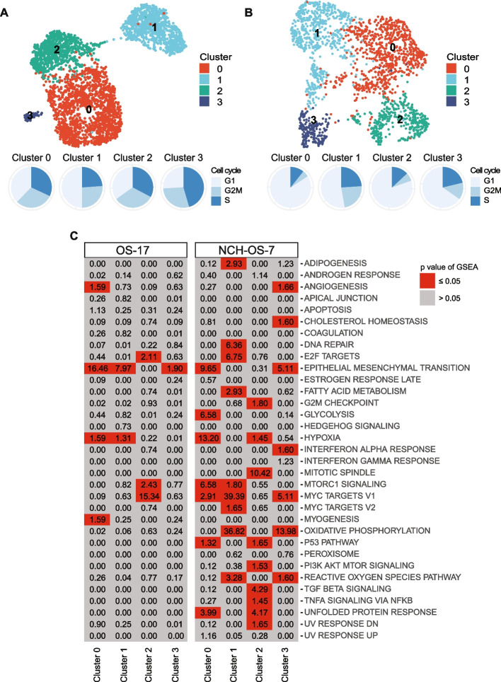
Recent work has shown that osteosarcoma tumors collected directly from patients exhibit significant transcriptional heterogeneity [16, 17]. However, it is unclear if this heterogeneity is maintained within cell line and PDX models of disease [6]. We investigated phenotypic heterogeneity within two in vitro and in vivo models of osteosarcoma by performing scRNA-seq. We generated transcriptomic libraries from a cell line (OS-17) and a PDX (NCH-OS-7; see Table 1, Additional file 1: Table S1, for full characteristics as per PDX Minimal Information standards) grown in cell culture or as subcutaneous flank tumors (>2500 cells per model). To distinguish tumor cells from murine host cells, we separated cells with reads that mapped to the human genome from those that mapped to mouse. Principal component analysis identified that the majority of the variation in gene expression of tumor cells was dominated by cell cycle related genes, a common finding in proliferative malignant tissues [18] (see Additional file 2: Figure S1). Scaling out cell-cycle-related genes unmasked underlying heterogeneity that did not correspond to variation arising solely from cells being in different states of cell cycle (see Additional file 2: Figure S1, Additional file 3: S2A, B, and Fig. 1A, B). Using genes that were differentially upregulated in each cluster relative to the remaining cells in each sample, we identified pathways enriched in these subsets of cells as well as those pathways differentially downregulated in each cluster (Fig. 1C, see Additional file 4: Figure S3A). While subsets within each model shared some enriched gene sets—for example, OS-17 cells in cluster 0 and cluster 1 showed enrichment for epithelial to mesenchymal transition (EMT) genes—none of the subsets had completely identical profiles, suggesting that these subsets are indeed transcriptionally distinct. Interestingly, while EMT transcription factors play an important role in promotion of osteosarcoma cell invasion and metastasis [19], expression of EMT-related genes was associated with certain, distinct subsets. Furthermore, individual subsets had differentially expressed genes that were associated with multiple gene sets. For example, OS-17 cells in cluster 2 were enriched for E2F targets, G2M checkpoint target genes, and MYC target genes, while NCH-OS-7 cells in cluster 0 were enriched for genes associated with EMT, glycolysis, hypoxia, and MYC target genes. This identification of gene sets exclusive to specific clusters of cells validated that these cell line and PDX models demonstrate intra-tumor phenotypic heterogeneity.
Table 1.
Characteristics of osteosarcoma models used in this study
| Model | Species | Type | Host | Endpoint | Characteristics |
|---|---|---|---|---|---|
| OS-17 | Human | Cell Line | Icr-scid | 70 days | Low passage, very well-characterized, reliable metastases |
| 143B | Human | Cell Line | Icr-scid | 65 days | Widely utilized, very well-characterized, reliable metastases |
| NCH-OS-2 | Human | PDX | Icr-scid | 200 days | Very low-passage, highly penetrant, from an untreated synchronous metastasis |
| NCH-OS-7 | Human | PDX | Icr-scid | 184 days | Very low-passage, highly penetrant, from a heavily-pretreated, multiply-relapsed metastasis |
Fig. 1.
Cell line and PDX models of osteosarcoma display transcriptional heterogeneity. A UMAP analysis of OS-17 cells (n = 3327) grown in cell culture. Cell cycle distribution of cells (n = 3327) is visualized as pie charts. B UMAP analysis of NCH-OS-7 cells (n = 1998) grown as a subcutaneous flank tumor. Cell cycle distribution of cells is visualized as pie charts. C Pathway enrichment analysis for hallmark gene sets associated with genes upregulated in distinct clusters identified in the two osteosarcoma models. P values were adjusted for multiple comparisons. Boxes in gray identify non-significant enrichments, whereas boxes in red identify statistically significant enrichments. MTORC1, mechanistic target of rapamycin (mTOR) complex 1. PI3K, Phosphoinositide 3-kinase. AKT, Protein kinase B. TGF, Transforming growth-factor. TNF, Tumor necrosis factor. NFKB, Nuclear Factor kappa-light-chain-enhancer of activated B cells
Transcriptional signatures associated with colonization of bone and lung microenvironments
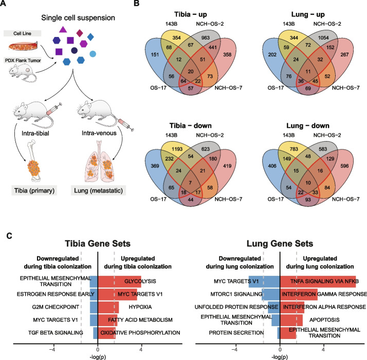
Microenvironment cues play a critical role in shaping transcriptional plasticity [20]. We hypothesized that transcriptional signatures shared across models upon colonization of tibia or lung microenvironments will inform characteristics that are microenvironment-specific rather than tumor-specific. Single-cell suspensions of two cell lines and two PDXs were injected intra-tibially or intravenously to generate orthotopic models of bone-colonizing and lung-colonizing lesions (Fig. 2A schematic). Both the PDXs included in this study (NCH-OS-2, NCH-OS-7) are early passage (passage 3, passage 5) and were obtained from patient lung metastases.
Fig. 2.
Osteosarcoma cells adopt distinct transcriptional profiles as they colonize tibia and lung microenvironments. A Schematic of study design depicting generation of orthotopic tibia- and lung-colonizing tumors. Tumors were harvested for scRNA-seq when mice reached endpoint. B Venn diagrams showing overlap of differentially expressed genes that are up- or downregulated in tibia or lung lesions relative to corresponding starting population of cells (cell culture or PDX flank tumors). Regions outlined in red identify differentially expressed genes shared across at least three models. C Pathway enrichment analysis with adjusted p values for hallmark gene sets associated with these shared genes
To study the transcriptional profiles associated with tibia and lung colonization, we performed a differential expression analysis of the pseudo-bulked scRNA-seq data from bone-colonizing tumors or lung-colonizing lesions relative to their corresponding starting population (cell culture/flank tumors). Most of the differentially regulated genes were unique to each model, with only 0.3–0.8% of genes shared across all four models (Fig. 2B). Using genes that were either up- or downregulated in at least three of the four models (2.7–5.6% of genes), we performed pathway enrichment analysis to identify pathways associated with colonization of tibia or lung tissues (Fig. 2C). We verified the shared pathways between these human osteosarcoma samples and mouse osteosarcoma models (K7M2, F420) to determine shared pathways between xenograft and syngeneic models (see Additional file 5: Figure S4). Genes that were significantly upregulated upon tibia colonization included those associated with glycolysis, hypoxia, MYC targets, fatty acid metabolism, and oxidative phosphorylation. None of the identified downregulated gene sets were statistically significant. Gene sets that were most significantly upregulated with lung colonization included those associated with TNFα signaling via NFκB and EMT, whereas those significantly downregulated included MYC targets and MTORC1 signaling. Overall, we identified common pathways that were differentially regulated across the four models upon tibia or lung colonization.
Transcriptional heterogeneity of individual cells within a tumor
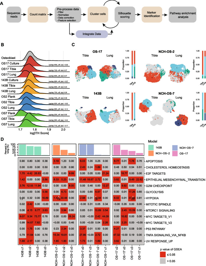
We analyzed single-cell transcriptional profiles of the above datasets using our scRNA-seq bioinformatics workflow to study the role of intra-tumor heterogeneity in tissue colonization (Fig. 3A). To estimate the degree of intra-tumor heterogeneity in our models, we computed intra-tumor heterogeneity scores (ITH scores) from the average Euclidian distance between each cell and every other cell in the same dataset [18]. In essence, this is a measure of how similar any particular cell is to the other cells within a sample. Since osteosarcoma can arise from transformed progenitor cells with osteoblastic differentiation and osteoid production [6], we compared the heterogeneity scores in osteosarcoma cells relative to human primary osteoblast cells grown in cell culture. We found that most osteosarcoma samples exhibit somewhat higher ITH scores than do normal osteoblast cultures (p <0.001, Fig. 3B), though there remained a high degree of overlap in the distributions of phenotypic similarity (ITH scores) within the populations of all three cell cultures. Indeed, cells from osteoblast cultures exhibit several distinct phenotypes that readily cluster away from each other, consistent with previous reports [21] (see Additional file 6: Figure S5) and similar to that observed in the osteosarcoma samples. We have included overlap scores [22] in the figure, which gives a more appropriate statistical assessment of the proposed hypothesis than a p value, given the type of data [23] (i.e., we are not evaluating whether two populations containing thousands of cells are identical, rather evaluating the degree of similarity vs difference in their ITH Score distribution).
Fig. 3.
Osteosarcoma cells retain phenotypic heterogeneity despite adaptive changes in response to changing microenvironments. A Schematic outlining scRNA-seq bioinformatics workflow. B Osteosarcoma cells maintained overall heterogeneity with a high degree of overlap between conditions. The ridge plot shows ITH scores, which represent the gene expression “distance” between each tumor cell within a sample and all of the other tumor cells from that same sample. The overlap statistic describes the total percentage of overlap in the observed distributions between two samples. C UMAP analysis for merged tibia- and lung-colonizing tumor samples in each of the four models. Cells in gray represent the remaining cells in the merged sample. Cluster enrichment analysis shows the distribution of cells in each cluster in the two microenvironment conditions (tibia, lung). While some cells in the tibia and lung lesions adopted shared phenotypes, others adopted distinct phenotypes. D Metabolic heterogeneity in glycolysis activation. We used a pathway enrichment analysis for hallmark gene sets using genes differentially upregulated in each cluster relative to every other cluster within the same model. P values were adjusted for multiple comparisons. Boxes in gray identify non-significant pathway enrichments, whereas boxes in red identify statistically significant enrichments. Bar plot shows the percentage of cells identified per cluster in lung lesions. In B–D, samples subset to equal number of cells to allow inter-model comparison (n=1500 per condition)
We hypothesized that adaptation to distinct microenvironmental conditions would create selective pressures that would cause a narrowing of the transcriptional phenotypes of the tumor cells as the process would select for cells expressing genes that endow them with an environment-specific fitness phenotype. Surprisingly, we found that osteosarcoma cells grown in tibia and lung microenvironments showed no or little decrease in their ITH score distributions relative to those grown in vitro or as flank tumors (Fig. 3B, see Additional file 7: Figure S6A), though they do show a broadening of the cell-cell similarity distribution, with small populations of cells emerging that have increasingly distinct phenotypes (higher ITH scores). These results suggest that osteosarcoma cells are at least as transcriptionally heterogeneous as non-malignant osteoblast cells and hint that adaptation to new tissues might drive the emergence of minor subpopulations. At a high level, these data support the concept that these cells retain the programs of the tissues they derive from, and that phenotypic diversity (as defined by transcriptional heterogeneity) may be a fundamental property that is retained as they colonize the tibia and lung microenvironments.
To determine if bone-colonizing tumors and lung-colonizing lesions display similar phenotypes, we attempted to use an integrative approach across models (see Additional file 8: Figure S7). However, cells predominantly clustered by tumor model of origin, a phenomenon reported in other cancers including small cell lung cancers [18]. This indicates a high degree of inter-tumor heterogeneity, a well-known characteristic of osteosarcoma tumors [24]. We then compared bone-colonizing and lung-colonizing lesions within the same model (OS-17) and characterized the transcriptional profile of each cluster (see Additional file 9: Figure S8, Additional file 3: Figure S2C-F). Using a cluster distribution analysis, we calculated the relative percentages of cells in each cluster to compare cells that colonized the tibia or the lung (see Additional file 1: Table S2). We identified a significant overlap in the transcriptional profiles of cells that colonized the two microenvironments, while certain cells demonstrated profiles specific to each microenvironment (Fig. 3C, see Additional file 7: Figure S6B). For instance, both primary and metastatic OS-17 lesions had cells that exhibited the phenotype associated with cluster 0 (47% in the tibia and 42% in the lung); however, cells with the phenotype associated with cluster 2 were significantly enriched in metastatic lesions (1% in tibia and 40% in lung). Using the pathway enrichment analysis outlined in Fig. 1, we investigated the transcriptional profiles associated with each cluster (Fig. 3D, see Additional file 4: Figure S3B, Additional file 7: S6C). This cluster enrichment-based strategy allowed us to overcome the challenges presented by inter-tumoral heterogeneity and instead focus on the characteristics of subsets that show differential fitness within the bone and lung microenvironments. Pathways enriched in each cluster were unique, with high heterogeneity in the activation of individual pathways across clusters within the same tumor (see Additional file 10: Figure S9 – S12, Additional file 11: Figure S13 – S18). For example, while OS-17 cells in cluster 0 and cluster 3 were associated with MTORC1 signaling, only cells in cluster 3 were enriched for glycolysis-related genes. Our results suggested that the maintenance of subpopulations with distinct phenotypes is reproducible and likely important for the biology of lung colonization. To ensure that these results were indicative of generalizable properties of osteosarcoma tumors (and not simply an artifact of our chosen model system), we validated the presence of these intratumoral subsets defined by glycolysis, hypoxia, epithelial-mesenchymal transition, and TNFα signaling via NFKB within scRNA-seq datasets generated directly from patient tumors (see Additional file 12: Figure S19 – S22, Additional file 13: Figure S23 – S28). Our results suggest that colonization of lung tissue requires contributions from tumor cell subsets exhibiting distinct metabolic behaviors.
Metabolic heterogeneity in osteosarcoma tumors
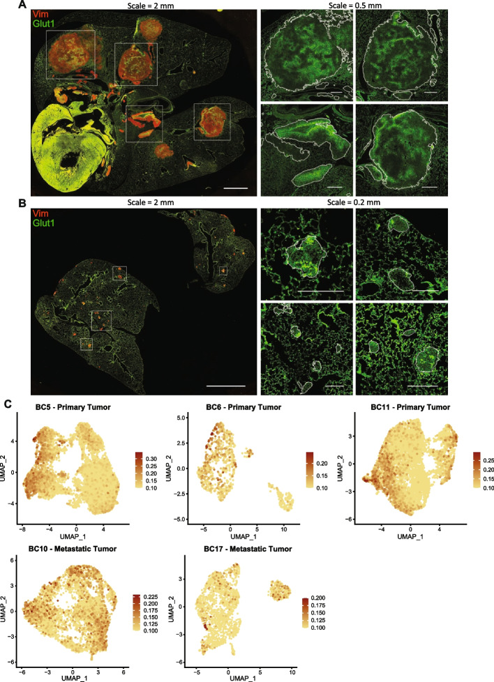
To validate our observations suggesting changes in gene expression related to metabolic heterogeneity at the protein level, we performed immunofluorescence staining for a known activation marker of glycolysis, glucose transporter 1 (GLUT1, a primary component of the observed gene signature), in lung lesions from two models (OS-17 and 143B). Sections were co-stained for vimentin to distinguish osteosarcoma cells from lung parenchyma. We observed variable staining for GLUT1 protein within individual lung lesions, proving that the observed heterogeneity is relevant at the protein level and not an artifact resulting from collections of homogenous, spatially isolated lesions that stained differently from one another (Fig. 4A, B). Interestingly, this heterogeneity was evident even in the smallest lesions, suggesting that this metabolic heterogeneity either represents a fundamental property intrinsic to subpopulations when exposed to the lung environment or provides some survival and/or growth advantage for metastatic lung lesions.
Fig. 4.
Heterogeneity in glycolysis activation identified within lung-colonizing osteosarcoma lesions. A, B Immunofluorescence staining of mouse lungs bearing OS-17 and 143B lung-colonizing tumors, respectively, for GLUT1 (green; a marker of glycolysis) and vimentin (red; marker to identify osteosarcoma cells). Magnified GLUT1 staining is shown for the boxed regions on the whole-section images. Lesion edges are indicated by white outlines in the magnified regions. Tumors showed a high degree of intra-tumor variation in GLUT1 staining intensity. C FeaturePlots for the glycolysis module score in patient primary and metastatic tumor datasets. The module score for Glycolysis was calculated using the “AddModuleScore” function in Seurat with MSigDB HALLMARK_GLYCOLYSIS genes as input features for the expression program
We validated this metabolic heterogeniety at the patient level using a gene expression module for glycolysis in patient primary and metastatic datasets (Fig. 4C, Additional file 12: Figure S19). To confirm that the heterogeneity observed within this representative pathway results from the activation of gene expression modules within the cells and not simply from stochastic variations in gene expression, we stained for other members of the glycolysis gene set (hexokinase 2–HK2, carbonic anhydrase 9–CA9, monocarboxylate transporter 4–MCT4) [25] and a known upstream regulator of glycolysis, MYC [26] to evaluate the degree of concordance within individual cells. We measured the intensity of GLUT1 fluorescence of individual pixels, along with fluorescence of other markers, within areas positive for vimentin. We noted a high correlation in staining for GLUT1 and the other markers within 143B and OS-17 metastatic lesions (see Additional file 14: Figure S29). Immunofluorescence staining for markers of glycolysis in bone tumors from these models identified a high degree of correlation for high, low, and intermediate staining for all of these targets (see Additional file 14: Figure S29A, Additional file 15: Figure S30). The same staining of these cells in culture indicated that heterogeneous expression is present in the culture (see Additional file 16: Figure S31). This staining validated our sequencing-based finding of heterogeneity in glycolysis activation at the protein level and confirmed that this heterogeneity was not the result of stochastic gene expression changes.
Tumor cell clones with diverse transcriptional phenotypes expand during lung colonization
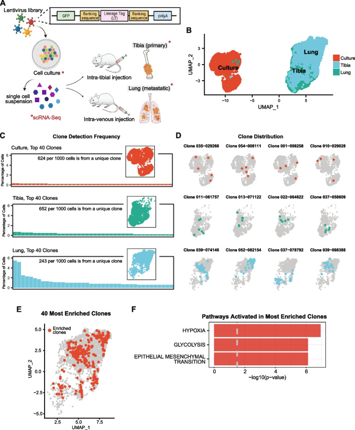
To evaluate the contributions of clonal selection to phenotypic adaptation and heterogeneity as osteosarcoma cells colonize tibia and lung microenvironments, we combined expressed-tag lineage tracing with single-cell transcriptomics (Fig. 5A). Lentivirus infection introduced a very high diversity, heritable, and 3’ capture compatible lineage tag library in osteosarcoma cells in culture. After reserving a representative sample of the uniquely tagged cells, we generated bone and lung lesions. Single-cell libraries of tibia tumors, lung tumors, and cells in culture were processed bioinformatically to overlay phenotype and lineage information for each cell, and cluster transcriptional profiles were determined (see Additional file 3: Figure S2G). At the level of phenotype, we noted that cells in the tibia and lung cluster away from cells in cell culture, suggesting tumor-level adaptation to tibia or lung microenvironments (Fig. 5B). Since the lineage tags are passed from parent to progeny as each cell divides, we used a frequency distribution analysis for lineage tags in the tibia and lung tumors to determine if tibia or lung colonization drove enrichment for specific clonal populations. Lineage barcode enrichment analysis for cells in cell culture identified 85% of cells to have a unique lineage tag, confirming that our starting population received lineage tags at high diversity. Surprisingly, cells from orthotopic primary tumors showed very little evidence of clonal selection, suggesting that bone represents a relatively permissive colonizing environment (Fig. 5C). In contrast, cells from lung lesions demonstrated a higher level of enrichment for specific barcode, suggesting either an environment selecting for clones with specific properties or the activation of programs driving proliferation within a subset of tumor cells (Fig. 5C). We confirmed that this pattern of clonal selection was reproducible in two independent replicates (see Additional file 17: Figure S32A; Additional file 1: Table S3 and Table S4, for lentivirus distribution in cell culture). Importantly, datasets from OS-17 cells from distinct biological replicates (different lots of cells, different lot of virus, different litter of mice) overlaid completely, even without batch correction, suggesting that intra-tumor transcriptional heterogeneity of these osteosarcoma cells is not random or stochastic and that our decision to avoid using batch correction techniques is biologically appropriate (see Additional file 17: Figure S32B).
Fig. 5.
Lung colonization bottleneck enriches for clonal populations with a common transcriptional phenotype. A Schematic of experimental set-up. B UMAP analysis of lineage-tagged OS-17 cells in culture, implanted into the tibia, or inoculated to generate lung-colonizing tumors. These lineage tags are heritable, allowing us to track the transcriptional profiles of daughter cells originating from the same ancestor. Lung- and tibia-colonizing tumor samples share many cells with similar phenotypes, though cultured cells are dramatically different (n = 2800 per condition). C Frequency distribution of clones identified in each of the conditions. A high level of clonal diversity is maintained in the tibia-colonizing tumors (78% unique clones in tibia-colonizing tumor compared to 86% unique clones in cell culture), while dominant clones emerge in the lung (only 33% unique clones). D Overlay of cells sharing a common ancestor onto the dimensional reduction plots in the top four enriched clonal families. E UMAP visualization of cells belonging to top ten enriched clonal families highlighted in red from the lung-colonizing tumor sample. F Pathway enrichment analysis for hallmark gene sets comparing enriched clones relative to the remaining lung-colonizing tumor cells identifies hypoxia, glycolysis, and EMT signaling-related genes to be significantly upregulated
We then overlaid lineage information on phenotypic profiles to identify the transcriptional phenotypes most permissive to clonal expansion in the lung. Interestingly, while we found that cells within given enriched clonal families clustered together, suggesting that overarching transcriptional states were passed from parent to progeny, we also found that these expanding clones emerged from parents exhibiting phenotypes across the transcriptional spectrum (Fig. 5D, see Additional file 18: Figure S33). This suggested that the process leading to clonal expansion might come from the triggering of specific gene programs rather than the selection of cells with any particularly fit phenotype. To identify gene programs that might be activated within these expanding clones, we performed pathway enrichment analysis using the genes differentially upregulated in the top 10 expanded clones. This analysis showed that genes associated with hypoxia, glycolysis, EMT, and MTORC1 signaling were upregulated within the expanding clones, suggesting a potential mechanism for the metabolic heterogeneity noted earlier, whereas differentially downregulated genes were associated with interferon response pathways (Fig. 5E, F; see Additional file 1: Table S5, for differentially upregulated genes; see Additional file 4: Figure S3C for downregulated pathways).
Discussion
These studies provide insight into the roles that intra-tumor heterogeneity and clonal evolution play as osteosarcoma tumors adapt to bone and lung microenvironments. Using an orthotopic cell line and PDX models of osteosarcoma, we show that these tumor cells maintain transcriptional heterogeneity as they respond to changing microenvironment conditions while also plastically adapting their transcriptional profiles. We identified that tibia and lung tumors demonstrate phenotypic heterogeneity where subsets of cells upregulate genes associated with distinct pathways, including glycolysis, hypoxia, EMT, and TNFα signaling via NFκB. We validated this functional heterogeneity for the identified pathways in publicly available patient primary tumor datasets. Furthermore, we showed that intratumoral heterogeneity within at least one metabolically relevant pathway was reproducible using immunofluorescent staining for key proteins, suggesting heterogeneity in glycolytic metabolism within individual metastatic lesions, irrespective of lesion size. Finally, by combining lineage tracing and single-cell transcriptomic analysis, we showed that rapidly expanding clones emerge from cells across the transcriptional landscape, each of which adopts a growth-associated sub-phenotype, which is at least associated with the activation of pathways also associated with metabolism. Together, our studies suggest a tumor-intrinsic mechanism that allows for the maintenance of phenotypic diversity, despite clonal selection, while undergoing adaptive transcriptional changes during lung colonization (Fig. 6). These observations implicate several transcriptional programs in the process of lung metastasis, including several pathways linked to energy metabolism. Most interestingly, this data suggests potential cooperation between distinct tumor subpopulations, which could be a driver for the maintenance of heterogeneity and lung tropism. Further study will be necessary to determine the functional importance of these pathways.
Fig. 6.
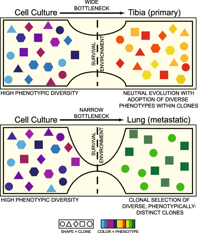
Schematic of tumor cell adaptation to changing microenvironments. Osteosarcoma cells demonstrate high transcriptional heterogeneity (colors) in cell culture, orthotopic tibia, and lung microenvironments. Tumors that grow in the orthotopic tibia lesions follow dynamics of neutral evolution where different cancer clones (shapes) co-exist and propagate simultaneously. The selective pressure of lung colonization results in enrichment of clonal families with different phenotypes (colors), where cells belonging to the same family (shape) tend to have similar phenotypes (color)
Developmental processes in normal cells have been shown to require and maintain a fundamental level of phenotypic heterogeneity [27, 28]. Congruently, osteoblast cells grown in cell culture demonstrated a baseline level of transcriptional heterogeneity. In our studies, osteosarcoma cells maintained an even higher degree of transcriptional heterogeneity, whether grown in mice or cell culture, and maintained that same degree of heterogeneity when exposed to selective pressures of distinct tissue environments. While more samples are needed to capture the true heterogeneity in osteosarcoma, one might speculate that the normal developmental programs that support normal bone growth are maintained (or even amplified) in tumor cells and that these cooperative mechanisms could produce a survival advantage in a transformed malignant state. Indeed, studies published using patient tumor tissues support this hypothesis [16, 17] and analysis of those published data sets identifies heterogeneity in the same pathways that we identified in our mouse models.
While pseudo-bulk analyses identified shared pathways associated with tibia and lung colonization across the models, single-cell analysis identified that each tumor was composed of phenotypically distinct subsets associated with unique combinations of activated pathways. In this study, we validated findings of heterogeneity in glycolytic activation from our sequencing experiments by identifying correlated staining patterns for four distinct markers of glycolysis activation, namely GLUT1, HK2, CA9, MCT4 [25], along with a known upstream regulator of GLUT1, MYC [26]. We showed that this heterogeneity in glycolysis activation is intrinsic to individual metastatic lesions and not an artifact created by spatially isolated, metabolically distinct lesions. Strong correlation between stains for each of these five markers confirmed that the observed heterogeneity did not arise from stochastic gene expression changes. These findings also suggest that a transition to glycolytic metabolism occurs within subsets within a tumor. Reliance on glycolytic metabolism is a deeply studied phenomenon in cancer cells, where rapid energy production can support the abnormally high needs of malignant transformation [29, 30]. Our findings align with those reported in recent single-cell sequencing studies in other cancers, which also showed heterogeneous activation of glycolytic pathways [31]. This may suggest that tumor cells with distinct phenotypes leverage some type of metabolic cooperation, with each cell specializing to perform a specific set of necessary tasks while relying on the activity of cells with different phenotypes for needs that they cannot meet alone. Indeed, many examples of metabolic cooperation between tumor cells and the surrounding stroma have been documented. Evidence within the remote literature hints that similar processes may be active within cultures of tumor cells [32, 33]. Whatever the biologic advantage provided by this heterogeneity, these tumors clearly resemble something more like a developing tissue than a simple clonal proliferation of malignant cells. While tumors have long been described as complex organs, based on the myriad essential interactions between tumor and host cells within the microenvironment [34], studies presented here lend support for the idea that interactions between tumor cells with distinct phenotypes may also be critical for tumor development.
Combining the power of scRNA-seq with lineage tracing allowed us to make inferences about the roles of clonal selection and transcriptional plasticity in the emergence of traits that facilitate lung colonization. We showed that lung colonization enriches for multiple clonal families, wherein clonally related cells tend to have similar transcriptional profiles. In contrast, tumor cells retained clonal diversity on tibia colonization, suggesting that this environment is relatively permissive—at least to cells previously growing in culture.
It should be noted that PDXs and patient-derived cell lines may not fully recapitulate the entire spectrum of heterogeneity within a patient tumor. However, studies evaluating the rate of evolution within PDX models over time suggest that the low passage PDXs utilized in this study should retain a high degree of fidelity to the original tissue used to generate the models [35, 36].
We acknowledge that the experimental metastasis model chosen for these studies produces results that are agnostic of the multi-step process required to effect metastasis in a patient. This choice was deliberate and designed to very simply ask whether the capacity to colonize these different tissues is contained within a few rare clones with a pre-existing phenotype or whether diverse groups of cells have the capacity to adapt to stresses imposed by the tibia or lung environment. Our results suggest that both answers have some truth. The drastic shift in phenotype from cell culture or flank tumors to bone or lung tumors endorses the strong role that microenvironmental cues play in shaping tumor cell behavior.
However, lung colonization enriched for specific clonal populations that all demonstrated a similar growth-associated phenotype. Genes differentially upregulated in the top ten enriched clonal phylogenies overlapped mostly with genes related to hypoxia and glycolysis, a finding that may inform the interpretation of the staining patterns seen in our gene signature validation studies. Indeed, activation of pathways associated with hypoxia and glycolysis is closely linked and may be important in generating the energy and building blocks needed to support the rapid proliferation of these tumor cells. While our results cannot definitively show whether the enriched clones composed individual lung-colonized lesions, or whether the same clones colonized multiple lesions independently, the immunofluorescence stains for glycolysis-related gene performed earlier suggest that this metabolic heterogeneity is a phenomenon intrinsic to this process that likely occurs diffusely. Whether this transition represents a developmental step that occurs within every lesion as it begins to expand rapidly, what changes (likely epigenetic) constitute the trigger that facilitates clonal expansion, and whether the conservation of transcriptional profiles in cells that originated from a common ancestor has an underlying genetic or epigenetic basis all remain points of active study.
While the overall differential expression analysis identified genes associated with TNFα signaling via NFκB and EMT to be most significantly upregulated upon lung colonization (Fig. 2C), the comparison between the most enriched clones and non-expanded clones identified a distinct transcriptional program associated with activation of glycolysis and hypoxia-related genes. These data may suggest that lung colonization follows a two-step process—the first characterized by survival and niche establishment, with a second occurring through rapid expansion of a subset of cells that adopt a growth-associated phenotype distinct from the remaining bulk of the lung-colonizing tumor cells.
Conclusions
Overall, we show that osteosarcoma tumors maintain a high level of intra-tumoral transcriptional heterogeneity, even while experiencing broad transcriptional changes as tumors adapt to tibia and lung microenvironments. Distinct tumors maintain subpopulations of cells characterized by the activation of gene expression pathways that demonstrate similarities across models within each environment. The process of lung colonization enriches for distinct clonal populations. Interestingly, while a particular transcriptional signature appears common to rapidly expanding clonal phylogenies, these arise from groups of cells with diverse genome-wide transcriptional profiles that are maintained within each phylogeny. Additionally, maintenance of the range/degree of intra-tumor heterogeneity endorses the role of cell-autonomous mechanisms in holding up a basal evolutionary advantage associated with maintaining diverse transcriptional states [37]. How this transcriptional heterogeneity is regulated in osteosarcoma cells and whether underlying genomic heterogeneity engenders transcriptional heterogeneity remain unanswered questions.
Methods
Experimental model—PDXs, cell lines, and murine studies
Patient-derived xenografts (PDXs)
OS-17 PDX tissue was obtained from a primary femur biopsy performed at St. Jude’s Children’s Research Hospital in Memphis and was a gift from Peter Houghton [38]. Patient-derived samples, NCH-OS-2 and NCH-OS-7, were obtained from patients who consented under an Institutional Review Board (IRB)-approved protocol IRB11-00478 at Nationwide Children’s Hospital (Human Subject Assurance Number 00002860).
Cell lines
OS-17 cells were derived from an early passage of the OS-17 PDX (see above). These cells were grown in RPMI (Corning, 10-040-CV) supplemented with 10% fetal bovine serum (FBS). 143B cells were obtained from the American Type Culture Collection (ATCC, CRL-8303) and grown in Dulbecco’s modified Eagle medium (DMEM) (Corning, 10-013-CV) supplemented with 10% FBS. Human osteoblast cells (hFOB 1.19) were obtained from the American Type Culture Collection (ATCC, CRL-11372) and grown in DMEM/F12 1:1 (Gibco, 21041-025) supplemented with 0.3 mg/ml G418 and 10% FBS at 34°C. All cell lines were tested annually for both short tandem repeat (STR) genotyping and mycoplasma contamination using testing services provided by Genetica.
Murine studies
All animal studies were approved by Nationwide Children’s Hospital Institutional Animal Care and Use Committee (IACUC) (protocols AR15-00022 and AR14-00045). Flank tumors: Cryopreserved viable tissue pieces from OS-17, 143B, NCH-OS-2, and NCH-OS-7 PDX tumors were placed in the right flank of recipient C.B-17/IcrHsd-Prkdcscid mice (Envigo, Frederick, MD). These subcutaneous tumors were allowed to grow to 300 mm3 before excision. These were then prepped for scRNA-seq. Orthotopic primary tumors: Single-cell suspensions of 5×10⁵ cells of OS-17, 143B, NCH-OS-2, and NCH-OS-7 were injected intra-tibially in C.B-17/IcrHsd-Prkdcscid mice as per IACUC guidelines. Primary tumors were excised once they grew to 800 mm3 and prepped for scRNA-seq. Experimental metastasis: Single-cell suspensions of 1×10⁶ cells of OS-17, 143B, NCH-OS-2, and NCH-OS-7 were injected intravenously in C.B-17/IcrHsd-Prkdcscid mice. Lungs were harvested once these mice reached endpoint. Endpoint criteria for euthanasia were defined as weight loss of >10% or a body condition score (BCS) of <10. For fluorescence immunohistochemistry studies, tumors were placed in 10% formalin for 24 h at 4°C, then moved to phosphate-buffered saline (PBS) for at least 1 h at 4°C. Tissues were then placed in tissue cassettes and placed in fresh PBS. Tissues were embedded in paraffin.
Fluorescent immunohistochemistry
Primary antibodies against glucose transporter 1 (Abcam, ab15309, 1:200), hexokinase 2 (Abcam, [3D3] ab104836, 1:10), carbonic anhydrase 9 (Abcam, [2D3] ab107257, 1:100), c-myc (Invitrogen, [9E10] MA1-980, 1:100), monocarboxylate transporter 4 (Santa Cruz Biotechnology, [D-1] sc-376140, 1:50), and vimentin (Cell Signaling Technology, [D21H3] 5741, 1:100 and Abnova, [SRL33] MAB9596, 1:200) were used. Secondary antibodies and counterstain used were donkey anti-rabbit Alexa Fluor 488 (Invitrogen, A21206, 1:500), donkey anti-mouse Alexa Fluor 488 (Invitrogen, A21202, 1:500), donkey anti-rabbit Alexa Fluor 568 (Invitrogen, A10042, 1:500), donkey anti-mouse Alexa Fluor 568 (Invitrogen, A10037, 1:500), and 4, 6-diamidino-2-phenylindole dihydrochloride (DAPI) (Invitrogen, D1306, 1:500).
Paraffin-embedded tissues were cut into 4 µm sections and placed on glass slides. The sections were deparaffinized with xylene and rehydrated. Sections were submerged into a Tris-Ethylenediaminetetraacetic acid (EDTA) solution (pH 9.0) or a citrate solution (pH 6.0) and heated for antigen retrieval. Sections were blocked and permeabilized with a solution of PBS + 0.2% triton + 2% bovine serum albumin for 1 h at room temperature. Primary antibodies were diluted in PBS + 0.2% triton + 2% bovine serum albumin. The blocking solution was removed from slides and primary antibodies were applied to the sections overnight at 4°C. Sections were washed three times with PBS + 0.2% triton. All secondary antibodies and DAPI were diluted in PBS + 0.2% triton + 2% bovine serum albumin and added to the samples for one hour at room temperature. Sections were washed three times with PBS + 0.2% triton, and once a final time with reverse osmosis water. Samples were mounted in an aqueous mountant (Invitrogen, 00-4958-02). Each tissue section was co-stained for vimentin and one of the other markers of interest.
Whole-section multichannel images were captured using a Nikon Ti2-E motorized microscope with a Lumencor SOLA LED light engine (at 50% power), a Hamamatsu ORCA Fusion camera, and Nikon Plan Apochromat Lambda objectives using Nikon NIS-Elements AR version 5.30 software. Each sample was imaged at 10× magnification with a final 16-bit image resolution of 0.64 μm/pixel. Identical imaging settings were used for all samples within the same staining and imaging set.
Images were analyzed using NikoNIS-Elements AR software version 5.30 with the General Analysis 3 module. Semi-automated tumor segmentation was performed based on vimentin staining, and the segmented regions were manually modified by an unbiased operator as needed to ensure specific and complete tumor selection. Images were then rescaled to 1.28 μm/pixel and the intensities of all pixels representing tumor tissue were recorded in a frequency table for each sample. For assessing the correlation between non-vimentin markers, image channels representing each marker of interest from different tissue sections were aligned using the multimodal image registration tool. Following alignment, an intensity profile line of 2–3 mm in length was plotted through tumor regions showing diverse signal landscapes, and the intensities of pixels along the profile line were recorded for each marker. All marker intensities were plotted by distance along the profile line in GraphPad Prism software version 9.0.0, and the curves were smoothed using a 2nd order polynomial based on 50 neighboring points (25 on each side) to account for minor mismatches in image channels from different tissue sections. The Pearson correlation of each pair of markers was then calculated from the smoothed XY curves.
Single-cell RNA-Seq
Tumors and lungs harvested from mice were processed using the human tumor dissociation kit (Miltenyi Biotec, 130-095-929) with a GentleMacs Octo Dissociator with Heaters (Miltenyi Biotec, 130-096-427). Single-cell suspensions in 0.04% BSA-PBS of cell lines, dissociated tumor and lung tissues were generated and run on the Chromium Single Cell 3′RNA-sequencing system (10x Genomics) with the Reagent Kit v3.1 (10XGenomics, PN-1000121) according to the manufacturer’s instructions. Briefly, cells were loaded into Chromium Next GEM Chip G Single Cell Kit (10x Genomics, PN-1000120) with a targeted cell recovery of 5000 cells per sample. After performing cDNA purification, amplification, and library construction as instructed, we sequenced sample libraries on a half lane of HS4000 (Illumina) to yield (after quality control) about 65,000 paired-end reads per cell. For samples that contained lineage tracing barcodes 0.9 uL (100 µM) of Cellecta FSeqRNA-BC14 primer was added into the sample index pcr reaction.
Cellular barcoding
Barcoded lentivirus libraries were synthesized using CloneTracker XP™ 10M Barcode-3' Library with Venus-Puro (plasmid) (Cellecta, BCXP10M3VP-P) as described in Wang and McManus 2009 [39] with polyethylenimine (PEI) (Alfa Aesar, 43896) as the transfection reagent. OS-17 cells were infected with in-house prepared virus library in the presence of Polybrene (8 μg/ml) (MilliporeSigma, TR1003G). The barcoded plasmids contain ~10M 38-bp semi-random oligonucleotide sequence that is captured on the Chromium Single Cell 3′RNA-sequencing system. Forty-eight hours post-infection, OS-17 cells were used to generate orthotopic primary and metastatic tumor models as described above.
Cellular barcoding computational analysis
Raw sequencing data was pre-processed to extract the lineage tag (LT) using known flanking sequences, and the matching cell ID (CID). Lines without high-confidence CID assignment were removed and redundant reads were removed. The extracted LT barcode reads were matched against the known Cellecta barcode library. Reads with barcodes that did not match the Cellecta library were eliminated. The LT barcode for each matching CID was integrated into the Seurat object metadata to allow for further analysis (see below).
Single-cell RNA-Seq analysis
Cell Ranger version 3.0.2 (10x Genomics) was used to convert Illumina BCL files to FASTQ files. These FASTQ files were then de-multiplexed and aligned to the hg19 human reference genome, provided by 10X Genomics, to generate gene-cell matrices. We used the Seurat R package [40–42] for quality control, filtering, and analysis of the data. Cells were filtered to remove doublets (outliers with high count and high genes per cell), low-quality cells (outliers with low count and low genes per cell), and cells with high mitochondrial genes (indicative of cells with broken membrane). Cells with fewer than 800 expressed genes and genes expressed in fewer than 5 cells were filtered out. The total numbers of cells in each model after filtering were as follows: Osteoblasts (cell culture): 5735; OS-17 (cell culture): 3327; OS-17 (tibia): 4574; OS-17 (lung): 2849; 143B (cell culture): 3354; 143B (tibia): 6187; 143B (lung): 5134; NCH-OS-2 (flank): 6749; NCH-OS-2 (tibia): 4285; NCH-OS-2 (lung): 1645; NCH-OS-7 (flank): 1998; NCH-OS-7 (tibia): 2891; NCH-OS-7 (lung): 5133. In analyses that required comparison across conditions or models, samples were subset to contain equal numbers of cells and this number is specified in the figure legends. We transformed and normalized unique molecular identifier (UMI) counts using the “NormalizeData” function in Seurat package version 4.0.2 [43] with default parameters. We mitigated the effects of cell cycle heterogeneity by regressing out canonical G2/M- and S-Phase genes using the “ScaleData” function in Seurat. Principle component analysis (PCA) was performed using the top 2000 highly variable genes identified by the Seurat function “FindVariableFeatures” with default parameters. For each dataset, the first twenty principal components were selected based on the elbow plot for percentage explained variances, representing ~55.5–60.3% of total variances. The Uniform Manifold Approximation and Projection (UMAP) transformation [44] was performed on selected principal components using the “RunUMAP” function. We applied shared nearest neighbor (SNN) modularity optimization [45] for clustering. We calculated silhouette scores for different numbers of clusters (from two clusters to ten clusters) to measure how similar one cell was to other cells in its own cluster compared with other clusters using the silhouette function in the R package cluster. We determined the optimum number of clusters for each sample by maximizing the silhouette score. Datasets were integrated using four different approaches (FastMNN [46], SCTransform [47], Harmony [48], merge Seurat objects [49]) to determine the optimal method. PCA using the expression of cell cycle genes identified that integrating datasets with “merge” function in Seurat or application of Harmony gave consistent results where cells did not cluster by cell cycle. Marker genes were identified for each cluster relative to other clusters using the “FindMarkers” function, returning only positive markers, with “test.use” set to DESeq2 [50] which uses a negative binomial distribution. Pathway enrichment analysis was performed on these marker genes using the enricher function in the R package clusterProfiler [51] with default parameters with Molecular Signatures Database (MSigDB) Hallmark gene sets [52]. Pseudo-bulk analysis was performed by setting identities of each cell to the sample. We identified gene differentially regulated using the “FindMarkers” function for both positive and negative markers upon tibia or lung colonization. Pathway enrichment analysis was performed on these markers as described above. The ITH score was calculated as described in Stewart et al. [18]. Briefly, it was defined as the average Euclidean distance between each cell to every other cell in each sample, in terms of the selected principal components.
We downloaded validation datasets from Gene Expression Omnibus (GEO) for seven patient primary tumor tissues published in a recent study (GEO accession number-GSE152048) [16]. We used the Seurat R package [40–42] for quality control, filtering, and analysis of these gene expression matrices. Cells were filtered to remove doublets, low-quality cells, and cells with high mitochondrial genes as described above. Cells with fewer than 800 expressed genes and genes expressed in fewer than 5 cells were filtered out. We transformed and normalized UMI counts using the “NormalizeData” function in Seurat package version 4.0.2 [43] with default parameters. PCA was performed using the top 2000 highly variable genes identified by the Seurat function “FindVariableFeatures” with default parameters. Using the marker gene sets used in the paper [16], we separated tumor cells from patient host cells. For each sample, the first twenty principal components were selected based on the elbow plot for percentage explained variances, representing ~51.8–61.5% of total variances. The UMAP transformation [44] was performed on selected principal components using the “RunUMAP” function. Module scores for glycolysis, hypoxia, EMT, and TNFα signaling via NFκB were calculated using the “AddModuleScore” function in Seurat with corresponding Hallmark MSigDB gene sets as input features for the expression program.
Statistical analysis
Statistical analyses were performed using R software environment for statistical computing [53] and Prism 9 (GraphPad Software, Inc.). The packages used in R software are mentioned in the text in the “Methods” section. For differential gene expression analysis, a negative binomial model (DESeq2) [50] was used. For multiple testing, p values were adjusted using Benjamini Hochberg (BH) correction. For ITH score comparisons, data were subjected to one-way analysis of variance (ANOVA) followed by Šidák multiple comparisons test.
Supplementary Information
Additional file 1: Table S1. Characteristics of Patient-Derived Tumor Xenograft Models used in this study. Table S2. Relative percentage of cells distributed between clusters in each model between Tibia and Lung colonization condition. Table S3. Lineage tag distribution statistics for two biological replicates. Table S4. Lineage tag summary statistics for two biological replicates. Table S5. Table showing differentially upregulated genes in the top ten enriched clonal families relative to the remaining metastatic OS-17 cells.
Additional file 2: Figure S1. Majority of the variance in our osteosarcoma datasets is dominated by cell cycle associated genes.
Additional file 3: Figure S2. In-depth analysis of clusters utilized in analyses.
Additional file 4: Figure S3. Downregulated pathways across all cell line- and PDX-derived tumors.
Additional file 5: Figure S4. Mouse osteosarcoma models share distinct transcriptional profiles with human osteosarcoma models during tibia and lung colonization.
Additional file 6: Figure S5. Osteoblast cells demonstrate phenotypic heterogeneity.
Additional file 7: Figure S6. Additional mouse models of osteosarcoma retain phenotypic heterogeneity despite adaptive changes in response to changing microenvironments.
Additional file 8: Figure S7. High inter-tumor heterogeneity identified between models.
Additional file 9: Figure S8. Integrative analysis for within-model comparison of primary and metastatic lesions.
Additional file 10: Figure S9. Heterogeneous activation of glycolysis identified across multiple cell line and PDX tumor datasets. Figure S10. Heterogeneous activation of hypoxia related genes identified across multiple cell line and PDX tumor datasets. Figure S11. Heterogeneous activation of EMT identified across multiple cell line and PDX tumor datasets. Figure S12. Heterogeneous activation of ‘TNFα signaling via NFκB’ identified across multiple cell line and PDX tumor datasets.
Additional file 11: Figure S13. Correlation analysis for multiple cell line and PDX tumor datasets reveals strong positive correlation between glycolysis and hypoxia module scores. Figure S14. Correlation analysis for multiple cell line and PDX tumor datasets reveals minimal correlation between glycolysis and EMT module scores. Figure S15. Correlation analysis for multiple cell line and PDX tumor datasets reveals minimal correlation between glycolysis and ‘TNFα signaling via NFκB’ module scores. Figure S16. Correlation analysis for multiple cell line and PDX tumor datasets reveals mixed correlation between hypoxia and EMT module scores. Figure S17. Correlation analysis for multiple cell line and PDX tumor datasets reveals strong correlation between hypoxia and ‘TNFα signaling via NFκB’ module scores. Figure S18. Correlation analysis for multiple cell line and PDX tumor datasets reveals moderate correlation between EMT and ‘TNFα signaling via NFκB’ module scores.
Additional file 12: Figure S19. Heterogeneous activation of glycolysis identified across multiple patient primary and metastatic tumor datasets. Figure S20. Heterogeneous activation of hypoxia related genes identified across multiple patient primary and metastatic tumor datasets. Figure S21. Heterogeneous activation of EMT related genes identified across multiple patient primary and metastatic tumor datasets. Figure S22. Heterogeneous activation of ‘TNFα signaling via NFκB’ related genes identified across multiple patient primary and metastatic tumor datasets.
Additional file 13: Figure S23. Correlation analysis for multiple patient primary and metastatic tumor datasets reveals strong correlation between glycolysis and hypoxia module scores. Figure S24. Correlation analysis for multiple patient primary and metastatic tumor datasets reveals minimal correlation between glycolysis and EMT module scores. Figure S25. Correlation analysis for multiple patient primary and metastatic tumor datasets reveals minimal correlation between glycolysis and ‘TNFα signaling via NFκB’ module scores. Figure S26. Correlation analysis for multiple patient primary and metastatic tumor datasets reveals mixed correlation between hypoxia and EMT module scores. Figure S27. Correlation analysis for multiple patient primary and metastatic tumor datasets reveals strong correlation between hypoxia and ‘TNFα signaling via NFκB’ module scores. Figure S28. Correlation analysis for multiple patient primary and metastatic tumor datasets reveals moderate correlation between EMT and ‘TNFα signaling via NFκB’ module scores.
Additional file 14: Figure S29. High correlation between multiple markers of glycolysis activation in primary and metastatic osteosarcoma lesions.
Additional file 15: Figure S30. Primary tumors demonstrate heterogeneity in GLUT1 staining.
Additional file 16: Figure S31. Tumor cell cultures demonstrate heterogeneity in GLUT1 staining.
Additional file 17: Figure S32. Replicates demonstrate reproducible lineage tag enrichment profiles and consistent phenotypic profiles.
Additional file 18: Figure S33. Contingency table displaying the number of enriched clones per cluster in culture, tibia, and lung.
Acknowledgements
We would like to thank Dr. Kevin Flanigan for his contributions to imaging through his support of the Abigail Wexner Research Institute Microscopy Core.
Abbreviations
- PDX
Patient-derived xenograft
- ITH
Intra-tumor heterogeneity
- EMT
Epithelial to mesenchymal transition
- MTORC1
Mechanistic target of rapamycin (mTOR) complex 1
- PI3K
Phosphoinositide 3-kinase
- AKT
Protein kinase B
- TGF
Transforming growth-factor
- TNFα
Tumor necrosis factor α
- NFKB
Nuclear Factor kappa-light-chain-enhancer of activated B cells
Authors’ contributions
SR and RDR conceived and designed the study. SR and RDR developed the methodology. MC and ACG prepared libraries for next-generation sequencing. SR, EMF, CT, MW, MVC, and AO performed computation analysis. CAM performed immuno-fluorescence staining. TAV supported image acquisition and analysis of immunofluorescence experiments. SR and RDR interpreted all the consolidated data and wrote the manuscript. SR, EMF, CAM, and RDR edited the manuscript. SR, EMF, and RDR revised the manuscript. All authors read and approved the final manuscript.
Funding
We would like to thank our funding sources. This work was generously supported by funding provided by NIH/NCI (K08CA201638, RDR and T32CA269052, EMF), St Baldrick’s Foundation Scholar Award (RDR), Hyundai Hope on Wheels Young Investigator Award (RDR), CancerFree KIDS Foundation (RDR), Steps for Sarcoma Foundation (RDR), Sarcoma Foundation of America (RDR), a Pelotonia Fellowship (SR), a Nationwide Children’s Director’s Strategic Development Fund, and an NIH CTSA Grant UL1TR002733.
Availability of data and materials
The datasets supporting the conclusions of this article are available in the NCBI’s Gene Expression Omnibus (GEO) repository, GSE179681 [54] [https://www.ncbi.nlm.nih.gov/geo/query/acc.cgi?acc=GSE179681].
The previously published datasets analyzed in support of the conclusions of this article are available in the NCBI’s Gene Expression Omnibus (GEO) repository, GSE152048 [55] [https://www.ncbi.nlm.nih.gov/geo/query/acc.cgi?acc=GSE152048].
The bioinformatics analyses were performed using open-source software, including Seurat version 4.0.2 [43], clusterProfiler version 3.14.3 [51], and Cell Ranger version 3.0.2. All code generated and utilized during the current study is available on GitHub [56] [https://github.com/kidcancerlab/OSHetero2021].
Declarations
Ethics approval and consent to participate
Patient-derived samples were obtained from patients who consented under an Institutional Review Board (IRB)-approved protocol IRB11-00478 at Nationwide Children’s Hospital (Human Subject Assurance Number 00002860).
All animal studies were approved by Nationwide Children’s Hospital Institutional Animal Care and Use Committee (IACUC protocols AR15-00022 and AR14-00045).
Consent for publication
Not applicable.
Competing interests
The authors declare that they have no competing interests.
Footnotes
Publisher's Note
Springer Nature remains neutral with regard to jurisdictional claims in published maps and institutional affiliations.
Sanjana Rajan and Emily M. Franz are co-first authors.
Contributor Information
Sanjana Rajan, Email: SRajan11@its.jnj.com.
Emily M. Franz, Email: Emily.Franz@nationwidechildrens.org
Camille A. McAloney, Email: Camille.McAloney@nationwidechildrens.org
Tatyana A. Vetter, Email: Tatyana.Vetter@nationwidechildrens.org
Maren Cam, Email: Maren.Cam@nationwidechildrens.org.
Amy C. Gross, Email: Amy.Gross@nationwidechildrens.org
Cenny Taslim, Email: Cenny.Taslim@nationwidechildrens.org.
Meng Wang, Email: Meng.Wang@nationwidechildrens.org.
Matthew V. Cannon, Email: Matthew.Cannon@nationwidechildrens.org
Alexander Oles, Email: oles@musc.edu.
Ryan D. Roberts, Email: Ryan.Roberts@nationwidechildrens.org
References
- 1.Casali PG, Bielack S, Abecassis N, Aro HT, Bauer S, Biagini R, et al. Bone sarcomas: ESMO-PaedCan-EURACAN Clinical Practice Guidelines for diagnosis, treatment and follow-up. Ann Oncol. 2018;29:iv79–95. doi: 10.1093/annonc/mdy310. [DOI] [PubMed] [Google Scholar]
- 2.Savage SA, Mirabello L. Using epidemiology and genomics to understand osteosarcoma etiology. Sarcoma. 2011;2011:14–6. doi: 10.1155/2011/548151. [DOI] [PMC free article] [PubMed] [Google Scholar]
- 3.Lindsey BA, Markel JE, Kleinerman ES. Osteosarcoma Overview. Rheumatol Ther. 2017;4(1):25–43. doi: 10.1007/s40744-016-0050-2. [DOI] [PMC free article] [PubMed] [Google Scholar]
- 4.Geller DS, Gorlick R. Osteosarcoma: a review of diagnosis, management, and treatment strategies. Clin Adv Hematol Oncol. 2010;8(10):705–18. [PubMed] [Google Scholar]
- 5.Link MP, Goorin AM, Miser AW, Green AA, Pratt CB, Belasco JB, et al. The effect of adjuvant chemotherapy on relapse-free survival in patients with osteosarcoma of the extremity. N Engl J Med. 1986;314(25):1600–6. doi: 10.1056/NEJM198606193142502. [DOI] [PubMed] [Google Scholar]
- 6.Fan TM, Roberts RD, Lizardo MM. Understanding and modeling metastasis biology to improve therapeutic strategies for combating osteosarcoma progression. Front Oncol. Front Media S.A.; 2020;10:13. [DOI] [PMC free article] [PubMed]
- 7.Aljubran AH, Griffin A, Pintilie M, Blackstein M. Osteosarcoma in adolescents and adults: survival analysis with and without lung metastases. Ann Oncol. 2009;20(6):1136–41. doi: 10.1093/annonc/mdn731. [DOI] [PubMed] [Google Scholar]
- 8.Khanna C, Fan TM, Gorlick R, Helman LJ, Kleinerman ES, Adamson PC, et al. Toward a drug development path that targets metastatic progression in osteosarcoma. Clin Cancer Res. 2014;20(16):4200–9. doi: 10.1158/1078-0432.CCR-13-2574. [DOI] [PMC free article] [PubMed] [Google Scholar]
- 9.Gambera S, Abarrategi A, González-Camacho F, Morales-Molina Á, Roma J, Alfranca A, et al. Clonal dynamics in osteosarcoma defined by RGB marking. Nat Commun. 2018;9(1):3994. [DOI] [PMC free article] [PubMed]
- 10.Bruland ØS, Høifødt H, Sæter G, Smeland S, Fodstad Ø. Hematogenous micrometastases in osteosarcoma patients. Clin Cancer Res. 2005;11(13):4666–73. doi: 10.1158/1078-0432.CCR-05-0165. [DOI] [PubMed] [Google Scholar]
- 11.Vanharanta S, Massagué J. Origins of metastatic traits. Cancer Cell. 2013;24(4):410–21. doi: 10.1016/j.ccr.2013.09.007. [DOI] [PMC free article] [PubMed] [Google Scholar]
- 12.Valastyan S, Weinberg RA. Tumor metastasis: molecular insights and evolving paradigms. Cell. 2011;147(2):275–92. doi: 10.1016/j.cell.2011.09.024. [DOI] [PMC free article] [PubMed] [Google Scholar]
- 13.Turajlic S, Swanton C. Metastasis as an evolutionary process. Science. 2016;352(6282):169–75. doi: 10.1126/science.aaf2784. [DOI] [PubMed] [Google Scholar]
- 14.Sethi N, Kang Y. Unravelling the complexity of metastasis — molecular understanding and targeted therapies. Nat Rev Cancer. 2011;11(10):735–48. doi: 10.1038/nrc3125. [DOI] [PMC free article] [PubMed] [Google Scholar]
- 15.Cristofanilli M, Budd GT, Ellis MJ, Stopeck A, Matera J, Miller MC, et al. Circulating tumor cells, disease progression, and survival in metastatic breast cancer. N Engl J Med. 2004;351(8):781–91. doi: 10.1056/NEJMoa040766. [DOI] [PubMed] [Google Scholar]
- 16.Zhou Y, Yang D, Yang Q, Lv X, Huang W, Zhou Z, et al. Single-cell RNA landscape of intratumoral heterogeneity and immunosuppressive microenvironment in advanced osteosarcoma. Nat Commun. 2021;11(1):1–17. doi: 10.1038/s41467-020-20059-6. [DOI] [PMC free article] [PubMed] [Google Scholar]
- 17.Liu Y, Feng W, Dai Y, Bao M, Yuan Z, He M, et al. Single-cell transcriptomics reveals the complexity of the tumor microenvironment of treatment-naive osteosarcoma. Front Oncol. 2021;21:2818. doi: 10.3389/fonc.2021.709210. [DOI] [PMC free article] [PubMed] [Google Scholar]
- 18.Stewart CA, Gay CM, Xi Y, Sivajothi S, Sivakamasundari V, Fujimoto J, et al. Single-cell analyses reveal increased intratumoral heterogeneity after the onset of therapy resistance in small-cell lung cancer. Nat Cancer. 2020;1:423–36. [DOI] [PMC free article] [PubMed]
- 19.Yu X, Yustein JT, Xu J. Research models and mesenchymal/epithelial plasticity of osteosarcoma. Cell & Biosci. 2021;11(1):1–12. doi: 10.1186/s13578-021-00600-w. [DOI] [PMC free article] [PubMed] [Google Scholar]
- 20.Sun XX, Yu Q. Intra-tumor heterogeneity of cancer cells and its implications for cancer treatment. Acta Pharmacol Sin. 2015;36(10):1219–27. doi: 10.1038/aps.2015.92. [DOI] [PMC free article] [PubMed] [Google Scholar]
- 21.Yoshioka H, Okita S, Nakano M, Minamizaki T, Nubukiyo A, Sotomaru Y, et al. Single-cell RNA-sequencing reveals the breadth of osteoblast heterogeneity. JBMR Plus. 2021;5(6):e10496. [DOI] [PMC free article] [PubMed]
- 22.Ridout MS, Linkie M. Estimating overlap of daily activity patterns from camera trap data. J Agricultural Biol Environ Stat. 2009;14(3):322–37. doi: 10.1198/jabes.2009.08038. [DOI] [Google Scholar]
- 23.Lin M, Lucas HC, Shmueli G. Research Commentary—Too big to fail: large samples and the p-value problem. 2013 ;24(4):906–17. Cited 2022 Dec 4. Available from: https://pubsonline.informs.org/doi/abs/10.1287/isre.2013.0480. 10.1287/isre20130480.
- 24.Sayles LC, Breese MR, Koehne AL, Leung SG, Lee AG, Liu HY, et al. Genome-informed targeted therapy for osteosarcoma. Cancer Discov. 2019;9(1):46–63. doi: 10.1158/2159-8290.CD-17-1152. [DOI] [PMC free article] [PubMed] [Google Scholar]
- 25.Kim MJ, Kim DH, Jung WH, Koo JS. Expression of metabolism-related proteins in triple-negative breast cancer. Int J Clin Exp Pathol. 2014;7(1):301–12. [PMC free article] [PubMed] [Google Scholar]
- 26.Dang CV, Le A, Gao P. MYC-induced cancer cell energy metabolism and therapeutic opportunities. Clin Cancer Res. 2009;15(21):6479–83. doi: 10.1158/1078-0432.CCR-09-0889. [DOI] [PMC free article] [PubMed] [Google Scholar]
- 27.Altschuler SJ, Wu LF. Cellular heterogeneity: do differences make a difference? Cell. 2010;141:559–63. doi: 10.1016/j.cell.2010.04.033. [DOI] [PMC free article] [PubMed] [Google Scholar]
- 28.Chang HH, Hemberg M, Barahona M, Ingber DE, Huang S. Transcriptome-wide noise controls lineage choice in mammalian progenitor cells. Nature. 2008;453(7194):544–7. doi: 10.1038/nature06965. [DOI] [PMC free article] [PubMed] [Google Scholar]
- 29.DeBerardinis RJ, Lum JJ, Hatzivassiliou G, Thompson CB. The biology of cancer: metabolic reprogramming fuels cell growth and proliferation. Cell Metab. 2008;7(1):11–20. doi: 10.1016/j.cmet.2007.10.002. [DOI] [PubMed] [Google Scholar]
- 30.Hsu PP, Sabatini DM. Cancer cell metabolism: Warburg and beyond. Cell. 2008;134(5):703–7. doi: 10.1016/j.cell.2008.08.021. [DOI] [PubMed] [Google Scholar]
- 31.Xiao Z, Dai Z, Locasale JW. Metabolic landscape of the tumor microenvironment at single cell resolution. Nat Commun. 2019;10(1):1–12. doi: 10.1038/s41467-019-11738-0. [DOI] [PMC free article] [PubMed] [Google Scholar]
- 32.Gupta S, Roy A, Dwarakanath BS. Metabolic cooperation and competition in the tumor microenvironment: Implications for therapy. Front Oncol. 2017;7:68. doi: 10.3389/fonc.2017.00068. [DOI] [PMC free article] [PubMed] [Google Scholar]
- 33.Rattigan YI, Patel BB, Ackerstaff E, Sukenick G, Koutcher JA, Glod JW, et al. Lactate is a mediator of metabolic cooperation between stromal carcinoma associated fibroblasts and glycolytic tumor cells in the tumor microenvironment. Exp Cell Res. 2012;318(4):326–35. doi: 10.1016/j.yexcr.2011.11.014. [DOI] [PMC free article] [PubMed] [Google Scholar]
- 34.Egeblad M, Nakasone ES, Werb Z. Tumors as organs: complex tissues that interface with the entire organism. Dev Cell. 2010;18(6):884–901. doi: 10.1016/j.devcel.2010.05.012. [DOI] [PMC free article] [PubMed] [Google Scholar]
- 35.Rajan S, Zaccaria S, Cannon M v., Cam M, Gross AC, Raphael BJ, et al. Structurally complex osteosarcoma genomes exhibit limited heterogeneity within individual tumors and across evolutionary time. bioRxiv. 2022 [Cited 2022 Dec 4];2021.08.30.458268. Available from: https://www.biorxiv.org/content/10.1101/2021.08.30.458268v3. [DOI] [PMC free article] [PubMed]
- 36.Sun H, Cao S, Mashl RJ, Mo CK, Zaccaria S, Wendl MC, et al. Comprehensive characterization of 536 patient-derived xenograft models prioritizes candidates for targeted treatment. Nat Commun. 2021;12(1):1–20. doi: 10.1038/s41467-021-25177-3. [DOI] [PMC free article] [PubMed] [Google Scholar]
- 37.Marusyk A, Polyak K. Tumor heterogeneity: causes and consequences. Biochim Biophys Acta. 2010;1805(1):105–17. doi: 10.1016/j.bbcan.2009.11.002. [DOI] [PMC free article] [PubMed] [Google Scholar]
- 38.Houghton PJ, Morton CL, Tucker C, Payne D, Favours E, Cole C, et al. The pediatric preclinical testing program: description of models and early testing results. Pediatr Blood Cancer. 2007;49(7):928–40. [DOI] [PubMed]
- 39.Wang X, McManus M. Lentivirus production. J Vis Exp. 2009;(32):1499. [DOI] [PMC free article] [PubMed]
- 40.Satija R, Farrell JA, Gennert D, Schier AF, Regev A. Spatial reconstruction of single-cell gene expression data. Nat Biotechnol. 2015;33(5):495–502. doi: 10.1038/nbt.3192. [DOI] [PMC free article] [PubMed] [Google Scholar]
- 41.Haber AL, Biton M, Rogel N, Herbst RH, Shekhar K, Smillie C, et al. A single-cell survey of the small intestinal epithelium. Nature. 2017;551(7680):333–9. doi: 10.1038/nature24489. [DOI] [PMC free article] [PubMed] [Google Scholar]
- 42.Macosko EZ, Basu A, Satija R, Nemesh J, Shekhar K, Goldman M, et al. Highly parallel genome-wide expression profiling of individual cells using nanoliter droplets. Cell. 2015;161(5):1202–14. doi: 10.1016/j.cell.2015.05.002. [DOI] [PMC free article] [PubMed] [Google Scholar]
- 43.Stuart T, Butler A, Hoffman P, Hafemeister C, Papalexi E, Mauck WM, 3rd, et al. Comprehensive Integration of Single-Cell Data. Cell. 2019;177(7):1888–1902.e21. doi: 10.1016/j.cell.2019.05.031. [DOI] [PMC free article] [PubMed] [Google Scholar]
- 44.McInnes L, Healy J, Melville J. UMAP: uniform manifold approximation and projection for dimension reduction. 2018. [Google Scholar]
- 45.Waltman L, van Eck NJ. A smart local moving algorithm for large-scale modularity-based community detection. Eur Phys J B. 2013;86(11):1–14. doi: 10.1140/epjb/e2013-40829-0. [DOI] [Google Scholar]
- 46.Haghverdi L, Lun ATL, Morgan MD, Marioni JC. Batch effects in single-cell RNA-sequencing data are corrected by matching mutual nearest neighbors. Nat Biotechnol. 2018;36(5):421–7. doi: 10.1038/nbt.4091. [DOI] [PMC free article] [PubMed] [Google Scholar]
- 47.Hafemeister C, Satija R. Normalization and variance stabilization of single-cell RNA-seq data using regularized negative binomial regression. Genome Biol. 2019;20(1):1–15. doi: 10.1186/s13059-019-1874-1. [DOI] [PMC free article] [PubMed] [Google Scholar]
- 48.Korsunsky I, Millard N, Fan J, Slowikowski K, Zhang F, Wei K, et al. Fast, sensitive and accurate integration of single-cell data with Harmony. Nature Methods. 2019;16(12):1289–96. doi: 10.1038/s41592-019-0619-0. [DOI] [PMC free article] [PubMed] [Google Scholar]
- 49.merge: Merge Two Data Frames. [Cited 2021 Sep 6]. Available from: https://rdrr.io/r/base/merge.html.
- 50.Love MI, Huber W, Anders S. Moderated estimation of fold change and dispersion for RNA-seq data with DESeq2. Genome Biol. 2014;15(12):1–21. doi: 10.1186/s13059-014-0550-8. [DOI] [PMC free article] [PubMed] [Google Scholar]
- 51.Yu G, Wang LG, Han Y, He QY. ClusterProfiler: An R package for comparing biological themes among gene clusters. OMICS. 2012;16(5):284–7. doi: 10.1089/omi.2011.0118. [DOI] [PMC free article] [PubMed] [Google Scholar]
- 52.Liberzon A, Subramanian A, Pinchback R, Thorvaldsdóttir H, Tamayo P, Mesirov JP. Molecular signatures database (MSigDB) 3.0. Bioinformatics. 2011;27(12):1739–40. doi: 10.1093/bioinformatics/btr260. [DOI] [PMC free article] [PubMed] [Google Scholar]
- 53.Foundation TR. The R Project for Statistical Computing. [Cited 2020 Jun 5]. Available from: https://www.r-project.org/.
- 54.Rajan S, Cam M, Gross A, Cannon M, Roberts R. Osteosarcoma tumors maintain intratumoral heterogeneity, even while adapting to environmental pressures that drive clonal selection. 2021. [Google Scholar]
- 55.Hu H, Chen P, Pan X, Yao Y. Expression profiling by high throughput sequencing. NCBI GEO. https://www.ncbi.nlm.nih.gov/geo/query/acc.cgi?acc=GSE152048.
- 56.Rajan S, Franz E, Cannon M, Oles A, Roberts R. GitHub. 2023. https://github.com/kidcancerlab/OSHetero2021.10.5281/zenodo.7764115.
Associated Data
This section collects any data citations, data availability statements, or supplementary materials included in this article.
Supplementary Materials
Additional file 1: Table S1. Characteristics of Patient-Derived Tumor Xenograft Models used in this study. Table S2. Relative percentage of cells distributed between clusters in each model between Tibia and Lung colonization condition. Table S3. Lineage tag distribution statistics for two biological replicates. Table S4. Lineage tag summary statistics for two biological replicates. Table S5. Table showing differentially upregulated genes in the top ten enriched clonal families relative to the remaining metastatic OS-17 cells.
Additional file 2: Figure S1. Majority of the variance in our osteosarcoma datasets is dominated by cell cycle associated genes.
Additional file 3: Figure S2. In-depth analysis of clusters utilized in analyses.
Additional file 4: Figure S3. Downregulated pathways across all cell line- and PDX-derived tumors.
Additional file 5: Figure S4. Mouse osteosarcoma models share distinct transcriptional profiles with human osteosarcoma models during tibia and lung colonization.
Additional file 6: Figure S5. Osteoblast cells demonstrate phenotypic heterogeneity.
Additional file 7: Figure S6. Additional mouse models of osteosarcoma retain phenotypic heterogeneity despite adaptive changes in response to changing microenvironments.
Additional file 8: Figure S7. High inter-tumor heterogeneity identified between models.
Additional file 9: Figure S8. Integrative analysis for within-model comparison of primary and metastatic lesions.
Additional file 10: Figure S9. Heterogeneous activation of glycolysis identified across multiple cell line and PDX tumor datasets. Figure S10. Heterogeneous activation of hypoxia related genes identified across multiple cell line and PDX tumor datasets. Figure S11. Heterogeneous activation of EMT identified across multiple cell line and PDX tumor datasets. Figure S12. Heterogeneous activation of ‘TNFα signaling via NFκB’ identified across multiple cell line and PDX tumor datasets.
Additional file 11: Figure S13. Correlation analysis for multiple cell line and PDX tumor datasets reveals strong positive correlation between glycolysis and hypoxia module scores. Figure S14. Correlation analysis for multiple cell line and PDX tumor datasets reveals minimal correlation between glycolysis and EMT module scores. Figure S15. Correlation analysis for multiple cell line and PDX tumor datasets reveals minimal correlation between glycolysis and ‘TNFα signaling via NFκB’ module scores. Figure S16. Correlation analysis for multiple cell line and PDX tumor datasets reveals mixed correlation between hypoxia and EMT module scores. Figure S17. Correlation analysis for multiple cell line and PDX tumor datasets reveals strong correlation between hypoxia and ‘TNFα signaling via NFκB’ module scores. Figure S18. Correlation analysis for multiple cell line and PDX tumor datasets reveals moderate correlation between EMT and ‘TNFα signaling via NFκB’ module scores.
Additional file 12: Figure S19. Heterogeneous activation of glycolysis identified across multiple patient primary and metastatic tumor datasets. Figure S20. Heterogeneous activation of hypoxia related genes identified across multiple patient primary and metastatic tumor datasets. Figure S21. Heterogeneous activation of EMT related genes identified across multiple patient primary and metastatic tumor datasets. Figure S22. Heterogeneous activation of ‘TNFα signaling via NFκB’ related genes identified across multiple patient primary and metastatic tumor datasets.
Additional file 13: Figure S23. Correlation analysis for multiple patient primary and metastatic tumor datasets reveals strong correlation between glycolysis and hypoxia module scores. Figure S24. Correlation analysis for multiple patient primary and metastatic tumor datasets reveals minimal correlation between glycolysis and EMT module scores. Figure S25. Correlation analysis for multiple patient primary and metastatic tumor datasets reveals minimal correlation between glycolysis and ‘TNFα signaling via NFκB’ module scores. Figure S26. Correlation analysis for multiple patient primary and metastatic tumor datasets reveals mixed correlation between hypoxia and EMT module scores. Figure S27. Correlation analysis for multiple patient primary and metastatic tumor datasets reveals strong correlation between hypoxia and ‘TNFα signaling via NFκB’ module scores. Figure S28. Correlation analysis for multiple patient primary and metastatic tumor datasets reveals moderate correlation between EMT and ‘TNFα signaling via NFκB’ module scores.
Additional file 14: Figure S29. High correlation between multiple markers of glycolysis activation in primary and metastatic osteosarcoma lesions.
Additional file 15: Figure S30. Primary tumors demonstrate heterogeneity in GLUT1 staining.
Additional file 16: Figure S31. Tumor cell cultures demonstrate heterogeneity in GLUT1 staining.
Additional file 17: Figure S32. Replicates demonstrate reproducible lineage tag enrichment profiles and consistent phenotypic profiles.
Additional file 18: Figure S33. Contingency table displaying the number of enriched clones per cluster in culture, tibia, and lung.
Data Availability Statement
The datasets supporting the conclusions of this article are available in the NCBI’s Gene Expression Omnibus (GEO) repository, GSE179681 [54] [https://www.ncbi.nlm.nih.gov/geo/query/acc.cgi?acc=GSE179681].
The previously published datasets analyzed in support of the conclusions of this article are available in the NCBI’s Gene Expression Omnibus (GEO) repository, GSE152048 [55] [https://www.ncbi.nlm.nih.gov/geo/query/acc.cgi?acc=GSE152048].
The bioinformatics analyses were performed using open-source software, including Seurat version 4.0.2 [43], clusterProfiler version 3.14.3 [51], and Cell Ranger version 3.0.2. All code generated and utilized during the current study is available on GitHub [56] [https://github.com/kidcancerlab/OSHetero2021].


