Abstract
While protein–protein interaction is the first step of the SARS‐CoV‐2 infection, recent comparative proteomic profiling enabled the identification of over 11,000 protein dynamics, thus providing a comprehensive reflection of the molecular mechanisms underlying the cellular system in response to viral infection. Here we summarize and rationalize the results obtained by various mass spectrometry (MS)‐based proteomic approaches applied to the functional characterization of proteins and pathways associated with SARS‐CoV‐2‐mediated infections in humans. Comparative analysis of cell‐lines versus tissue samples indicates that our knowledge in proteome profile alternation in response to SARS‐CoV‐2 infection is still incomplete and the tissue‐specific response to SARS‐CoV‐2 infection can probably not be recapitulated efficiently by in vitro experiments. However, regardless of the viral infection period, sample types, and experimental strategies, a thorough cross‐comparison of the recently published proteome, phosphoproteome, and interactome datasets led to the identification of a common set of proteins and kinases associated with PI3K‐Akt, EGFR, MAPK, Rap1, and AMPK signaling pathways. Ephrin receptor A2 (EPHA2) was identified by 11 studies including all proteomic platforms, suggesting it as a potential future target for SARS‐CoV‐2 infection mechanisms and the development of new therapeutic strategies. We further discuss the potentials of future proteomics strategies for identifying prognostic SARS‐CoV‐2 responsive age‐, gender‐dependent, tissue‐specific protein targets.
Keywords: biomarkers, comparative proteomics, COVID‐19, kinase‐substrate signaling, post‐translational modifications, targeted proteomics, top‐down proteomics
Abbreviations
- PI3K‐Akt
phosphatidylinositol 3‐kinase‐ protein kinase B
- EGFR
epidermal growth factor receptor
- MAPK
mitogen‐activated protein kinase
- Rap1
ras‐related protein 1
- AMPK
AMP‐activated protein kinase
- BioID
proximitydependent biotin identification
- DDA
data‐dependent acquisition
- MEK
mitogen‐activated protein kinase kinase' SRC
protooncogene tyrosine‐protein kinase Src
1. INTRODUCTION
Severe acute respiratory syndrome coronavirus 2 (SARS‐CoV‐2) induced coronavirus disease 2019 (COVID‐19) has claimed over 2.1 million lives globally in 2020. Although initially it was thought that the respiratory system, particularly the lungs, are the primary target for the infectious coronavirus, an increasing number of COVID‐19 case studies showed a wide range of internal organs, including the heart and blood vessels, liver, kidneys, intestine, and the brain can also be severely damaged by this viral infection [1]. The COVID‐19 outbreak has not just impacted human health across the world; it has also resulted in a severe disruption of global daily life, leading researchers to develop a keen interest in resolving the effects of the pandemic. Particularly, scientists in biological fields are now exploring the possibilities of using various biological technologies in order to acquire more information regarding the pathogenic nature of this virus in the human body. The rate of new research on COVID‐19 produced by the scientific community has been enormous, with more than 32,000 papers on the subject being published within the last few months (Figure S1A). In comparison, SARS‐CoV and Middle East Respiratory Syndrome (MERS)‐CoV outbreaks only led to modest increases in literature output (Figure S1A). Furthermore, the vast majority of papers aimed either on the viral infection itself or on the genomic studies of the virus, while the application of proteomics platforms on viral research has not yet been fully explored (Figure S1B).
Among the cutting‐edge molecular technologies, mass spectrometry (MS)‐based proteomics has emerged as a promising biotechnological tool in the post‐genomic era particularly in clinical settings and system biology. The most relevant advantage of using MS‐based technology in clinical proteomics is the ability to obtain a wide range of proteomic information with high‐throughput capability, multiplexity, and high reproducibility in a dynamic quantitative fashion. Furthermore, unlike traditional biochemical assays for disease biomarkers, MS‐based approaches do not require affinity agents or antibodies and thus do not demand prior knowledge of the disease to deliver information regarding biomarkers and clinical indicators. Proteomic platforms have been utilized successfully for a better understanding of the infectious mechanisms of SARS‐CoV‐1 (Table S1) and SARS‐CoV‐2 (Table 1).
TABLE 1.
Mass‐spectrometry based proteomic platforms used for identification and/or characterization of the differentially abundant proteins/modification sites in response to SARS‐CoV‐2 pathogenesis
| Cell‐line/ tissue | Cell origin | Type of experiment | Proteomic platform | No. of Host proteins/ sites a | No of Virus Proteins/sites b | Reference |
|---|---|---|---|---|---|---|
| Cell lines | ||||||
| HuH7 | Liver | Comparative | TMT, RPLC‐MS/MS | 3562/‐ | ‐/‐ | Appelberg et al., 2020[ 2 ] |
| Caco‐2 | Colon | Comparative | TMT, LC‐MS/MS | 412/‐ | ‐/‐ | Bojkova et al., 2020[ 3 ] |
| Vero E6 # | Kidney | Comparative | SDS‐PAGE, LC‐MS/MS | 1401/‐ | ‐/‐ | Grenga et al., 2020[ 4 ] |
| Vero E6 | Kidney | Comparative/ Targeted | LC‐MS/MS, TMT, DDA, Label Free, PRM | 1499/‐ | ‐/‐ | Zecha et al., 2020[ 5 ] |
| A549 | Lung | Comparative | LC‐MS/MS, DIA, DDA | 1053/‐ | ‐/‐ | Stukalov et al., 2020[ 6 ] |
| A549 | Lung | Immuno Proteomics | AP‐MS/MS, | 1146/‐ | ‐/‐ | Stukalov et al., 2020[ 6 ] |
| HEK293 | Kidney | Immuno Proteomics | AP, LC‐MS/MS | 295/‐ | ‐/‐ | Li et al., 2020a[ 7 ] |
| HEK293 | Kidney | Immuno Proteomics | BioID, LC‐MS/MS | 2128/‐ | ‐/‐ | Laurent et al., 2020[ 8 ] |
| HEK293 | Kidney | Immuno Proteomics | BioID, LC‐MS/MS | 1010/‐ | ‐/‐ | St‐Germain et al., 2020[ 9 ] |
| HEK293T | Kidney | Immuno‐proteomic | AP, LC‐MS/MS | 332/‐ | ‐/‐ | Gordon et al., 2020[ 10 ] |
| A549 | Lung | Immuno Proteomics | BioID, LC‐MS/MS | 2242/‐ | ‐/‐ | Samavarchi‐Tehrani et al., 2020[ 11 ] |
| HEK293T | Kidney | Immuno Proteomics | AP, TMT, LC‐MS/MS | 51/‐ | ‐/‐ | Davies et al., 2020[ 12 ] |
| iAT2 | Lung | Comparative | TMT, LC‐MS/MS | 2872/‐ | ‐/‐ | Hekman et al., 2020[ 13 ] |
| Vero E6 | Kidney | Shotgun/Targeted | LC‐HR‐MS/MS | ‐/‐ | 6/‐ | Gouveia et al., 2020a[ 14 ] |
| Vero E6 | Kidney | Shotgun/Targeted | DDA, PRM, LC‐MS/MS | ‐/‐ | 9/‐ | Bezstarosti et al., 2020[ 15 ] |
| A549 | Lung | Phospho Proteomics | EasyPhos, LC‐MS/MS, | ‐/1483* | ‐/‐ | Stukalov et al., 2020[ 6 ] |
| A549 | Lung | Ubiquitination | LC‐MS/MS | ‐/884* | ‐/‐ | Stukalov et al., 2020[ 6 ] |
| Vero E6 | Kidney | Phospho proteomics | LC‐MS/MS, label‐free | 689/1524 | 5/25* | Bouhaddou et al., 2020[ 16 ] |
| iAT2 | Lung | Phospho proteomics | TMT, LC‐MS/MS | 1166/4688 | 2/11 | Hekman et al., 2020[ 13 ] |
| Caco‐2 | Colon | Phospho proteomics | TMT, LC‐SPS‐MS3, Fe‐NTA | ‐/2996* | 6/33 | Klann et al., 2020[ 17 ] |
| Vero E6 | Kidney | Phospho proteomics | SDS‐PAGE, TiO2, LC‐MS/MS | ‐/‐ | 7/44* | Davidson et al., 2020[ 18 ] |
| Tissue | ||||||
| FFPE Tissues | Lung, Heart, Liver, spleen, Kidney, Testis, Thyroid | Comparative | TMT, LC‐MS/MS | 5336/‐ | ‐/‐ | Nie et al., 2021[ 19 ] |
| Lung | Lung | Comparative | LC‐MS/MS, label‐free | 641/‐ | ‐/‐ | Leng et al., 2020[ 20 ] |
| Liver | Liver | Comparative | LC‐MS/MS, label‐free | 1043/‐ | ‐/‐ | Leng et al., 2021[ 21 ] |
| Plasma | Blood | Comparative | nUHPLC‐MS/MS/DDA | 91/‐ | ‐/‐ | Park et al., 2020[ 22 ] |
| RBC | Blood | Comparative | nUHPLC‐MS/MS | 78/‐ | ‐/‐ | Thomas et al., 2020[ 23 ] |
| Serum | Blood | Comparative | TMT, UPLC‐MS/MS | 105/‐ | ‐/‐ | Shen et al., 2020[ 24 ] |
| Serum | Blood | Comparative | DIA/SWATH, LC‐MS/MS | 35/‐ | ‐/‐ | Messner et al., 2020[ 25 ] |
| PBMC | Blood | Comparative | TMT, LC‐MS/MS | 726/‐ | ‐/‐ | Li et al., 2020a[ 7 ] |
| Serum | Blood | Comparative | nUHPLC‐MS/MS | 241/‐ | ‐/‐ | D'Alessandro et al.,2020[ 26 ] |
| Saliva | Saliva | Immuno Proteomics | LC‐MS/MS | 12/‐ | ‐/‐ | Zhou and Wu, 2020[ 27 ] |
| Gargle solution | Saliva | Shotgun/Targeted | Nano‐HPLC/Nano‐ESI‐Orbitrap‐MS/MS | ‐/‐ | 1/‐ | Ihling et al., 2020[ 28 ] |
| Naso ** | Nasal | Targeted | DDA, PRM, LC‐MS/MS | ‐/‐ | 8/‐ | Cardozo et al., 2020[ 29 ] |
| Naso | Nasal | Targeted | UPLC‐HR‐MS/MS, PRM | ‐/‐ | 3/‐ | Saadi et al., 2021[ 30 ] |
| Mucus | Lung | Targeted | UPLC‐HR‐MS/MS, PRM | ‐/‐ | 2/‐ | Cazares et al., 2020[ 31 ] |
| Naso | Nasal | Comparative | DIA LC‐MS/MS | 17/‐ | ‐/‐ | Akgun et al., 2020[ 32 ] |
| Naso | Nasal | Shotgun/Targeted | UPLC‐MS/MS | ‐/‐ | 3/‐ | Gouveia et al., 2020b[ 33 ] |
| Nasopharynx | Nasal | Shotgun/Targeted | UPLC‐MS/MS, PASEF | ‐/‐ | 1/‐ | Nikolaev et al., 2020[ 34 ] |
| Nasopharynx | Nasal | Shotgun/Targeted | LC‐MS/MS | ‐/‐ | 2/‐ | Schuster et al., 2020[ 35 ] |
| Extra Cellular | ||||||
| Urine | Urine | Comparative | LC‐MS/MS | 322/‐ | ‐/‐ | Li et al., 2020b[ 36 ] |
| Urine | Urine | Comparative | TIMS‐LC/MSMS, DIA | 1986/‐ | ‐/‐ | Tian et al., 2020[ 37 ] |
**Naso‐oropharyngeal
Number of significantly changed proteins/sites
Number of proteins/sites identified
indicates non‐human cell‐line; N/A ‐ not applicable
*PTM sites/peptides; ‐ targeted proteomics/quantification.
This article is being made freely available through PubMed Central as part of the COVID-19 public health emergency response. It can be used for unrestricted research re-use and analysis in any form or by any means with acknowledgement of the original source, for the duration of the public health emergency.
Early reports showed that a traditional gel‐based proteomic approach led to the successful identification and characterization of the SARS‐CoV spike (S), nucleocapsid (N), and membrane (M) proteins, and is considered one of the first descriptions of the actual proteins in the then‐novel coronavirus and provide a mechanism by which SARS‐CoV could induce host cell apoptosis [38]. Therefore, efforts in discovery and structural proteomics have been particularly important in the biochemical characterization of SARS‐CoV‐2. Regarding COVID‐19, there is currently a gold rush of proteomics research focusing on SARS‐CoV‐2 (Figure S1B). Recently, the feasibility of MS‐based proteomic approaches, including discovery proteomics [2‐7, 13,14, 19‐26], immuno‐proteomics [6‐12, 27] and targeted methods [5, 29‐35] have been employed using COVID‐19 patients’ samples and/or infected cell lines in investigations of the molecular mechanism of viral infection, potential viral protein targets, and/or the viral proteins in human cell samples (Figure S1B, Table 1). Post‐translational modifications on the viral proteins and those induced by SARS‐CoV‐2 infection in human cells have also been demonstrated [6, 13, 16‐18].
Despite early progress in the understanding of the biochemical basis of SARS‐CoV‐2 pathology, there is still much work to be done, particularly in analyzing the tissue‐specific proteome, proteoforms, and post‐translational modifications in an age‐dependent manner before a suitable, reliable therapeutic for COVID‐19 can be obtained. In the current review we discuss how various proteomics technologies could be an essential means to this end, using examples from previous work in this field. In addition, we performed a comparative analysis of the SARS‐CoV‐2 responsive proteins identified by the recent MS‐based proteomics studies which shows proteomics research on COVID‐19 is in early stage. We also point out where further research could be conducted in order to increase the information overlap between cell lines and tissue‐specific based discovery and quantitative proteomics studies.
2. COMPARATIVE QUANTITATIVE PROTEOMICS IN RESPONSE TO SARS‐COV‐2 INFECTION
Comparative quantitative proteomic analysis is considered as the basic step of any large‐scale systems biology analysis which not only offers the global expression pattern of thousands of proteins but also unveils the potential pathways regulated in the cellular system under certain conditions. For instance, comparison of proteomes between infected versus control cells/tissues could lead researchers significantly closer to a therapeutic target [3, 6, 10, 13, 16, 17].
Proteome profiling dataset obtained from recent studies revealed proteomic dynamics of >11,000 proteins, which were altered in their abundance levels, and/or post‐translationally modified or interacted with viral proteins upon SARS‐CoV‐2 infection (Table 1, Figure 1, Table S2). However, common set of proteins being identified might be important and informative to a wide range of researchers along with the unique and tissue‐specific responsive proteins in SARS‐CoV‐2 pathogenesis. While a re‐analysis or thorough exploration of the MS raw data of these current proteomic studies is beyond the scope and purpose of this review article. To compile our list, we have considered all proteins indicated in the selected studies as significantly changed in abundance (0.05 p‐value with 1.5‐fold up‐/downregulation), post‐translationally modified in response to SARS‐CoV‐2 infection or identified as potential interactor with the viral proteins were used for comparative analysis (Figure 1A, Table S2).
FIGURE 1.
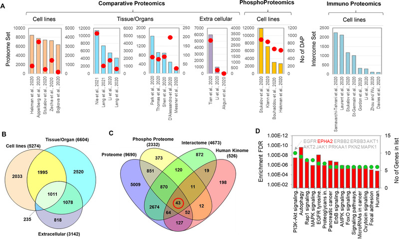
Proteomic platforms utilized for the analysis of SARS‐CoV‐2 and COVID‐19 samples. Datasets collected from 29 published papers were used in this comparative analysis (see details in Table S2). A, bar diagram of the total proteome, phosphoproteome and/or interactome datasets; number of differentially abundant proteins (DAP) identified in each study are indicated by red circles. B, Venn diagram analysis of unique and overlapped number of DAPs reported in five cell lines, nine tissue‐specific and three extracellular proteomic datasets showed in panel A. C, the common (red circle) and unique protein kinases identified in proteome, phosphoproteome and interactome datasets. D, the KEGG pathway analysis of the 43 kinases shared across the three different proteomic platforms
As expected, less than 50% of the differentially abundant proteins identified in tissue samples were found to be altered in the cell line samples (Figure 1B). Similarly, 30% of the altered proteins found in cell lines and extracellular proteome are unique compared to those identified from tissue samples (Figure 1B). Variability in protein abundance level among the cell lines/tissues with or without SARS‐CoV‐2 infection has been noticed in earlier studies [5, 13, 19]. In addition, proteomic dynamics even in the same tissue sample (e.g., plasma/serum) collected from COVID‐19 patients’ also show high variability, likely due to differences in age and other characteristics of the patients, variation in sample collection, multiplicity of infection (MOI), processing steps, and type of data analysis platforms [22]. Therefore, it is not surprising to observe inconsistency when datasets from different proteomic platforms are analyzed to identify a common set of SARS‐CoV‐2 responsive proteins (Figure 1B). Notably, the Venn diagram analysis showed over 78% of SARS‐CoV‐2 interacting proteins overlap with the differentially abundant proteins (Figure S2); over 44% of these shared proteins were identified as phosphorylated (Figure S2), suggesting a role of this modification in the molecular mechanism of SARS‐CoV‐2 pathogenesis, regardless of the infected tissue.
Recent studies demonstrated that several protein kinases play central role in viral entry and replication in host cells [39, 40, 41, 42]. Therefore, we compared data from the SARS‐CoV‐2 responsive proteome, phosphoproteome, and interactome dataset against the human kinome (https://www.uniprot.org/docs/pkinfam). The analysis revealed a total of 43 kinases shared across the three proteomic platforms (Figure 1C). The KEGG pathway analysis further demonstrated the enrichment of PI3K‐Akt signaling, autophagy, Rap1 signaling, MAPK signaling and EGFR tyrosine kinase inhibitor resistance pathways (Figure 1D). This supports the direct involvement of these kinases/pathways in SARS‐CoV‐2 pathogenesis, as previously reported [13, 17]. Heat map analysis of the collective SARS‐CoV‐2 differential proteome (>11,000 proteins) showed a total of 23 proteins were identified in at least 10 individual studies (Figure 2). In particular, ephrin receptor A2 (EPHA2), a tyrosine kinase, was identified by 11 studies including all proteomic platforms. It is important to note that, EPHA2 has been identified as one of the key host cell entry receptors for many RNA viruses [39, 40, 41, 42]. Therefore, alternative experimental strategies will be beneficial to demonstrate the potential role of this tyrosine kinase in the SARS‐CoV‐2 infection mechanisms.
FIGURE 2.
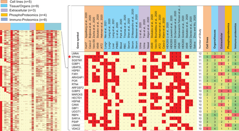
Heat map analysis of over 11,000 proteins identified as differentially abundant proteins (DAPs) and/or SARS‐CoV‐2 interacting proteins from a total of 29 individual proteomic studies. The right panel shows the list of 23 proteins identified in at least in 10 individual studies. Red star shows ephrin receptor A2 (EPHA2), a tyrosine kinase was identified by 11 studies including all three proteomic platforms (comparative, phosphoproteomics and immunoproteomics). See details in Table S2.
2.1. Feasibility of model cell lines for identification of SARS‐CoV‐2 responsive proteins
Model cell lines are the most commonly used tool in biological research particularly on transcriptomic and proteomics analysis and offer various advantages including but not limited to being cost effective, easy to use, continuous supply, and more importantly providing a pure population of cells, which enables sample consistency and reproducible results. In addition, different cell lines have been successfully used to determine the efficacy of many drugs/small molecules to inhibit SARS‐CoV‐2 replication [3, 5, 6, 16, 17], and development of mass‐spectrometry based COVID‐19 diagnostic assay [5, 14, 33]. Additionally, only low amount of proteins can be typically obtained from many of the organs/tissues critically impacted by SARS‐CoV‐2, and this is an important limitation for the enrichment of post‐translationally modified peptides/proteins. Conversely, cell lines offer higher amounts of protein within a short time, hence they are preferred for the characterization of various PTMs in translational research. Due to these advantages, many cell lines have been essential in efforts to analyze the base line proteome profiles in response to cellular perturbations including pathogenic responses of SARS‐CoV‐2 (Table 1). It is also important to note that, susceptibility of SARS‐CoV virus infection to natural human and non‐human model cell lines is highly variable, which is primarily associated with the expression level of angiotensin‐converting enzyme 2 (ACE2) receptor protein [43, 44, 45]. Therefore, compatible and permissive cell lines such as CaCo‐2, HuH7, Vero E6, or modified cell lines expressed hACE2 such as A549‐hACE2 were often used in all cell‐line‐based proteomic experiments to demonstrate the SARS‐CoV‐2 infection mechanisms (Table 1).
2.2. Tissue‐specific comparative proteomics offers potential biomarker
SARS‐CoV‐2 targets several organs throughout the human body, requiring researchers to move beyond the cell and consider the responses of tissues and organs to infection [1]. Cardiac injury is a prominent complication in many COVID‐19 patients [46]. A comparative proteomic profiling of ACE2 expression in adult human heart tissues helped demonstrate that ACE2 expression is significantly higher in patients with heart failure, making them more susceptible to SARS‐CoV‐2 [46].
Certainly, in vitro cell line‐based proteomic studies provide valuable information for the understanding of the molecular mechanisms of SARS‐CoV‐2 pathogenesis. However, in vitro samples may be insufficient to elucidate the mechanism of multiorgan failure in COVID‐19, indicating that we are far behind in the full understanding of SARS‐CoV‐2 induced in vivo mechanisms. Recently, Nie et al. [19] showed the real‐time architecture of the tissue‐specific proteome alternation wherein a total of 144 formalin fixed autopsy samples were collected from seven organs/tissues including lung, liver, spleen, kidney, heart, thyroid, and testis from 19 COVID‐19 patients. A comparative study among the tissues showed over 45% of the identified proteins were significantly dysregulated at least in one organ/time point indicating the devastating effect of SARS‐CoV‐2 infection and potential molecular pathogenesis of multi‐organ failure in COVID‐19 disease [19]. In addition, for the first time this study described cathepsin L1 (CTSL) as a new potential SARS‐CoV‐2 biomarker for lung. The potential role of CTSL was further supported by using CTSL inhibitor which effectively blocked the virus entry into the host cells [47]. Notably, CTSL was successfully identified as a differentially abundant protein also in cell lines‐based proteomic studies [3, 5] and as an interactor of SARS‐CoV‐2 proteins [11]. However, due to the lack of comparisons with other tissue‐specific proteome datasets, CTSL was not considered as lung biomarker for COVID‐19 in earlier studies [3, 5, 11].
2.3. Age‐ and gender‐dependent proteome analysis of COVID‐19 patients
The COVID‐19 shows a significant age‐dependent response in terms of severity of infection, illness and mortality rate [48] indicating that an age‐specific physiological and immunological response is crucial to the fight against viral infections. Thus, a proteomics‐based strategy was used to profile the influenza‐specific antibodies defining important epitope targets and uncovering the vaccination response in individuals of different ages against the viral infection. A semiquantitative comparative plasma proteomics workflow provides insight into factors that may explain age‐related differences in the incidence of severe sepsis in a community‐acquired pneumonia in the elderly [49].
Similarly, several epidemiologic reports from different countries support the fact that gender/sex has great influence on COVID‐19 outcomes. A recent scoping review including data from 59,254 COVID‐19 patients also showed the mortality rate was higher in men suggesting an increased susceptibility of male patients to SARS‐CoV‐2 pathogenesis [50]. While men and women have the same prevalence of coronavirus infection, infected men are at a higher risk for worse outcomes and death regardless of age, whereas women show a higher level of antibodies [51]. Since gender‐ and age‐specific response and susceptibility of COVID‐19 is apparent, global comparative proteome profiling of COVID‐19 male and female patients with a wide range of age groups could be a prudent choice to demonstrate the molecular response against SARS‐CoV‐2 pathogenesis and identify potential new diagnostic biomarkers.
3. IDENTIFICATION OF SARS‐COV‐2 INTERACTING PROTEINS BY MASS SPECTROMETRY‐BASED APPROACHES
An immediate challenge in developing drugs to treat COVID‐19 is the identification and characterization of protein–protein interactions between SARS‐CoV‐2 and the human proteome, which drive viral propagation and infection. Common methods to assess protein–protein interactions in host‐viral systems include yeast two‐hybrid (Y2H) analysis and affinity purification followed by mass spectrometry (AP‐MS). The sudden urgency for high‐throughput, discovery‐based research brought on by the COVID‐19 pandemic leads us to review methods used to detail coronavirus–host interaction maps with the goal of identifying therapeutic strategies. Although Y2H is considered a valuable tool to discover protein–protein interactions between host and virus, it is limited because it does not reconstitute a native, cellular context. This is a critical caveat in identifying druggable, context‐dependent targets which can be addressed using AP‐MS [6, 7, 10, 12, 27]. While AP‐MS remains the gold standard for studying virus‐host interactomics, application of novel techniques could increase the analysis throughput. Specially, proximity‐dependent biotin labeling, enabled by expression of a fusion protein between the protein of interest and a catalytically‐enhanced biotin ligase (commonly known as BioID), followed by MS identification of streptavidin‐enriched lysates [8, 9, 11]. A recent study extensively reviewed the potential challenges and limitations of the current methods used for identification of SARS‐CoV‐2 proteins interactome [52]. Despite the variability in the methods applied to the study of interactome and related findings, the identification of a common set of SARS‐CoV‐2 interacting proteins could be used to identify the potential targets, for further analysis and future development of therapeutic agents.
As several kinase inhibitors have proven to inhibit viral replication in cells [6, 13, 16, 17], we compared current SARS‐CoV‐2 interactome datasets against the entire human kinome (Figure S4A). BioID‐based studies covered over 22% of the total human kinome (corresponding to 115 kinases), while 8% (44 kinases) was identified by AP‐MS. Among the 44 kinases identified by AP‐MS based method, 50% of them (22 kinases) were also identified by BioID based method indicating as potential interactors of SARS‐CoV‐2 proteins (Figure S4). Cellular mechanisms involving these 22 kinases includes axon guidance (EPHA2, EPHB2, EPHB4, MET, BMPR1B, BMPR2), EGFR tyrosine kinase inhibitor resistance (ERBB2, JAK1, MET, AXL), PI3K‐Akt signaling pathway (EPHA2, ERBB2, JAK1, MET), cytokine‐cytokine receptor interaction (BMPR1B, BMPR2, TGFBR1, ACVR1B), and Epstein–Barr virus, immunodeficiency virus 1 and cytomegalovirus infection response (TBK1, JAK1, RIPK1).
Furthermore, the 22 kinases were compared against the SARS‐CoV‐2 responsive proteome and phosphoproteome datasets (Figure S4B). Interestingly, EPHA2, ERBB2, JAK1, MARK2, PRKDC, and TBK1 kinases were identified in at least seven studies indicating further studies suggesting a relevant role in SARS‐CoV‐2 pathogenesis. Notably, the BioID method let to identification not only of many of the cross‐validated kinases (e.g., EGFR, AKT1, AKT2, GSK3B, SRC, and MAPKs) but also to that multiple downstream kinases associated to the same signaling pathways (Figure S5). For instance, five kinases associated with axon guidance were identified by both AP‐MS and BioID based methods; however, 13 additional kinases belonging to this pathway were exclusively by BioID (Figure S5). Collectively, this comparative analysis suggests that complementary methods are essential for expanding our understanding of SARS‐CoV‐2 interacting protein networks.
4. POST‐TRANSLATIONAL MODIFICATIONS (PTMS) OF SARS‐COV‐2 PROTEINS AND HOST CELL PROTEOME
Post‐translational modifications (PTMs) have been implicated in the regulation of viral proteins and can have numerous effects on viral activity [53, 54, 55]. Importantly, a comparative global phosphoproteomic analysis of SARS‐CoV‐2 infected samples not only offers the exploration the phosphoproteome expression pattern of the host cells but can also determine the phosphorylation state of many viral proteins (Figure 3).
FIGURE 3.
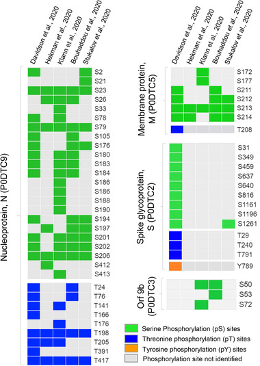
Phosphorylation of SARS‐CoV‐2 proteins. The heat map shows the phosphorylation sites identified by five large‐scale phosphoproteomic analyses. Proteins identified as phosphorylated in at least two studies were included. Green, blue and orange correspond to pS, pT and pY modifications, respectively. Gray indicates sites were not identified
A total of 34 unique phosphorylation sites were identified in nucleoprotein N (P0DTC9) by multiple phosphoproteomic analyses, indicating that N protein is one of the hyperphosphorylated protein among the SARS‐CoV‐2 proteins (Figure 3). Several phosphorylation sites of N protein such as S23, S79, S206, T198, and T417 were identified by all five phosphoproteomic studies suggesting these sites are main regulators of N protein bioactivity and particularly its interaction with RNA to modulate gene transcription [13, 56]. Collectively, these studies successfully identified many novel phosphorylation sites of viral proteins that could be considered as drug targets[57]. However, each phosphoproteomic study also identified a number of unique phosphorylation sites for many of the viral proteins (Figure 3) indicating that host cell lines physiology, infection period, phosphopeptides enrichment method and downstream analytic process could be associated with the alternation and identification of the viral protein phosphosites. The general consensus is that viral proteins utilize a dynamic mix of functionally important PTMs (Figure 4) that can efficiently altered their structures and binding with other proteins [54, 56, 58], with relevant effects on COVID‐19 pathogenesis.
FIGURE 4.
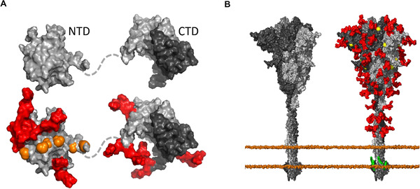
Post‐translational modifications on the surface of two SARS‐CoV‐2 proteins. (A) The N‐terminal domain (NTD) and C‐terminal domain (CTD) of the N protein; and (B) The S protein are depicted without and with posttranslational modifications (gray). Glycans are shown in red, phosphorylation in orange, palmitoylation in green, and methylation in yellow. Methylation and phosphorylation are represented as spheres. The domains of the N protein are built from two structures, PDB ID: 6YI3 and 6ZCO, and glycosylation sites were modeled after Supekar et al. [59]. The NTD and CTD contain 3 and 2 glycans, respectively; the NTD contains a total of 8 modeled phosphorylation sites. The CTD exists as a dimer, which are depicted with different shades of gray. The non‐glycosylated structure of the S protein based on PDB‐ID 6VXX is available through CHARMM‐GUI [60] and Jo et al. [61], and methylation was modeled based on Sun et al. [62]. The S protein contains 23 glycosylation sites, 2 palmitoylation sites, and 5 methylation sites on each chain of the trimer. The phosphate heads of the membrane that the spike is embedded in are shown as spheres
For example, the glycosylation on the surface of the S protein shields epitopes from detection by the immune system [55, 60, 63]. Other modifications, such as the palmitoylation in the transmembrane region, are common to keep membrane proteins, anchored to the membrane [64]. In addition, proteolytic cleavages of viral proteins by both viral and host were also found to be important [65]. Furthermore, Meyer et al. [66], employed the N‐terminomics based proteomic approach to determine the proteolysis and the resulting proteolytic proteoforms during SARS‐CoV‐2 infection which led to the identification of many novel cleavage sites in multiple viral proteins including S and N proteins, and potential host‐cell substrates of the main and papain‐like proteases. Further validation with in vitro assay, siRNA depletion, and drugs targeting of these proteins showed a significant reduction of SARS‐COV‐2 replication in cells, suggesting potential therapeutic strategies to develop to inhibit SARS‐CoV‐2 infection [66].
The combined analysis of various phosphoproteome studies based on the use of different cell lines infected with SARS‐CoV‐2 provides a deep understanding of the multi‐level signaling pathways altered during the infection. These include the activation/inactivation of many kinases associated with cell cycle arrest, stress and DNA damage response, regulation of transcription and cell junction organization [6, 13, 16, 17]. Identification of many differentially abundant tyrosine phosphorylation sites (0.2% of the total phosphoproteome) suggests that tyrosine kinases may play a potential role in SARS‐CoV‐2 pathogenesis [66, 67]. In accordance, tyrosine kinase inhibitors have been identified as a potential drug to prevent viral replication in cells [16, 66, 68] and could be used a potential therapeutic strategy in severe COVID‐19 [16, 66, 67, 68].
Previous studies demonstrated substantial differences in protein abundance levels among cell lines and related tissues [5, 69, 70]. In accordance, here we showed a number of kinases including EPHA2, CDK13, ERBB2, BUB1B, and PKMYT1 were identified as differentially abundant proteins and/or phosphoproteins in only cell lines‐based proteomic studies (Figure S3). This result suggests that abundance level of these kinases in tissue samples could be very low or these kinases were mostly regulated by post‐translational level. Notably, phosphoproteome profiling of COVID‐19 tissue samples remains unknown, as there are no publications at the time of this comparative analysis. Similarly, several metabolism related proteins including but not limited to LDHB, GNS, LRG1, TMX1, STOML2, NDUFS3, FLOT1, and TBK1 were primarily identified in tissue‐specific proteomic studies or interactomic analyses (Figure S3). Taken together, these comparative analyses indicate that our knowledge in proteome profile alternation in response to SARS‐CoV‐2 infection is still incomplete and the tissue‐specific response to SARS‐CoV‐2 infection can probably not be recapitulated efficiently by in vitro experiments [19].
Stukalov et al. [6] further demonstrated that together with the changes in phosphorylation, also several hundreds of ubiquitination sites (884) were altered in SARS‐CoV‐2 infected A549 cells. This large‐scale systematic multiple PTMs profiling of SARS‐CoV‐2 responsive proteins further revealed an interplay between phosphorylation and ubiquitination on both host and viral proteins [6]. Thus, mapping of these regulatory modifications on viral and host proteins could favor the identification of both potential therapeutic targets and diagnostic biomarkers, as well as help the elucidation of SARS‐CoV‐2 infection and proliferation mechanisms [6].
5. MS‐BASED TARGETED PROTEOMIC METHODS OFFERS HIGHLY REPRODUCIBLE MULTIPLEXED DIAGNOSTIC TOOLS FOR COVID‐19
Currently, nucleic acid‐based assays (e.g., PCR) are being used as a gold standard for diagnostic testing of SARS‐CoV‐2 and other viruses in clinical platforms. However, many recent studies showed MS‐based assay has the potential to supplement the diagnostic testing of COVID‐19 [5,14, 28‐35]. Therefore, the development of rapid, sensitive, and multiplexed LC‐MS/MS methods could be an attractive alternative in the COVID‐19 testing field. Recently, many studies applied targeted MS‐based methods for detection of SARS‐CoV‐2 proteins (Table 1). One of the earliest reports by the Armengaud group [14] identified the ideal set of viral peptides to use in subsequent assay by employing the discovery proteomic platform to detect SARS‐CoV‐2 peptides in complex biological samples. The efficacy of those viral peptides was further validated by developing a 3‐min MS‐based targeted assay using clinical samples [33]. Another study used parallel reaction monitoring (PRM), which employs a high‐resolution Orbitrap mass analyzer for peptide detection instead of a quadrupole analyzer like in multiple‐reaction monitoring (MRM), to detect SARS‐CoV‐2 proteins from virus‐infected Vero cells [5]. This study led to the identification of SARS‐CoV‐2 nucleocapsid protein in the attomolar range, which corresponds to the signal roughly 10,000 SARS‐CoV‐2 particles. This finding is significant, as clinical samples are often dilute and have a low viral load. More recently, multiple MS‐based targeted protocols have been developed and successfully applied to detect SARS‐CoV‐2 proteins including N, S, and M proteins from various clinical samples [28‐35]. Despite these successful pilot studies, potential challenges and technical limitations of virus diagnostics using MS‐based method from clinical samples exist and have been recently discussed in a review by Grossegesse et al. [71].
Collectively, these emerging studies demonstrate promising alternative methods for SARS‐CoV‐2 diagnostic testing using targeted MS. Moreover, targeted LC‐MS/MS not only display greater sensitivity than some nucleic acid‐based tests, but also requires shorter time (∼30 min compared to ∼4 h).
6. SUMMARY AND FUTURE STRATEGIES
Despite the proteomic investigations that have already been conducted on COVID‐19 patient samples and SARS‐CoV‐2 infected cell lines, it is obvious that a great deal of research (Figure 5), still needs to be performed in order to develop a more complete picture of how the human proteome responds to COVID‐19.
FIGURE 5.
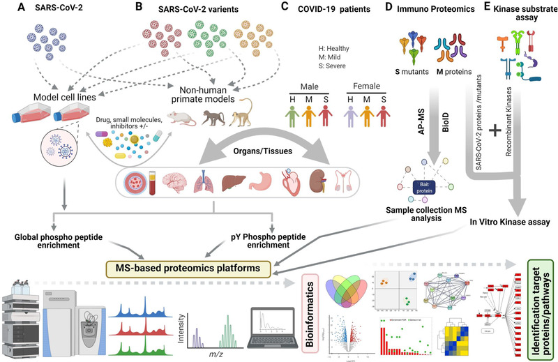
Potential bottom‐up proteomics platforms for elucidation of the molecular mechanisms of SARS‐CoV‐2 infection. A, cell‐line based phosphoproteomic approach with or without potential drugs treatments to SARS‐CoV‐2 and mutant variants. B, study of non‐human primate models infected by SARS‐CoV‐2 and mutant variants with or without drug response for large‐scale tissue‐specific proteome and phosphoproteome analysis. C, tissue‐specific proteomic analysis of COVID‐19 patient samples from diverse cohorts. D, identification of SARS‐CoV‐2 mutant proteins interacting partners using complementary methodologies. E, identification and verification of kinase‐specific targets and modification sites using kinase substrate assay followed by mass spectrometry analysis. This figure is created using BioRender.com
It is our notion that the proposed following strategies will improve our understanding of the molecular mechanisms of SARS‐CoV‐2 pathogenesis and will help to develop efficient, multiplexed tissue‐specific diagnostic assays and potential therapeutics.
Tissue‐Specific global phosphoproteomics analysis:
Results from recent proteomics dataset confirmed that several tissue‐specific mechanisms functioned in various organs/tissues of COVID‐19 patients [19]. However, very limited information is available on phosphoproteome profiling of COVID‐19 patient tissue samples. Furthermore, comparison of the differentially abundant proteins so far identified among the cell lines and tissues revealed both unique and overlapping sets of proteins dysregulated by SARS‐CoV‐2 pathogenesis (Figure 1B), indicating the importance of global phosphoproteome analysis of SARS‐CoV‐2 infected tissue samples (Figure 5C).
-
b.
Enrichment of phospho tyrosine (pY) proteome of SARS‐CoV‐2 infected samples:
Gene expression analysis of CD4+ T‐cells from moderate and severe COVID‐19 patients showed differential expression of T‐Cell receptor (TCR), EGFR, and P53 signaling pathways genes [17, 26]. It is well documented that TCR, EGFR, P53, and many other proteins involved in these signaling pathways are regulated by tyrosine phosphorylation (pY). More importantly, it has been shown that many compounds including receptor tyrosine kinase inhibitor successfully inhibit the phosphorylation of the downstream target proteins of these pathways, thus preventing the viral replication in the host cells [16, 17, 66, 67, 68]. Therefore, together with the global phosphoproteome, a specific enrichment of phosphotyrosine proteome of SARS‐CoV‐2 infected samples would be of great interest (Figure 5A). Due to the low abundance of pY sites, enrichment of pY peptides/proteins by using global phosphoproteomics methods (e.g., TiO2‐based) is always challenging. However, this issue could be resolved by several optimized MS‐based pY enrichment protocols [72‐76].
-
c.
Feasibility of SARS‐CoV‐2 infected non‐human primate models:
Several studies have shown non‐human primate models could develop mild to moderate respiratory disease, with clinical and hematologic findings consistent with those documented for COVID‐19 [77, 78]. Thus, non‐human models have successfully been used to evaluate the efficacy of potential drugs and/or vaccines and therapies to many other RNA viruses [79‐81]. We posit that model animals could be a great resource for large‐scale tissue‐specific proteomic analysis. For instance, tissue‐specific pY proteomics analysis in response to different MOI stages of COVID‐19 requires high amount of materials which is difficult to obtain from patient's biopsies.
A recent study revealed that naturally occurring mutations on viral spike protein can reduce or enhance viral cell entry via ACE2 and TMPRSS2 proteins [82]. This study provides a great clue of SARS‐CoV‐2 genetic variations associated with the infection mechanism; however, host genetic factors remain elusive. In this regard, systematic tissue‐specific proteome analysis of non‐human models infected by SARS‐CoV‐2 spike protein variants could enhance our understanding of the pathogenesis of the mutants (Figure 5B).
-
d.
Interactome analysis of SARS‐CoV‐2 mutant proteins:
While mutations and post‐translational modifications frequently occur throughout the SARS‐CoV‐2 proteins, further studies showed these mutations also lead to alterations in secondary/tertiary protein structure that might contribute to remodeling protein functions [83]. AP and proximity‐based interactome analysis successfully identified thousands of interacting partners of SARS‐CoV‐2 proteins (Table 1). However, how PTMs and mutations of these viral proteins affect their interacting partners remains unclear. Therefore, further, interactome analyses of these mutants and post‐translationally modified versions of SARS‐CoV‐2 proteins will improve our understanding of how these sources of structural variation could alter the host cellular mechanisms (Figure 5D).
-
e.
Identification upstream kinases responsible for phosphorylation of SARS‐CoV‐2 proteins:
Host cell kinases often regulate phosphorylation of coronavirus proteins playing an essential in viral replication [53]. Like other coronaviruses, most of the SARS‐CoV‐2 proteins are also phosphorylated at multiple positions. However, the responsible kinases remain unknown [17]. Although, a bioinformatic analysis of immuno proteomic dataset could predict some potential target kinases such as those of the CMGC family kinases (including casein kinase II, CK2), their actual role needs to be experimentally validated [17]. On the other hand, it has been demonstrated that inhibitors and drugs targeting many kinases such as phosphatidylinositol 3‐kinase (PI3K), protein kinase B (AKT), SRC and MEK are capable to arrest viral replication in host cell lines [16, 17, 66, 68] suggesting that these kinases could also be a potential upstream target for phosphorylation of viral proteins responsible for replication.
Together with the above‐mentioned kinases, our comparative study showed a total of six additional kinases identified by both AP‐MS and BiOID‐based studies also altered their abundance level at proteome or phosphoproteome in response to SARS‐CoV‐2 infection (Figure S4B). In this regard, the in vitro kinase assay with the potential kinases and recombinant SARS‐CoV‐2 proteins followed by MS analysis could potentially represent an alternative strategy (Figure 5E) for the identification of kinase‐specific phosphosites of SARS‐CoV‐2 proteins [13].
-
f.
Feasibility of top‐down proteomics:
Intact protein analysis, also known as “top‐down” strategy, could complement bottom‐up proteomics results. The gas‐phase analysis of proteoforms and their complexes could take two routes: (1) the most common is a structural biology application in the form of targeted studies performed under native‐like ionization conditions, focused on either whole viral particles [84] or (2) protein–protein and protein–nucleic acid complexes between biomolecules of SARS‐CoV‐2 and host (i.e., an application typically referred to as native top‐down MS) [85]. While the study of large intact proteins (and particularly membrane proteins) is still challenging, we believe that such structural investigations could play an important role in deciphering differences between SARS‐CoV‐2 strains (some of which have already been reported) in interacting with the host, and elucidate the mechanisms of viral infection and replication with great molecular detail. A pioneering study from the Robinson research group has investigated the interaction of SARS‐CoV‐2 nucleocapsid protein with RNA and antibodies at the proteoform level, underlying the role played by proteolysis in modulating these interactions [86]. Additionally, native top‐down MS could also be applied to the study of viral capsid assembly, determining differences in capsid's stability among SARS‐CoV‐2 strains [87]. As an alternative to native MS, scientists could also perform large‐scale top‐down analyses of host proteoforms under denaturing conditions to identify those altered in their abundances [88] and/or modification patterns upon SARS‐CoV‐2 infection [89]. A proteoform‐level investigation could identify potential PTMs required for certain viral‐host protein–protein interactions and could provide insight on the complex PTM dynamics that help mediate viral entry and host response. Top‐down proteomics could also elucidate PTM crosstalk [90] and with that facilitate the understanding of the fine molecular mechanisms of viral infection.
-
g.
Single cell proteomics in understanding of COVID‐19 biology:
As an emerging direction, proteomic studies of single cells, the most basic components of life, become increasingly popular. Despite the success of conventional LC‐MS platforms, data acquired from these studies are regarded as the average results from populations of cells; however, molecular information representing the cell‐to‐cell difference (i.e., cell heterogeneity) is lost. Although a number of MS‐based single cell proteomics methods, such as nanoPOTs (nanodroplet processing in one pot for trace samples) [91] and SCoPE (Single‐Cell ProtEomics) [92], have been developed for studies of broad ranges of proteins, research focusing on COVID‐19 has not yet been reported to this date. While, mass cytometry, which is one of the popular single cell MS proteomics methods [93], has been successfully used for characterizing immune cell responses to COVID‐19 [94‐99]. It is expected that other single cell proteomic platforms can be developed and applied to understand the underlying mechanisms operated by immune cells in response to SARS‐CoV‐2 infection.
CONFLICT OF INTEREST
The authors declare no conflict of interest.
AUTHORS CONTRIBUTION
Nagib Ahsan conceptually outlined the manuscript. All authors contributed their designated sections and revised the entire manuscript.
Supporting information
Supplementary information
Supplementary information
Supplementary information
Supplementary information
Supplementary information
Supplementary information
Supplementary information
ACKNOWLEDGMENTS
N.A., L.F., and S.B.F. gratefully acknowledges the initial funding support from the OU VPRP Office for the establishment of the Proteomics Core Facility. D.K. is supported by the NIH and NIGM grants R01GM133840 and R01GM123055. MRA is supported by the NIH grant R01HL133624. Z.Y. is supported by NIH and NSF grants (R01GM116116 and OCE‐1634630).
Ahsan, N. , Rao, R S. P. , Wilson, R. S. , Punyamurtula, U. , Salvato, F. , Petersen, M. , Ahmed, M. K. , Abid, M. R. , Verburgt, J. C. , Kihara, D. , Yang, Z. , Fornelli, L. , Foster, S. B. , & Ramratnam, B. (2021). Mass spectrometry‐based proteomic platforms for better understanding of SARS‐CoV‐2 induced pathogenesis and potential diagnostic approaches. Proteomics. 21:e2000279. 10.1002/pmic.202000279
REFERENCES
- 1. Wadman, M. , Couzin‐Frankel, J. , Kaiser, J. , & Matacic, C. (2020). A Rampage Through the Body. Science, 368, 356–360. [DOI] [PubMed] [Google Scholar]
- 2. Appelberg, S. , Gupta, S. , Akusjärvi, S. S. , Ambikan, A. T. , Mikaeloff, F. , Saccon, E. , Végvári, Á. , Benfeitas, R. , Sperk, M. , Ståhlberg, M. , Krishnan, S. , Singh, K. , Penninger, J. M. , Mirazimi, A. , & Neogi, U. (2020). Dysregulation in Akt/mTOR/HIF‐1 signaling identified by proteo‐transcriptomics of SARS‐CoV‐2 infected cells. Emerg Microbes Infect, 9, 1748–1760. [DOI] [PMC free article] [PubMed] [Google Scholar]
- 3. Bojkova, D. , Klann, K. , Koch, B. , Widera, M. , Krause, D. , Ciesek, S. , Cinatl, J. , & Münch, C. (2020). Proteomics of SARS‐CoV‐2‐Infected Host Cells Reveals Therapy Targets. Nature, 583, 469–472. [DOI] [PMC free article] [PubMed] [Google Scholar]
- 4. Grenga, L. , Gallais, F. , Pible, O. , Gaillard, J. C. , Gouveia, D. , Batina, H. , Bazaline, N. , Ruat, S. , Culotta, K. , Miotello, G. , Debroas, S. , Roncato, M. A. , Steinmetz, G. , Foissard, C. , Desplan, A. , Alpha‐Bazin, B. , Almunia, C. , Gas, F. , Bellanger, L. , & Armengaud, J. (2020). Shotgun proteomics analysis of SARS‐CoV‐2‐infected cells and how it can optimize whole viral particle antigen production for vaccines. Emerg Microbes Infect, 9, 1712–1721. [DOI] [PMC free article] [PubMed] [Google Scholar]
- 5. Zecha, J. , Lee, C. Y. , Bayer, F. P. , Meng, C. , Grass, V. , Zerweck, J. , Schnatbaum, K. , Michler, T. , Pichlmair, A. , Ludwig, C. , & Kuster, B. (2020). Data, reagents, assays and merits of proteomics for SARS‐CoV‐2 research and testing. Molecular & Cellular Proteomics, 19, 1503–1522. [DOI] [PMC free article] [PubMed] [Google Scholar]
- 6. Stukalov, A. , Girault, V. , Grass, V. , Bergant, V. , Karayel, O. , Urban, C. , Haas, D. A. , Huang, Y. , Oubraham, L. , Wang, A. , Hamad, S. M. , Piras, A. , Tanzer, M. , Hanzen, F. M. , Enghleitner, T. , Reinecke, M. , Lavacca, T. M. , Ehmann, R. , Wölfel, R. , … Pichlmair, A. (2020). Multi‐level proteomics reveals host‐perturbation strategies of SARS‐CoV‐2 and SARS‐CoV. bioRxiv, 10.1101/2020.06.17.156455. [DOI] [PubMed] [Google Scholar]
- 7. Li, J. , Guo, M. , Tian, X. , Wang, X. , Yang, X. , Wu, P. , Liu, C. , Xiao, Z. , Qu, Y. , Yin, Y. , Wang, C. , Zhang, Y. , Zhu, Z. , Liu, Z. , Peng, C. , Zhu, T. , Liang, Q. (2020). Virus‐Host Interactome and Proteomic Survey Reveal Potential Virulence Factors Influencing SARS‐CoV‐2 Pathogenesis. Med (N Y). 10.1016/j.medj.2020.07.002. [DOI] [PMC free article] [PubMed] [Google Scholar]
- 8. Laurent, E. M. N. , Yorgos Sofianatos, Y. , Komarova, A. , Gimeno, J. , Payman Samavarchi‐Tehrani, P. , Kim, D. , Abdouni, H. , Duhamel, M. , Cassonnet, P. , Knapp, J. J. , Kuang, D. , Chawla, A. , Sheykhkarimli, D. , Rayhan, A. , Li, R. , Pogoutse, O. , Hill, D. E. , Calderwood, M. A. , Falter‐Braun, P. , … Coyaud, E. (2020). Global BioID‐based SARS‐CoV‐2 proteins proximal interactome unveils novel ties between viral polypeptides and host factors involved in multiple COVID19‐associated mechanisms. bioRxiv, 10.1101/2020.08.28.272955. [DOI] [Google Scholar]
- 9. St‐Germain, J. , Astori, A. , Samavarchi‐Tehrani, P. , Abdouni, H. , Macwan, V. , Kim, D. , Knapp, J. J. , Roth, F. P. , Gingras, A. , & Raught, B. (2020). A SARS‐CoV‐2 BioID‐based virus‐host membrane protein interactome and virus peptide compendium: New proteomics resources for COVID‐19 research. bioRxiv, 10.1101/2020.08.28.269175. [DOI] [Google Scholar]
- 10. Gordon, D. E. , Jang, G. M. , Bouhaddou, M. , Xu, J. , Obernier, K. , White, K. M. , O'Meara, M. J. , Rezelj, V. V. , Guo, J. Z. , Swaney, D. L. , Tummino, T. A. , Hüttenhain, R. , Kaake, R. M. , Richards, A. L. , Tutuncuoglu, B. , Foussard, H. , Batra, J. , Haas, K. , Modak, M. , … Krogan, N. J. (2020). A SARS‐CoV‐2 protein interaction map reveals targets for drug repurposing. Nature, 583, 459–468. [DOI] [PMC free article] [PubMed] [Google Scholar]
- 11. Samavarchi‐Tehrani, P. , Abdouni, H. , Knight, J. D. R. , Astori, A. , Samson, R. , Lin, Z. , Kim, D. , Knapp, J. J. , St‐Germain, J. , Go, C. D. , Larsen, B. , Wong, C. J. , Cassonnet, O. , Demeret, C. , Jacob, Y. , Roth, F. P. , & Gingras, A. C. (2020). A SARS‐CoV‐2—Host proximity interactome. bioRxiv, 10.1101/2020.09.03.282103. [DOI] [Google Scholar]
- 12. Davies, J. P. , Almasy, K. M. , McDonald, E. F. , & Plate, L. (2020). Comparative multiplexed interactomics of SARS‐CoV‐2 and homologous coronavirus non‐structural proteins identifies unique and shared host‐cell dependencies. ACS Infectious Diseases, 6, 3174–3189. [DOI] [PMC free article] [PubMed] [Google Scholar]
- 13. Hekman, R. M. , Hume, A. J. , Goel, R. K. , Abo, K. M. , Huang, J. , Blum, B. C. , Werder, R. B. , Suder, E. L. , Paul, I. , Phanse, S. , Youssef, A. , Alysandratos, K. D. , Padhorny, D. , Ojha, S. , Mora‐Martin, A. , Kretov, D. , Ash, P. E. A. , Verma, M. , Zhao, J. , … Emili, A. (2020). Actionable Cytopathogenic Host Responses of Human Alveolar Type 2 Cells to SARS‐CoV‐2. Molecular Cell, 80(6), 1104–1122.e9. [DOI] [PMC free article] [PubMed] [Google Scholar]
- 14. Gouveia, D. , Grenga, L. , Gaillard, J.‐C. , Gallais, F. , Bellanger, L. , Pible, O. , & Armengaud, J. (2020). Shortlisting SARS‐CoV‐2 Peptides for Targeted Studies from Experimental Data‐Dependent Acquisition Tandem Mass Spectrometry Data. Proteomics, 20, e2000107. [DOI] [PMC free article] [PubMed] [Google Scholar]
- 15. Bezstarosti, K. , Lamers, M. M. , Haagmans, B. L. , & Demmers, J. A. A. (2020). Targeted Proteomics for the Detection of SARS‐CoV‐2 Proteins. bioRxiv, DOI: 10.1101/2020.04.23.057810. [DOI] [Google Scholar]
- 16. Bouhaddou, M. , Memon, D. , Meyer, B. , White, K. M. , Rezelj, V. V. , Correa Marrero, M. , Polacco, B. J. , Melnyk, J. E. , Ulferts, S. , Kaake, R. M. , Batra, J. , Richards, A. L. , Stevenson, E. , Gordon, D. E. , Rojc, A. , Obernier, K. , Fabius, J. M. , Soucheray, M. , Miorin, L. , … Krogan, N. J. (2020). The Global Phosphorylation Landscape of SARS‐CoV‐2 Infection. Cell, 182(3), 685–712.e19. [DOI] [PMC free article] [PubMed] [Google Scholar]
- 17. Klann, K. , Bojkova, D. , Tascher, G. , Ciesek, S. , Münch, C. , & Cinatl, J. (2020). Growth Factor Receptor Signaling Inhibition Prevents SARS‐CoV‐2 Replication. Molecular Cell, 80(1), 164–174.e4. [DOI] [PMC free article] [PubMed] [Google Scholar]
- 18. Davidson, A. D. , Williamson, M. K. , Lewis, S. , Shoemark, D. , Carroll, M. W. , Heesom, K. J. , Zambon, M. , Ellis, J. , Lewis, P. A. , Hiscox, J. A. , & Matthews, D. A. (2020). Characterisation of the transcriptome and proteome of SARS‐CoV‐2 reveals a cell passage induced in‐frame deletion of the furin‐like cleavage site from the spike glycoprotein. Genome Medicine, 12, 68. [DOI] [PMC free article] [PubMed] [Google Scholar]
- 19. Nie, X. , Qian, L. , Sun, R. , Huang, B. , Dong, X. , Xiao, Q. , Zhang, Q. , Lu, T. , Yue, L. , Chen, S. , Li, X. , Sun, Y. , Li, L. , Xu, L. , Li, Y. , Yang, M. , Xue, Z. , Liang, S. , Ding, X. , … Guo, T. (2021). Multi‐organ proteomic landscape of COVID‐19 autopsies. Cell, 184(3), 775–791.e14. [DOI] [PMC free article] [PubMed] [Google Scholar]
- 20. Leng, L. , Cao, R. , Ma, J. , Mou, D. , Zhu, Y. , Li, W. , Lv, L. , Gao, D. , Zhang, S. , Gong, F. , Zhao, L. , Qiu, B. , Xiang, H. , Hu, Z. , Feng, Y. , Dai, Y. , Zhao, J. , Wu, Z. , Li, H. , & Zhong, W. (2020). Pathological features of COVID‐19‐associated lung injury: a preliminary proteomics report based on clinical samples. Signal Transduct Target Ther, 5(1), 240. [DOI] [PMC free article] [PubMed] [Google Scholar]
- 21. Leng, L. , Cao, R. , Ma, J. , Lv, L. , Li, W. , Zhu, Y. , Wu, Z. , Wang, M. , Zhou, Y. , & Zhong, W. (2021). Pathological features of COVID‐19‐associated liver injury‐a preliminary proteomics report based on clinical samples. Signal Transduct Target Ther, 6(1), 9. [DOI] [PMC free article] [PubMed] [Google Scholar]
- 22. Park, J. , Kim, H. , Kim, S. Y. , Kim, Y. , Lee, J. S. , Dan, K. , Seong, M. W. , & Han, D. (2020). In‐depth blood proteome profiling analysis revealed distinct functional characteristics of plasma proteins between severe and non‐severe COVID‐19 patients. Scientific Reports, 10(1), 22418. [DOI] [PMC free article] [PubMed] [Google Scholar]
- 23. Thomas, T. , Stefanoni, D. , Dzieciatkowska, M. , Issaian, A. , Nemkov, T. , Hill, R. C. , Francis, R. O. , Hudson, K. E. , Buehler, P. W. , Zimring, J. C. , Hod, E. A. , Hansen, K. C. , Spitalnik, S. L. , & D'Alessandro, A. (2020) Evidence of Structural Protein Damage and Membrane Lipid Remodeling in Red Blood Cells from COVID‐19 Patients. Journal of Proteome Research, 10.1021/acs.jproteome.0c00606. [DOI] [PMC free article] [PubMed] [Google Scholar]
- 24. Shen, B. , Yi, X. , Sun, Y. , Bi, X. , Du, J. , Zhang, C. , Quan, S. , Zhang, F. , Sun, R. , Qian, L. , Ge, W. , Liu, W. , Liang, S. , Chen, H. , Zhang, Y. , Li, J. , Xu, J. , He, Z. , Chen, B. , … Guo, T. ( 2020). Proteomic and Metabolomic Characterization of COVID‐19 Patient Sera. Cell, 182, 59–72.e15. [DOI] [PMC free article] [PubMed] [Google Scholar]
- 25. Messner, C. B. , Demichev, V. , Wendisch, D. , Michalick, L. , White, M. , Freiwald, A. , Textoris‐Taube, K. , Vernardis, S. I. , Egger, A. S. , Kreidl, M. , Ludwig, D. , Kilian, C. , Agostini, F. , Zelezniak, A. , Thibeault, C. , Pfeiffer, M. , Hippenstiel, S. , Hocke, A. , von Kalle, C. , … Ralser, M. (2020). Ultra‐High‐Throughput Clinical Proteomics Reveals Classifiers of COVID‐19 Infection. Cell Systems, 11, 11–24.e4. [DOI] [PMC free article] [PubMed] [Google Scholar]
- 26. D'Alessandro, A. , Thomas, T. , Dzieciatkowska, M. , Hill, R. C. , Francis, R. O. , Hudson, K. E. , Zimring, J. C. , Hod, E. A. , Spitalnik, S. L. , & Hansen, K. C. ( 2020). Serum Proteomics in COVID‐19 Patients: Altered Coagulation and Complement Status as a Function of IL‐6 Level. Journal of Proteome Research, 19(11), 4417–4427. [DOI] [PMC free article] [PubMed] [Google Scholar]
- 27. Zhou, D. , & Wu, C. (2020). Saliva Glycoproteins Bind to Spike Protein of SARS‐CoV‐2. Preprints, DOI: 10.20944/preprints202005.0192.v1. https://www.preprints.org/manuscript/202005.0192/v1 [DOI] [Google Scholar]
- 28. Ihling, C. , Tänzler, D. , Hagemann, S. , Kehlen, A. , Hüttelmaier, S. , Arlt, C. , & Sinz, A. (2020). Mass Spectrometric Identification of SARS‐CoV‐2 Proteins from Gargle Solution Samples of COVID‐19 Patients. Journal of Proteome Research, 19(11), 4389–4392. [DOI] [PubMed] [Google Scholar]
- 29. Cardozo, K. H. M. , Lebkuchen, A. , Okai, G. G. , Schuch, R. A. , Viana, L. G. , Olive, A. N. , Lazari, C. S. , Fraga, A. M. , Granato, C. F. H. , & Carvalho, V. M. (2020). Establishing a mass spectrometry‐based system for rapid detection of SARS‐CoV‐2 in large clinical sample cohorts. Nature communications, 11(1 ), 6201. [DOI] [PMC free article] [PubMed] [Google Scholar]
- 30. Saadi, J. , Oueslati, S. , Bellanger, L. , Gallais, F. , Dortet, L. , Roque‐Afonso, A. M. , Junot, C. , Naas, T. , Fenaille, F. , & Becher, F. (2021) Quantitative Assessment of SARS‐CoV‐2 Virus in Nasopharyngeal Swabs Stored in Transport Medium by a Straightforward LC‐MS/MS Assay Targeting Nucleocapsid, Membrane, and Spike Proteins. Journal of Proteome Research, 20(2), 1434–1443. [DOI] [PubMed] [Google Scholar]
- 31. Cazares, L. H. , Chaerkady, R. , Samuel Weng, S. H. , Boo, C. C. , Cimbro, R. , Hsu, H. E. , Rajan, S. , Dall'Acqua, W. , Clarke, L. , Ren, K. , McTamney, P. , Kallewaard‐LeLay, N. , Ghaedi, M. , Ikeda, Y. , & Hess, S. (2020). Development of a Parallel Reaction Monitoring Mass Spectrometry Assay for the Detection of SARS‐CoV‐2 Spike Glycoprotein and Nucleoprotein. Analytical Chemistry, 92(20), 13813–13821. [DOI] [PubMed] [Google Scholar]
- 32. Akgun, E. , Tuzuner, M. B. , Sahin, B. , Kilercik, M. , Kulah, C. , Cakiroglu, H. N. , Serteser, M. , Unsal, I. , & Baykal, A. T. (2020). Proteins associated with neutrophil degranulation are upregulated in nasopharyngeal swabs from SARS‐CoV‐2 patients. Plos One, 15, e0240012. [DOI] [PMC free article] [PubMed] [Google Scholar]
- 33. Gouveia, D. , Miotello, G. , Gallais, F. , Gaillard, J. C. , Debroas, S. , Bellanger, L. , Lavigne, J. P. , Sotto, A. , Grenga, L. , Pible, O. , & Armengaud, J. (2020). Proteotyping SARS‐CoV‐2 Virus from Nasopharyngeal Swabs: A Proof‐of‐Concept Focused on a 3 Min Mass Spectrometry Window. Journal of Proteome Research, 19(11), 4407–4416. [DOI] [PubMed] [Google Scholar]
- 34. Nikolaev, E. N. , Indeykina, M. I. , Brzhozovskiy, A. G. , Bugrova, A. E. , Kononikhin, A. S. , Starodubtseva, N. L. , Petrotchenko, E. V. , Kovalev, G. I. , Borchers, C. H. , & Sukhikh, G. T. (2020). Mass‐Spectrometric Detection of SARS‐CoV‐2 Virus in Scrapings of the Epithelium of the Nasopharynx of Infected Patients via Nucleocapsid N Protein. Journal of Proteome Research, 19(11), 4393–4397. [DOI] [PubMed] [Google Scholar]
- 35. Schuster, O. , Zvi, A. , Rosen, O. , Achdout, H. , Ben‐Shmuel, A. , Shifman, O. , Yitzhaki, S. , Laskar, O. , & Feldberg, L. (2021). Specific and Rapid SARS‐CoV‐2 Identification Based on LC‐MS/MS Analysis. ACS Omega., 6(5), 3525–3534. [DOI] [PMC free article] [PubMed] [Google Scholar]
- 36. Li, Y. , Wang, Y. , Liu, H. , Sun, W. , Ding, B. , Zhao, Y. , Chen, P. , Zhu, L. , Li, Z. , Li, N. , Chang, L. , Wang, H. , Bai, C. , & Xu, P. (2020). Urine Proteome of COVID‐19 Patients. medRxiv, 10.1101/2020.05.02.20088666. [DOI] [PMC free article] [PubMed] [Google Scholar]
- 37. Tian, W. , Zhang, N. , Jin, R. , Feng, Y. , Wang, S. , Gao, S. , Gao, R. , Wu, G. , Tian, D. , Tan, W. , Chen, Y. , Gao, G. F. , & Wong, C. C. L. (2020). Immune suppression in the early stage of COVID‐19 disease. Nature communications, 11(1), 5859. [DOI] [PMC free article] [PubMed] [Google Scholar]
- 38. Ying, W. , Hao, Y. , Zhang, Y. , Peng, W. , Qin, E. , Cai, Y. , Wei, K. , Wang, J. , Chang, G. , Sun, W. , Dai, S. , Li, X. , Zhu, Y. , Li, J. , Wu, S. , Guo, L. , Dai, J. , Wang, J. , Wan, P. , … He, F. (2004). Proteomic Analysis on Structural Proteins of Severe Acute Respiratory Syndrome Coronavirus. Proteomics, 4, 492–504. [DOI] [PMC free article] [PubMed] [Google Scholar]
- 39. Lupberger, J. , Zeisel, M. B. , Xiao, F. , Thumann, C. , Fofana, I. , Zona, L. , Davis, C. , Mee, C. J. , Turek, M. , Gorke, S. , Royer, C. , Fischer, B. , Zahid, M. N. , Lavillette, D. , Fresquet, J. , Cosset, F. L. , Rothenberg, S. M. , Pietschmann, T. , Patel, A. H. , … Baumert, T. F. (2011). EGFR and EphA2 are host factors for hepatitis C virus entry and possible targets for antiviral therapy. Nature Medicine, 17(5), 589–95. [DOI] [PMC free article] [PubMed] [Google Scholar]
- 40. Chakraborty, S. , Veettil, M. V. , Bottero, V. , Chandran, B. (2012). Kaposi's sarcoma‐associated herpesvirus interacts with EphrinA2 receptor to amplify signaling essential for productive infection. Proceedings of the National Academy of Sciences of the United States of America, 109(19), E1163‐72. [DOI] [PMC free article] [PubMed] [Google Scholar]
- 41. Chen, J. , Sathiyamoorthy, K. , Zhang, X. , & Schaller, S. , Perez White, B. E. , Jardetzky, T. S. , Longnecker, R. (2018). Ephrin receptor A2 is a functional entry receptor for Epstein‐Barr virus. Nature Microbiology, 3(2), 172–180. [DOI] [PMC free article] [PubMed] [Google Scholar]
- 42. Zhang, H. , Li, Y. , Wang, H. B. , Zhang, A. , Chen, M. L. , Fang, Z. X. , Dong, X. D. , Li, S. B. , Du, Y. , Xiong, D. , He, J. Y. , Li, M. Z. , Liu, Y. M. , Zhou, A. J. , Zhong, Q. , Zeng, Y. X. , Kieff, E. , Zhang, Z. , Gewurz, B. E. , … Zeng, M. S. (2018). Ephrin receptor A2 is an epithelial cell receptor for Epstein‐Barr virus entry. Nature Microbiology, 3(2), 1–8. [DOI] [PubMed] [Google Scholar]
- 43. Hattermann, K. , Müller, M. A. , Nitsche, A. , Wendt, S. , Donoso Mantke, O. , & Niedrig, M. (2005). Susceptibility of different eukaryotic cell lines to SARS‐coronavirus. Arch. Virol, 150, 1023–1031. [DOI] [PMC free article] [PubMed] [Google Scholar]
- 44. Mossel, E. C. , Huang, C. , Narayanan, K. , Makino, S. , Tesh, R. B. , & Peters, C. J. (2005). Exogenous ACE2 expression allows refractory cell lines to support severe acute respiratory syndrome coronavirus replication. Journal of Virology, 79, 3846–3850. [DOI] [PMC free article] [PubMed] [Google Scholar]
- 45. Hoffmann, M. , Kleine‐Weber, H. , Schroeder, S. , Krüger, N. , Herrler, T. , Erichsen, S. , Schiergens, T. S. , Herrler, G. , Wu, N. H. , Nitsche, A. ; Müller, M. A. , Drosten, C. , & Pöhlmann, S. (2020). SARS‐CoV‐2 Cell Entry Depends on ACE2 and TMPRSS2 and Is Blocked by a Clinically Proven Protease Inhibitor. Cell, 181, 271–280.e8. [DOI] [PMC free article] [PubMed] [Google Scholar]
- 46. Chen, L. , Li, X. , Chen, M. , Feng, Y. , & Xiong, C. (2020). The ACE2 Expression in Human Heart Indicates New Potential Mechanism of Heart Injury among Patients Infected with SARS‐CoV‐2. Cardiovascular Research, 116, 1097–1100. [DOI] [PMC free article] [PubMed] [Google Scholar]
- 47. Ou, X. , Liu, Y. , Lei, X. , Li, P. , Mi, D. , Ren, L. , Guo, L. , Guo, R. , Chen, T. , & Hu, J. (2020). Characterization of spike glycoprotein of SARS‐CoV‐2 on virus entry and its immune cross‐reactivity with SARS‐CoV. Nature communications, 11, 1620. [DOI] [PMC free article] [PubMed] [Google Scholar]
- 48. Davies, N. G. , Klepac, P. , Liu, Y. , Prem, K. , Jit, M. , CMMID COVID‐19 working group; Eggo, R. M. (2020). Age‐Dependent Effects in the Transmission and Control of COVID‐19 Epidemics. Nature Medicine, 26(8), 1205–1211. [DOI] [PubMed] [Google Scholar]
- 49. Cao, Z. , Yende, S. , Kellum, J. A. , Angus, D. C. , & Robinson, R. A. S. (2014). Proteomics Reveals Age‐Related Differences in the Host Immune Response to Sepsis. Journal of Proteome Research, 13, 422–432. [DOI] [PMC free article] [PubMed] [Google Scholar]
- 50. Borges do Nascimento, I. J. , Cacic, N. , Abdulazeem, H. M. , von Groote, T. C. , Jayarajah, U. , Weerasekara, I. , Esfahani, M. A. , Civile, V. T. , Marusic, A. , Jeroncic, A. , Carvas Junior, N. , Pericic, T. P. , Zakarija‐Grkovic, I. , Guimarães, S. M. M. , Bragazzi, L. N. , Bjorklund, M. , Sofi‐Mahmudi, A. , Altujjar, M. , Tian, M. , … Marcolino, M. S. (2020). Novel Coronavirus Infection (COVID‐19) in Humans: A Scoping Review and Meta‐Analysis. Journal of Clinical Medicine, 9, 941. [DOI] [PMC free article] [PubMed] [Google Scholar]
- 51. Zeng, F. , Dai, C. , Cai, P. , Wang, J. , Xu, L. , Li, J. , Hu, G. , Wang, Z. , Zheng, F. , & Wang, L. (2020). A Comparison Study of SARS‐CoV‐2 IgG Antibody between Male and Female COVID‐19 Patients: A Possible Reason Underlying Different Outcome between Sex. Journal of Medical Virology, 92(10), 2050–2054. [DOI] [PMC free article] [PubMed] [Google Scholar]
- 52. Terracciano, R. , Preianò, M. , Fregola, A. , Pelaia, C. , Montalcini, T. , & Savino, R. (2021) Mapping the SARS‐CoV‐2‐Host Protein‐Protein Interactome by Affinity Purification Mass Spectrometry and Proximity‐Dependent Biotin Labeling: A Rational and Straightforward Route to Discover Host‐Directed Anti‐SARS‐CoV‐2 Therapeutics. International Journal of Molecular Sciences, 22(2), 532. 10.3390/ijms22020532. [DOI] [PMC free article] [PubMed] [Google Scholar]
- 53. Wu, C. H. , Yeh, S. H. , Tsay, Y. G. , Shieh, Y. H. , Kao, C. L. , Chen, Y. S. , Wang, S. H. , Kuo, T. J. , Chen, D. S. , & Chen, P. J. (2009). Glycogen synthase kinase‐3 regulates the phosphorylation of severe acute respiratory syndrome coronavirus nucleocapsid protein and viral replication. Journal of Biological Chemistry, 284(8), 5229–39. [DOI] [PMC free article] [PubMed] [Google Scholar]
- 54. Tung, H. Y. L. , & Limtung, P. (2020). Mutations in the phosphorylation sites of SARS‐CoV‐2 encoded nucleocapsid protein and structure model of sequestration by protein 14‐3‐3. Biochemical and Biophysical Research Communications, 532(1), 134–138. [DOI] [PMC free article] [PubMed] [Google Scholar]
- 55. Watanabe, Y. , Allen, J. D. , Wrapp, D. , McLellan, J. S. , & Crispin, M. (2020). Site‐Specific Glycan Analysis of the SARS‐CoV‐2 Spike. Science, 369, 330–333. [DOI] [PMC free article] [PubMed] [Google Scholar]
- 56. Carlson, C. R. , Asfaha, J. B. , Ghent, C. M. , Howard, C. J. , Hartooni, N. , Safari, M. , Frankel, A. D. , & Morgan, D. O. (2020). Phosphoregulation of Phase Separation by the SARS‐CoV‐2 N Protein Suggests a Biophysical Basis for its Dual Functions. Molecular Cell, 80(6), 1092–1103.e4. [DOI] [PMC free article] [PubMed] [Google Scholar]
- 57. Kang, S. , Yang, M. , Hong, Z. , Zhang, L. , Huang, Z. , Chen, X. , He, S. , Zhou, Z. , Zhou, Z. , Chen, Q. , Yan, Y. , Zhang, C. , Shan, H. , & Chen, S. (2020). Crystal structure of SARS‐CoV‐2 nucleocapsid protein RNA binding domain reveals potential unique drug targeting sites. Acta Pharm. Sin. B., 10(7), 1228–1238. [DOI] [PMC free article] [PubMed] [Google Scholar]
- 58. Uslupehlivan, M. , & Sener, E. (2020). Computational Analysis of SARS‐CoV‐2 S1 Protein O‐Glycosylation and Phosphorylation Modifications and Identifying Potential Target Positions against CD209L‐Mannose Interaction to Inhibit Initial Binding of the Virus. bioRxiv, 10.1101/2020.03.25.007898. [DOI] [Google Scholar]
- 59. Supekar, N. T. , Shajahan, A. , Gleinich, A. S. , et al. (2020). SARS‐CoV‐2 Nucleocapsid protein is decorated with multiple N‐ and O‐glycans. bioRxiv, https://www.biorxiv.org/content/10.1101/2020.08.26.269043v1 [Google Scholar]
- 60. Woo, H. , Park, S. ‐ J. , Choi, Y. K. , et al. (2020). Developing a fully glycosylated full‐length SARS‐CoV‐2 spike protein model in a viral membrane. The Journal of Physical Chemistry B, 124, 7128–7137. [DOI] [PMC free article] [PubMed] [Google Scholar]
- 61. Jo, S. , Kim, T. , Iyer, V. G. , & Im, W. (2008). CHARMM‐GUI: A web‐based graphical user interface for CHARMM. Journal of Computational Chemistry, 29, 1859–1865. [DOI] [PubMed] [Google Scholar]
- 62. Sun, Z. , Ren, K. , Zhang, X. , et al. ( 2020). Mass spectrometry analysis of newly emerging coronavirus HCoV‐19 spike protein and human ACE2 reveals camouflaging glycans and unique post‐translational modifications. Engineering, 10.1016/j.eng.2020.07.014. [DOI] [PMC free article] [PubMed] [Google Scholar]
- 63. Zhang, Y. , Zhao, W. , Mao, Y. , Chen, Y. , Wang, S. , Zhong, Y. , Su, T. , Gong, M. , Du, D. , Lu, X. , Cheng, J. , & Yang, H. (2020). Site‐specific N‐glycosylation Characterization of Recombinant SARS‐CoV‐2 Spike Proteins. Molecular & Cellular Proteomics, 10.1074/mcp.RA120.002295. [DOI] [PMC free article] [PubMed] [Google Scholar]
- 64. Fung, T. S. , & Liu, D. X. (2018). Post‐translational modifications of coronavirus proteins: roles and function. Future Virology., 13(6), 405–430. [DOI] [PMC free article] [PubMed] [Google Scholar]
- 65. Koudelka, T. , Boger, J. , Henkel, A. , Schönherr, R. , Krantz, S. , Fuchs, S. , Rodríguez, E. , Redecke, L. , & Tholey, A. (2021). N‐Terminomics for the Identification of In Vitro Substrates and Cleavage Site Specificity of the SARS‐CoV‐2 Main Protease. Proteomics, 21(2), e2000246. [DOI] [PMC free article] [PubMed] [Google Scholar]
- 66. Meyer, B. , Chiaravalli, J. , Brownridge, P. , Bryne, D. P. , Daly, L. A. , Agou, F. , Eyers, C. E. , Eyers, P. A. , Vignuzzi, M. , & Emmott, E. (2020). Characterisation of protease activity during SARS‐CoV‐2 infection identifies novel viral cleavage sites and cellular targets for drug repurposing. bioRxiv, https://www.biorxiv.org/content/10.1101/2020.09.16.297945v1 [Google Scholar]
- 67. Roschewski, M. , Lionakis, M. S. , Sharman, J. P. , Roswarski, J. , Goy, A. , Andrew, M. , Roshon, M. , Wrzesinski, S. H. , Desai, J. V. , Zarakas, M. A. , Collen, J. , Rose, K. , Hamdy, A. , Izumi, R. , Wright, G. W. , Chung, K. K. , Baselga, J. , Louis, M. , & Wilson, W. H. (2020). Inhibition of Bruton Tyrosine Kinase in Patients with Severe COVID‐19. Science Immunology, 5, eabd0110. [DOI] [PMC free article] [PubMed] [Google Scholar]
- 68. Dong, W. , Xie, W. , Liu, Y. , Sui, B. , Zhang, H. , Liu, L. , Tan, Y. , Tong, X. , Fu, Z. F. , Yin, P. , Fang, L. , & Peng, G. (2020). Receptor tyrosine kinase inhibitors block proliferation of TGEV mainly through p38 mitogen‐activated protein kinase pathways. Antiviral Research, 173, 104651. [DOI] [PMC free article] [PubMed] [Google Scholar]
- 69. Hikmet, F. , Méar, L. , Edvinsson, Å. , Micke, P. , Uhlén, M. , & Lindskog, C. (2020). The protein expression profile of ACE2 in human tissues. Molecular Systems Biology, 16(7), e9610. [DOI] [PMC free article] [PubMed] [Google Scholar]
- 70. Aguiar, J. A. , Tremblay, B. J. , Mansfield, M. J. , Woody, O. , Lobb, B. , Banerjee, A. , Chandiramohan, A. , Tiessen, N. , Cao, Q. , Dvorkin‐Gheva, A. , Revill, S. , Miller, M. S. , Carlsten, C. , Organ, L. , Joseph, C. , John, A. , Hanson, P. , Austin, R. C. , McManus, B. M. , … Hirota, J. A. (2020). Gene expression and in situ protein profiling of candidate SARS‐CoV‐2 receptors in human airway epithelial cells and lung tissue. European Respiratory Journal, 56(3), 2001123. [DOI] [PMC free article] [PubMed] [Google Scholar]
- 71. Grossegesse, M. , Hartkopf, F. , Nitsche, A. , Schaade, L. , Doellinger, J. , & Muth, T. (2020). Perspective on proteomics for virus detection in clinical samples. Journal of Proteome Research, 19, 4380–4388. [DOI] [PubMed] [Google Scholar]
- 72. Bian, Y. , Li, L. , Dong, M. , Liu, X. , Kaneko, T. , Cheng, K. , Liu, H. , Voss, C. , Cao, X. , Wang, Y. , Litchfield, D. , Ye, M. , Li, S. S. , & Zou, H. (2016). Ultra‐deep tyrosine phosphoproteomics enabled by a phosphotyrosine superbinder. Nature Chemical Biology, 12(11), 959–966. [DOI] [PubMed] [Google Scholar]
- 73. Ahsan, N. , Salomon, A. R. (2017). Quantitative Phosphoproteomic Analysis of T‐Cell Receptor Signaling. Methods in Molecular Biology, 1584, 369–382. [DOI] [PMC free article] [PubMed] [Google Scholar]
- 74. Ahsan, N. , Belmont, J. , Chen, Z. , Clifton, J. G. , & Salomon, A. R. (2017). Highly reproducible improved label‐free quantitative analysis of cellular phosphoproteome by optimization of LC‐MS/MS gradient and analytical column construction. Journal of Proteomics, 165, 69–74. [DOI] [PMC free article] [PubMed] [Google Scholar]
- 75. Yao, Y. , Wang, Y. , Wang, S. , Liu, X. , Liu, Z. , Li, Y. , Fang, Z. , Mao, J. , Zheng, Y. , & Ye, M. (2019). One‐Step SH2 Superbinder‐Based Approach for Sensitive Analysis of Tyrosine Phosphoproteome. Journal of Proteome Research, 18(4), 1870–1879. [DOI] [PubMed] [Google Scholar]
- 76. Chua, X. Y. , Mensah, T. , Aballo, T. , Mackintosh, S. G. , Edmondson, R. D. , & Salomon, A. R. (2020). Tandem Mass Tag Approach Utilizing Pervanadate BOOST Channels Delivers Deeper Quantitative Characterization of the Tyrosine Phosphoproteome. Molecular & Cellular Proteomics, 19(4), 730–743. [DOI] [PMC free article] [PubMed] [Google Scholar]
- 77. Johnston, S. C. , Ricks, K. M. , Jay, A. , Raymond, J. L. , Rossi, F. , Zeng, X. , Scruggs, J. , Dyer, D. , Frick, O. , Koehler, J. W. , Kuehnert, P. A. , Clements, T. L. , Shoemaker, C. J. , Coyne, S. R. , Delp, K. L. , Moore, J. , Berrier, K. , Esham, H. , Shamblin, J. , … Nalca, A. (2021). Development of a coronavirus disease 2019 nonhuman primate model using airborne exposure. Plos One, 16(2), e0246366. [DOI] [PMC free article] [PubMed] [Google Scholar]
- 78. Singh, D. K. , Singh, B. , Ganatra, S. R. , Gazi, M. , Cole, J. , Thippeshappa, R. , Alfson, K. J. , Clemmons, E. , Gonzalez, O. , Escobedo, R. , Lee, T. H. , Chatterjee, A. , Goez‐Gazi, Y. , Sharan, R. , Gough, M. , Alvarez, C. , Blakley, A. , Ferdin, J. , Bartley, C. , … Kaushal, D. (2021). Responses to acute infection with SARS‐CoV‐2 in the lungs of rhesus macaques, baboons and marmosets. Nature Microbiology, 6(1), 73–86. [DOI] [PMC free article] [PubMed] [Google Scholar]
- 79. Abbink, P. , Larocca, R. A. , De La Barrera, R. A. , Bricault, C. A. , Moseley, E. T. , Boyd, M. , Kirilova, M. , Li, Z. , Ng'ang'a, D. , Nanayakkara, O. , Nityanandam, R. , Mercado, N. B. , Borducchi, E. N. , Agarwal, A. , Brinkman, A. L. , Cabral, C. , Chandrashekar, A. , Giglio, P. B. , Jetton, D. , … Barouch, D. H. (2016). Protective efficacy of multiple vaccine platforms against Zika virus challenge in rhesus monkeys. Science, 353(6304), 1129–32.. [DOI] [PMC free article] [PubMed] [Google Scholar]
- 80. Pardy, R. D. , Rajah, M. M. , Condotta, S. A. , Taylor, N. G. , Sagan, S. M. , & Richer, M. J. (2017). Analysis of the T Cell Response to Zika Virus and Identification of a Novel CD8+ T Cell Epitope in Immunocompetent Mice. Plos Pathogens, 13(2), e1006184. [DOI] [PMC free article] [PubMed] [Google Scholar]
- 81. Haese, N. N. , Broeckel, R. M. , Hawman, D. W. , Heise, M. T. , Morrison, T. E. , Streblow, D. N. (2016). Animal Models of Chikungunya Virus Infection and Disease. Journal of Infectious Diseases, 214(suppl 5):S482‐S487. [DOI] [PMC free article] [PubMed] [Google Scholar]
- 82. Ozono, S. , Zhang, Y. , Ode, H. , Sano, K. , Tan, T. S. , Imai, K. , Miyoshi, K. , Kishigami, S. , Ueno, T. , Iwatani, Y. , Suzuki, T. , & Tokunaga, K. (2021). SARS‐CoV‐2 D614G spike mutation increases entry efficiency with enhanced ACE2‐binding affinity. Nature communications, 12(1), 848. [DOI] [PMC free article] [PubMed] [Google Scholar]
- 83. Azad, G. K. , & Khan, P. K. (2021). Variations in Orf3a protein of SARS‐CoV‐2 alter its structure and function. Biochemistry and Biophysics Reports, 26, 100933. [DOI] [PMC free article] [PubMed] [Google Scholar]
- 84. Snijder, J. , van de Waterbeemd, M. , Damoc, E. , Denisov, E. , Grinfeld, D. , Bennett, A. , Agbandje‐McKenna, M. , Makarov, A. , & Heck, A. J. (2014). Defining the stoichiometry and cargo load of viral and bacterial nanoparticles by Orbitrap mass spectrometry. Journal of the American Chemical Society, 136(20), 7295–9. [DOI] [PMC free article] [PubMed] [Google Scholar]
- 85. Liko, I. , Allison, T. M. , Hopper, J. T. , & Robinson, C. V. (2016). Mass spectrometry guided structural biology. Current Opinion in Structural Biology, 40, 136–144. [DOI] [PubMed] [Google Scholar]
- 86. Lutomski, C. A. , El‐Baba, T. J. , Bolla, J. R. , & Robinson, C. V. (2020). Proteoforms of the SARS‐CoV‐2 nucleocapsid protein are primed to proliferate the virus and attenuate the antibody response. bioRxiv, 10.1101/2020.10.06.328112. [DOI] [Google Scholar]
- 87. Shoemaker, G. K. , van Duijn, E. , Crawford, S. E. , Uetrecht, C. , Baclayon, M. , Roos, W. H. , Wuite, G. J. , Estes, M. K. , Prasad, B. V. , & Heck, A. J. (2010). Norwalk virus assembly and stability monitored by mass spectrometry. Molecular and Cellular Proteomics, 9(8), 1742–51. [DOI] [PMC free article] [PubMed] [Google Scholar]
- 88. Durbin, K. R. , Fornelli, L. , Fellers, R. T. , Doubleday, P. F. , Narita, M. , & Kelleher, N. L. (2016). Quantitation and Identification of Thousands of Human Proteoforms below 30 kDa. Journal of Proteome Research 15(3), 976–82. [DOI] [PMC free article] [PubMed] [Google Scholar]
- 89. Toby, T. K. , Fornelli, L. , & Kelleher, N. L. (2016). Progress in Top‐Down Proteomics and the Analysis of Proteoforms. Annu Rev Anal Chem (Palo Alto Calif), 9(1), 499–519. [DOI] [PMC free article] [PubMed] [Google Scholar]
- 90. Ntai, I. , Fornelli, L. , DeHart, C. J. , Hutton, J. E. , Doubleday, P. F. , LeDuc, R. D. , van Nispen, A. J. , Fellers, R. T. , Whiteley, G. , Boja, E. S. , Rodriguez, H. , & Kelleher, N. L. (2018). Precise characterization of KRAS4b proteoforms in human colorectal cells and tumors reveals mutation/modification cross‐talk. Proceedings of the National Academy of Sciences of the United States of America, 115(16), 4140–4145. [DOI] [PMC free article] [PubMed] [Google Scholar]
- 91. Zhu, Y. , Piehowski, P. D. , Zhao, R. , Chen, J. , Shen, Y. , Moore, R. J. , Shukla, A. K. , Petyuk, V. A. , Campbell‐Thompson, M. , Mathews, C. E. , Smith, R. D. , Qian, W. J. , & Kelly, R. T. (2018). Nanodroplet processing platform for deep and quantitative proteome profiling of 10–100 mammalian cells. Nature communications, 9(1):882. [DOI] [PMC free article] [PubMed] [Google Scholar]
- 92. Slavov, N. (2020). Unpicking the proteome in single cells. Science, 367(6477):512‐513. [DOI] [PMC free article] [PubMed] [Google Scholar]
- 93. Bandura, D. R. , Baranov, V. I. , Ornatsky, O. I. , Antonov, A. , Kinach, R. , Lou, X. D. , Pavlov, S. , Vorobiev, S. , Dick, J. E. , & Tanner, S. D. (2009). Mass Cytometry: Technique for Real Time Single Cell Multitarget Immunoassay Based on Inductively Coupled Plasma Time‐of‐Flight Mass Spectrometry. Analytical Chemistry, 81(16), 6813–22. [DOI] [PubMed] [Google Scholar]
- 94. Hadjadj, J. , Yatim, N. , Barnabei, L. , Corneau, A. , Boussier, J. , Smith, N. , Pere, H. , Charbit, B. , Bondet, V. , Chenevier‐Gobeaux, C. , Breillat, P. , Carlier, N. , Gauzit, R. , Morbieu, C. , Pene, F. , Marin, N. , Roche, N. , Szwebel, T. A. , Merkling, S. H. , … Terrier, B. (2020). Impaired type I interferon activity and inflammatory responses in severe COVID‐19 patients. Science, 369(6504), 718.. [DOI] [PMC free article] [PubMed] [Google Scholar]
- 95. Kuri‐Cervantes, L. , Pampena, M. B. , Meng, W. Z. , Rosenfeld, A. M. , Ittner, C. A. G. , Weisman, A. R. , Agyekum, R. S. , Mathew, D. , Baxter, A. E. , Vella, L. A. , Kuthuru, O. , Apostolidis, S. A. , Bershaw, L. , Dougherty, J. , Greenplate, A. R. , Pattekar, A. , Kim, J. , Han, N. , Gouma, S. , … Betts, M. R. (2020). Comprehensive mapping of immune perturbations associated with severe COVID‐19. Science Immunology, 5(49). 10.1126/sciimmunol.abd7114. [DOI] [PMC free article] [PubMed] [Google Scholar]
- 96. Mathew, D. , Giles, J. R. , Baxter, A. E. , Oldridge, D. A. , Greenplate, A. R. , Wu, J. E. , Alanio, C. , Kuri‐Cervantes, L. , Pampena, M. B. , D'Andrea, K. , Manne, S. , Chen, Z. Y. , Huang, Y. J. , Reilly, J. P. , Weisman, A. R. , Ittner, C. A. G. , Kuthuru, O. , Dougherty, J. , Nzingha, K. , … Unit, U. C. P. (2020). Deep immune profiling of COVID‐19 patients reveals distinct immunotypes with therapeutic implications. Science, 369(6508), 1209. [DOI] [PMC free article] [PubMed] [Google Scholar]
- 97. Wang, W. J. , Su, B. , Pang, L. J. , Qiao, L. X. , Feng, Y. M. , Ouyang, Y. B. , Guo, X. H. , Shi, H. B. , Wei, F. L. , Su, X. G. , Yin, J. M. , Jin, R. H. , & Chen, D. X. (2020). High‐dimensional immune profiling by mass cytometry revealed immunosuppression and dysfunction of immunity in COVID‐19 patients. Cellular & Molecular Immunology, 17(6), 650–2. [DOI] [PMC free article] [PubMed] [Google Scholar]
- 98. Zheng, Y. F. , Liu, X. X. , Le, W. Q. , Xie, L. H. , Li, H. , Wen, W. , Wang, S. , Ma, S. , Huang, Z. H. , Ye, J. G. , Shi, W. , Ye, Y. X. , Liu, Z. P. , Song, M. S. , Zhang, W. Q. , Han, J. D. J. , Belmonte, J. C. I. , Xiao, C. L. , Qu, J. , … Su, W. R. ( 2020). A human circulating immune cell landscape in aging and COVID‐19. Protein Cell, 11(10), 740–70. [DOI] [PMC free article] [PubMed] [Google Scholar]
- 99. Shi, W. , Liu, X. , Cao, Q. , Ma, P. , Le, W. , Xie, L. , Ye, J. , Wen, W. , Tang, H. , Su, W. , Zheng, Y. , & Liu, Y. (2021). High‐dimensional single‐cell analysis reveals the immune characteristics of COVID‐19. American Journal of Physiology. Lung Cellular and Molecular Physiology, 320(1), L84‐L98. [DOI] [PMC free article] [PubMed] [Google Scholar]
Associated Data
This section collects any data citations, data availability statements, or supplementary materials included in this article.
Supplementary Materials
Supplementary information
Supplementary information
Supplementary information
Supplementary information
Supplementary information
Supplementary information
Supplementary information


