Abstract
Endogenous retroviruses (ERVs) found in vertebrate genomes are remnants of retroviral invasions of their ancestral species. ERVs thus represent molecular fossil records of ancient retroviruses and provide a unique opportunity to study viral-host interactions, including cross-species transmissions, in deep time. While most ERVs contain the mutated remains of the original retrovirus, on rare occasions evolutionary selection pressures lead to the co-option/exaptation of ERV genes for a host function. Here, we report the identification of two ancient related non-orthologous ERV env genes, ARTenvV and CARenvV, that are preserved with large open reading frames (ORFs) in the mammalian orders Artiodactyla and Carnivora, respectively, but are not found in other mammals. These Env proteins lack a transmembrane motif, but phylogenetic analyses show strong sequence preservation and positive selection of the env surface ORF in their respective orders, and transcriptomic analyses show a broad tissue expression pattern for both ARTenvV and CARenvV, suggesting that these genes may be exapted for a host function. Multiple lines of evidence indicate that ARTenvV and CARenvV were derived from an ancient ancestral exogenous gamma-like retrovirus that was independently endogenized in two mammalian orders more than 60 million years ago, which roughly coincides with the K-Pg mass extinction event and subsequent mammalian diversification. Thus, these findings identify the oldest known retroviral cross-ordinal transmission of a gamma-like retrovirus with no known extant infectious counterpart in mammals, and the first discovery of the convergent co-option of an ERV gene derived from the same ancestral retrovirus in two different mammalian orders.
Author summary
A unique feature of retrovirus replication results in the integration of the viral genome into host chromosomes. These insertions can be preserved as part of the host genome. These relics of infections by ancient retroviruses are termed endogenous retroviruses (ERVs). In this study, we identified two independently acquired ERVs in the genomes of even-toed ungulates and carnivores both of which contain related retroviral envelope genes. Phylogenetic analyses and the broad tissue expression of these genes suggest that they may be domesticated for a host function. Analyses of their sequence and genomic locations indicate that these ERVs were acquired by the ancestors of these two mammalian orders as a result of an ancient cross-species transmission event at least 60 million years ago. These findings identify the oldest cross-ordinal retroviral transmission event in mammals and the first evidence of independent domestication of an envelope gene from the same parental retrovirus.
Introduction
The retroviral replication cycle includes an obligatory step that is unique among animal viruses in which the reverse transcribed DNA copy of the viral RNA genome is integrated into host chromosomes and becomes a permanent part of the genomic DNA of the infected cell [1]. While exogenous retroviruses spread via horizontal transmission between individuals following the infection of somatic cells, when a retrovirus infects a germline cell or one of its precursors, this provirus can be vertically transmitted to the progeny of that individual [2,3]. These endogenous retroviruses (ERVs) make up a significant portion of vertebrate genomes (E.g., 8–10% of the human and mouse genomes) [4,5]. Despite being derived from rapidly evolving viruses, once fixed in the genome of a host, ERVs are subject to genetic drift and the neutral mutation rate of the host species. Over time, without selective forces to keep ERVs intact, but in the face of evolutionary pressures to remove or disable them, the vast majority of ERVs undergo successive deletions and other mutations leaving behind only remnants that identify the original exogenous retrovirus. Comparative genomics has shown that the ERV remains found in vertebrate genomes represent valuable molecular “fossil” records of ancient retroviruses and offer a window into the history of extinct retroviruses and their ancient hosts in deep time [6–10].
Cross-species transmission of viruses presents a significant threat to both human and animal health. Most human viral diseases are caused by zoonotic viruses that have jumped the species barrier to infect humans including rabies, influenza, Nipah, SARS and Ebola viruses [11–16]. In the case of retroviruses, the pandemic caused by HIV-1 group M arose following the transmission of SIVcpz from chimpanzees to humans in the early 20th century [17–19]. Similar cross-species transmission events are thought to have occurred between humans and various primates leading to the emergence of other disease-causing retroviruses in humans including HIV-2 and HTLVs [20–22]. Although the known infectious human retroviruses have jumped from closely related primate species [17,20,21,23], retroviral cross-species transmission events between other mammalian species have been reported and some of these jumps have resulted in endogenization [24–27]. ERVs thus provide a unique opportunity to study cross-species transmissions that occurred during the evolution of mammals [8,28–30].
While most mammalian ERVs are mutationally degraded or truncated, on rare occasions, when an ERV gene is beneficial to the host, it can be co-opted for a physiological function with evolutionary pressures keeping it intact for millions of years [3]. The most well-documented examples of this phenomenon are the fusogenic ERV envelope-derived genes in mammals called syncytins that are highly expressed in placental syncytiotrophoblasts and function in the formation of this temporary organ at the materno-fetal interface [31–33]. The various mammalian species rely on different syncytins, all of which were co-opted millions of years ago from different ERV insertions representing an extraordinary example of convergent evolution [34–42]. In addition to syncytins, a number of other ERV-derived genes have been co-opted for antiviral as well as other functions, including the retroviral restriction factor Fv1 of rodents which was derived from the gag gene of an ancient ERV-L-like element [43–45] and a truncated ERV envelope gene called suppressyn found in primates that is proposed to interfere with the binding of syncytin to its receptor [46].
We recently described an ERV-derived gag gene, gagV1, in the human genome that is specifically expressed in the placenta and conserved in all lineages of simian primates [47]. The provirus that contains gagV1, HERV-V1, also encodes an env gene called envV1 [47–49]. This region of the human genome contains an additional related provirus, HERV-V2, with a highly similar, intact env gene called envV2 [47–49]. In the present study, we report the discovery of two ERV env genes that are related to primate envV1 and envV2 in the mammalian orders Artiodactyla (even-toed ungulates) and Carnivora. Phylogenomic analyses show that these non-orthologous env genes, here named ARTenvV and CARenvV, contain highly conserved ORFs in all lineages of their respective orders and are part of ERVs that became independently fixed in the genomes of the common ancestors of extant members of Artiodactyla and Carnivora. Both ARTenvV and CARenvV produce transcripts derived from the splicing of two exons and are expressed in a variety of tissues suggesting that they are co-opted for a host function. While ARTenvV and CARenvV ORFs show high sequence conservation in their respective orders, we also found evidence of positive selection in a few codons of both of these genes. Despite being separated by more than 60 million years of evolution, nucleotide sequences of ARTenvV and CARenvV show remarkably high similarity in the surface (SU) subunit of the env gene suggesting that they were derived from the same exogenous retrovirus in an extraordinary example of an ancient cross-species transmission event.
Results
A conserved env ORF in Carnivora related to the HERV genes envV1 and envV2
We performed an in silico search of the annotated mammalian genomes to identify ERVs related to HERV-V using tBLASTn. As expected, the vast majority of the hits are the envV1 and envV2 primate orthologs. Outside primates, we found three hits in the Ursidae family (bears) in the order Carnivora that showed up to 45% identity to EnvV2. An iterative BLAST search of other Carnivora species using the DNA sequence of the Ursidae hits revealed highly similar sequences in the rest of the Carnivora species.
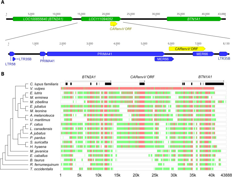
A closer look at the genomic location of this ERV in the domestic dog (Canis lupus familiaris) positions it between the genes BTN1A1 and BTN2A1 (Fig 1A). There is an open reading frame (ORF) of 1344 bp towards the 3’ end of this ERV that is upstream of the putative 3’ LTR (Fig 1A). A search of the PANTHER protein domain database [50] showed that the putative protein encoded by this ORF is closely related to the retroviral envelope protein subfamily (PTHR10424:SF74) that includes EnvV1 and EnvV2 [51]. This env ORF is also predicted to be part of a gene and a transcript annotated as “endogenous retrovirus group V member 2 Env polyprotein-like” in the NCBI RefSeq database. Hence, we named this gene CARenvV.
Fig 1. Genomic location and conserved synteny of CARenvV.
A. (Upper panel) Chromosomal context of the CARenvV locus is shown for the C. lupus familiaris (domestic dog) genome (ROS_Cfam_1.0, chromosome 35). Refseq annotated genes are shown in green, and the CARenvV ORF is in yellow. (Lower panel) ERV derived repeat elements identified by the Dfam repeat database for the indicated region are annotated in blue. B. Genomic segments containing the CARenvV ORF and the BTN1A1 and BTN2A1 genes were extracted from the NCBI genome database for each of the indicated species and aligned using the MultiPipMaker alignment tool [108]. The CARenvV ORF and the BTN1A1 and BTN2A1 exons are shown in black boxes in the C. lupus familiaris reference assembly. For the other species, regions with more than 75% identity are shown as red boxes while regions with 50–75% identity are shown as green boxes. On the left is a cladogram representing the phylogenetic relationships for the species analyzed that was generated using TimeTree [82].
Further analysis of CARenvV using the Dfam repeat database revealed remnants of gag and pol genes with similarity to an ERV group named Prima41 (Fig 1A), while its C-terminal portion shows similarity to the ERV group MER66 (Fig 1A) [52].
This provirus also contains remnants of 5’ and 3’ LTRs (Fig 1A) [52]. Since retroviral insertion into the host genome generates identical 5’ and 3’ LTRs, the divergence between the LTRs of an ERV can be used to estimate its age [53,54]. Because the CARenvV ERV in C. lupus familiaris contains a partial deletion of the 5’ LTR, we used the CARenvV ortholog of F. catus for this analysis. Comparison of these 5’ and 3’ LTRs (S1 File) determined this ERV to be ancient (91–121 million years old) putting its endogenization before the divergence of placental mammals [55,56].
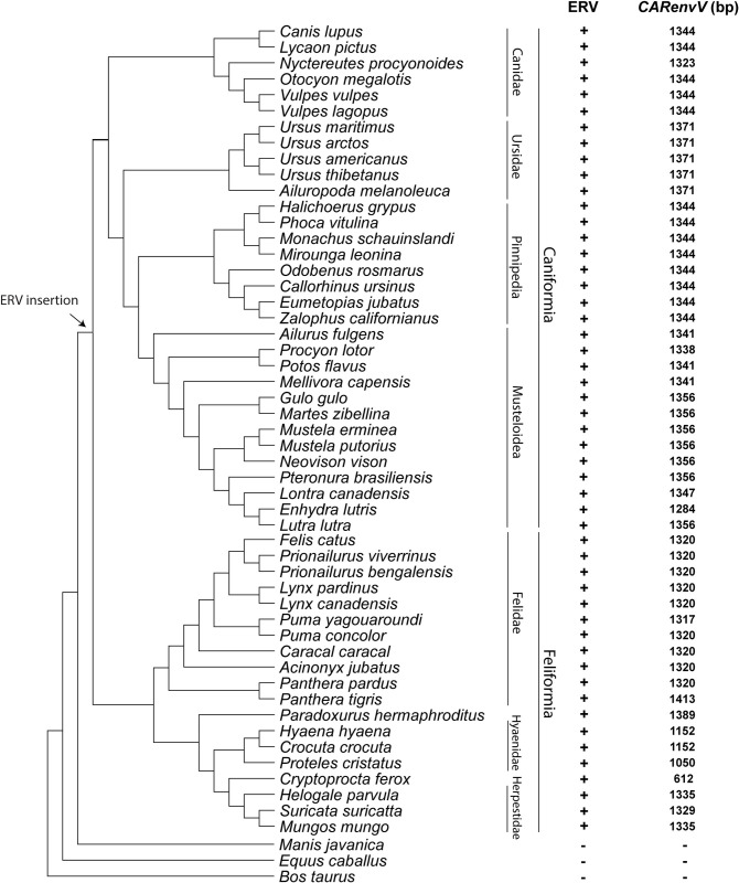
The age of an ERV can also be estimated from the orthology of its genomic location since retroviral integration is not site specific [54,57,58]. We extracted and aligned orthologous segments from the genomes of 15 Carnivora species and from 5 species in sister orders within the superorder Laurasiatheria (S1 Table). We observed high levels of sequence conservation of the CARenvV ORF among Carnivora as well as sequence conservation in most exons of the linked BTN1A1 and BTN2A1 genes in all the species examined confirming the orthology of this region (Fig 1B). In addition, the region that encompasses the CARenvV ORF, as well as the rest of the provirus, is missing in mammalian species outside of Carnivora (Fig 1B). CARenvV and its large ORF are conserved in individual Carnivora species in all lineages of this order, although a few species, including three members of the subfamily Hyaenidae, contain early stop codons in the CARenvV gene generating shorter ORFs (Fig 2). These results indicate that CARenvV was inherited from the common ancestor of the order Carnivora at least 54 mya [59] contradicting the older age estimate from the LTR dating analysis. LTR analyses can produce these older age estimates due to differential selective pressure on ERV LTRs or recombination with similar LTRs. Therefore, these findings collectively indicate that this ancient ERV was fixed in the common ancestor of the extant members of the order Carnivora at least 54 million years ago [59].
Fig 2. Conservation of CARenvV ORF in the lineages of Carnivora.
A cladogram illustrates the evolutionary relationships of species with assembled genomes in the order Carnivora along with three species from sister orders of Carnivora. The tree was generated with TimeTree [82]. Suborder and family classifications are shown on the right. + indicates the presence and - indicates the absence of ERV orthologs that contain CARenvV as identified via BLAST searches of each genome assembly. The size of the CARenvV ORF found in the orthologous location at each genome assembly is indicated. An arrow marks the likely insertion and fixation of the ERV that contains CARenvV, based on the orthology analysis.
An ancient ERV with an intact SUenv domain in Artiodactyla
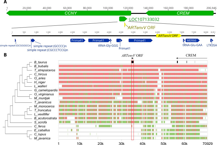
Next, we examined whether other mammalian orders contain similar ERVs. To this end, we used the ORF of the C. lupus familiaris CARenvV as a probe to search other sequenced mammalian genomes. This search revealed a highly similar sequence (>75% identity) in various members of the order Artiodactyla (even-toed ungulates). In Bos taurus (cattle) this sequence is located between the genes CREM and CCNY (Fig 3A) so it is not orthologous to CARenvV. Analysis of this ERV using the Dfam repeat database demonstrated that, like CARenvV, remnants of the gag and pol genes show significant similarity to the ERV group Prima41 while the ERV portion immediately upstream of the predicted 3’ LTR showed similarity to the ERV group MER66 (Fig 3A). Also, like CARenvV, this ERV contains a large ORF identified by computer annotation as “endogenous retrovirus group V member 1 Env”. Although there is an identifiable LTR at the 3’ end of the ERV there are no remnants of the 5’ LTR as the region immediately upstream of the annotated gene is dominated by GC-rich repeats (Fig 3A). This ERV is therefore related to CARenvV but is not orthologous, so we designated this gene ARTenvV.
Fig 3. Genomic location and conserved synteny of ARTenvV.
A. (Upper panel) The chromosomal context of the ARTenvV locus is shown for B. taurus (ARS-UCD1.2, chromosome 13). Refseq annotated genes are shown in green with the location of the ARTenvV ORF in yellow. (Lower panel) ERV derived repeat elements that are identified by the Dfam repeat database for the indicated region are annotated in blue. B. Genomic segments containing ARTenvV ORF and the CREM gene were extracted from the NCBI genome database for each of the indicated species and aligned using the MultiPipMaker alignment tool [108]. ARTenvV ORF and the exons of CREM are shown in black boxes in the B. taurus reference assembly. Regions with more than 75% identity are shown as red boxes while regions with 50–75% identity are shown as green boxes. On the left is a cladogram representing the phylogenetic relationships for the analyzed species generated using TimeTree [82].
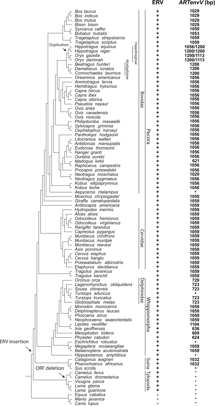
To determine whether this ERV is found in a region of conserved synteny in other members of Artiodactyla, we extracted and aligned the genomic region surrounding the ARTenvV ORF from 17 species of Artiodactyla as well as three other species from the sister orders Perissodactyla (odd-toed ungulates), Pholidota (pangolins) and Carnivora [59,60]. This region is highly conserved in species in the ruminant infraorder Pecora and shows high sequence conservation in the exons of CREM confirming the orthology of this genomic region in these orders (Fig 3B). This alignment also shows high conservation of ARTenvV in all lineages of Artiodactyla except for the Bactrian camel (Camelus ferus) (Fig 3B). The ERV containing ARTenvV is also absent in the horse (Equus caballus), dog (Canis lupus familiaris) and pangolin (Manis javanica) genomes (Fig 3B). To describe the evolutionary history of this ERV and confirm the conservation of the ARTenvV ORF, we individually inspected this genomic region in all Artiodactyla species that have a genome assembly (S1 Table). Remnants of this ERV are found in all Artiodactyla lineages, and the vast majority of these species also contained >1000 bp of the ARTenvV ORF (Fig 4). A few species, including members of the family Delphinidae (oceanic dolphins), contain early stop codons in ARTenvV (Fig 4). Notably, all members of the suborder Tylopoda (camels and llamas) have orthologous remnants of this ERV, although none of them contain an env ORF as part of this ERV (Fig 4). Detailed comparative analysis of the aligned region containing the ARTenvV ERV from B. taurus and C. dromaderius (dromedary camel) shows high sequence identity in the first exon of the CCNY gene immediately upstream of the ARTenvV ERV as well as the regions corresponding to the gag and pol portions of this ERV (S1 Fig). However, the Dfam repeat annotation reveals the insertion of a Long Interspersed Nuclear Element-1 (LINE-1) retrotransposon (DF0000255.4) at the location corresponding to the ARTenvV ORF in C. dromaderius (S1 Fig). Hence, these findings indicate that this ERV was fixed in the common ancestor of all extant Artiodactyla which diverged at least 64 million years ago, but that the ARTenvV ORF was likely lost in the common ancestor of the extant members of the suborder Tylopoda [60,61].
Fig 4. Conservation of ARTenvV ORF in the lineages of Artiodactyla.
Shown is a cladogram illustrating the evolutionary relationships for species with assembled genomes in the order Artiodactyla along with three species from sister orders of Artiodactyla. Suborder, family and subfamily classifications are shown on the right. The species tree was generated with TimeTree [82]. + indicates the presence and–indicates the absence of ERV orthologs that contain ARTenvV as identified via BLAST searches of each genome assembly. The size of the ARTenvV ORF found in the orthologous location at each genome assembly is indicated. * indicates a very early stop codon caused by an insertion or deletion. Arrows mark the insertion and fixation of the ERV that encodes ARTenvV, retroduplication of ARTenvV, and the deletion of the ARTenvV ORF.
Additional copies of ARTenvV and CARenvV related ERVs
These findings prompted us to determine whether there are other copies of these ERVs in Artiodactyla and Carnivora. Using the ORFs of these genes as probes we identified one additional ERV in both orders with high similarity (~ 70%) to their respective env ORFs. While neither of these ERVs contained an intact retroviral gene ORF, analysis of this sequence using the Dfam repeat database indicates that there are remnants of gag and pol that have the same Dfam designation (Prima41) as the comparable regions of the ERVs that contain ARTenvV and CARenvV (S2 Fig). To determine whether these ERVs are in a location of conserved synteny, we aligned the genomic region surrounding each ERV from 15 species of Artiodactyla or Carnivora as well as five other species from their sister orders in Laurasiatheria. Each ERV was only found in the same orthologous location in species of their respective orders (S3 Fig). These findings indicate that ERVs derived from a related retrovirus were independently fixed in the common ancestors of Carnivora and Artiodactyla at least 54 and 64 million years ago, respectively [59–61].
Retroduplication of ARTenvV
During the BLASTn search of Artiodactyla using the B. taurus ARTenvV ORF as a probe, we found an additional copy of this gene in the subfamily Hippotraginae (grazing antelopes). Alignment of the orthologous region of this additional copy showed that it is missing in other Artiodactyla including the Alcelaphinae subfamily which is most closely related to Hippotraginae (S4 Fig). Analysis of the DNA sequence of this copy in a member of this lineage, Oryx dammah, shows that it is > 97% identical to the computer annotated mRNA of the ARTenvV gene which is predicted to be the spliced product of two exons (S5 Fig). In addition, this extra copy is flanked by 14 bp target site duplications and contains a signature site required for insertion via LINE-1 (TTAAAA) suggesting that this copy represents a LINE-1-mediated retrotransposition. LINE-1 retrotransposons are autonomous non-LTR elements that encode an RNA binding protein and a protein with reverse transcriptase (RTase) and endonuclease function [62]. These elements can use a copy-paste mechanism of reverse transcription (RT) and insertion to recognize and replicate their own mRNAs as well as cellular mRNAs including mRNAs of co-opted ERV derived genes resulting in the insertion of intron-less copies of cellular mRNAs at random genomic locations [62–64]. To confirm the presence of this extra copy of ARTenvV we extracted DNA from O. dammah and two species of Bovidae. Using primer pairs in and around this ARTenvV copy, we generated PCR products with the expected size of ~ 700 base pairs (bp) from O. dammah but not from B. taurus and Ovis aries consistent with the absence of this retrotansposed copy (S6 Fig). Sequencing of these PCR products confirmed this conclusion. Collectively these findings indicate that ARTenvV was duplicated via LINE-1 mediated retrotransposition in the common ancestor of the subfamily Hippotraginae at least 7 million years ago [60,61].
Phylogenetic analysis identifies the ARTenvV and CARenvV proviruses as gamma-like ERVs
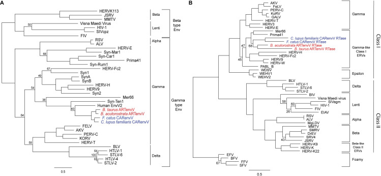
Retroviruses are classified into two subfamilies; orthoretroviruses and spumaretroviruses [65]. Orthoretroviruses are further subdivided into six genera; alpharetroviruses, betaretroviruses, gammaretroviruses, epsilonretroviruses, lentiviruses, and deltaretroviruses [65]. ERVs are classified based on their similarity to exogenous retroviruses into Class I ERVs (gamma and epsilon), Class II ERVs (alpha, beta, delta and lenti) and Class III ERVs (spuma) [66,67]. While the retroviral classification is based on the RTase domain of the pol gene, retroviral envelope proteins are classified into two subtypes, gamma-type and beta-type, based on the nature of the linkage (covalent or non-covalent) between the SU and TM subunits [68]. In a phylogenetic tree of endogenous and exogenous retroviral envelope proteins, CARenvV and ARTenvV cluster with proteins that belong to the gamma-type envelope subgroup with covalently linked SU and TM subunits, a subgroup that includes gamma, delta and alpha-retroviruses (Fig 5A and S2 File) [68]. CARenvV and ARTenvV group with each other and with human EnvV2 (Fig 5A). To identify the retroviral genus that includes the CARenvV and ARTenvV ERVs, we aligned the amino acid sequence of the pol gene RTase domain of exogenous and endogenous retroviruses (S2 Table and S3 File) and generated a phylogenetic tree. As shown in Fig 5B, the identifiable remnants of the RTase of the CARenvV and ARTenvV ERVs clustered with the exogenous gammaretroviruses and ancient mammalian gamma-like class I ERVs [66,67] together with Prima41 and Mer66 (Fig 5B).
Fig 5. Phylogeny of CARenvV and ARTenvV proteins.
A. The predicted amino acid sequence of CARenvV (in blue) and ARTenvV (in red) from the indicated species together with the amino acid sequence of selected endogenous and exogenous retroviral Env proteins (S2 Table) were aligned, and a maximum likelihood tree was generated using RaxML with 500 bootstraps. The tree was midrooted. B. Amino acid sequence of the predicted RTase domains of CARenvV ERV (in blue) and ARTenvV ERV (in red) from the indicated species together with the amino acid sequence of the RTase domain from the indicated endogenous and exogenous retroviruses (S2 Table) were aligned and a maximum likelihood tree was generated using RaxML with 500 bootstraps. The tree was rooted using foamy viruses. Bootstrap values that are less than 70 are shown at the relevant nodes. Scale bar represents 0.5 amino acid substitutions per site.
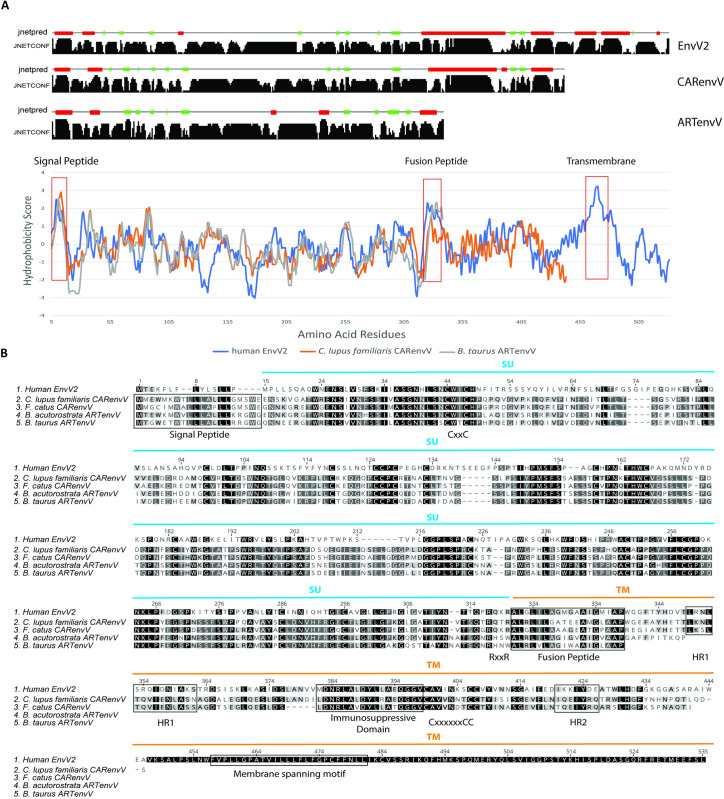
To determine whether ARTenvV and CARenvV contain features of retroviral envelope proteins we produced hydrophobicity plots of the ARTenvV and CARenvV proteins together with human EnvV2 and generated the predicted secondary structure of these proteins. Results revealed the presence of a putative N-terminal signal peptide that contains a hydrophobic region and an alpha helix, as well as a predicted fusion peptide in the C-terminal region that also contains a hydrophobic region and an alpha helix (Fig 6A). Amino acid alignment of human EnvV2 together with two orthologs of CARenvV and ARTenvV showed that both these proteins lack a fully intact TMenv, the more highly conserved Env domain. ARTenvV ends after the predicted TMenv fusion peptide and CARenvV is truncated just before the membrane-spanning domain near the env C-terminus (Fig 6B). The highest similarity between these Env proteins and human EnvV2 lies in the region immediately following the predicted signal peptide (Fig 6B). This alignment also identified the two conserved cysteine-containing motifs which are found at the same location as in EnvV2: CxxC in the SU domain of both ARTenvV and CARenvV and Cx6CC in the TM domain of CARenvV (Fig 6B). In addition, the RxxR furin cleavage motif (RQT/KR) is found right before the predicted fusion peptide in the CARenvV but is altered in ARTenvV (Fig 6B). CARenvV also contains an immunosuppressive domain and heptad repeats that show high similarity to the previously identified immunosuppressive domain and heptad repeats of EnvV2 (Fig 6B) [48,49]. The presence of the immunosuppressive domain and the Cx6CC motif in the TM domain of CARenvV are consistent with the grouping of this envelope protein with gamma-type envelopes of endogenous and exogenous retroviruses in a phylogenetic tree (Fig 5A) [68]. Taken together, these findings establish that ARTenvV and CARenvV originated from an ancient gamma-like retrovirus.
Fig 6. Structural features of CARenvV and ARTenvV.
A. Secondary structure predictions (top panels) and hydrophobicity profiles (lower graph) are shown for human EnvV2 (blue), C. lupus familiaris CARenvV (orange) and B. taurus ARTenvV (gray). Jnetconf shows the confidence levels for the secondary structure prediction while Jnetpred shows the consensus prediction. Red boxes indicate alpha helices and green boxes indicate beta sheets in the secondary structure prediction. The predicted locations of key structural features are indicated on the hydrophobicity profile graph. B. Amino acid alignment of human EnvV2, C. lupus familiaris, and F. catus CARenvV, and B. taurus and B. acutorostrata ARTenvV is shown. Predicted locations of the SU (blue) and TM (orange) domains, signal peptide, the fusion peptide, the immunosuppressive domain, membrane spanning motif, the furin cleavage site (RxxR) between the two domains of the Env, the CxxC and Cx6CC motifs involved in SU–TM interaction are indicated. Predicted heptad repeat regions are indicated as HR1 and HR2. Residues identical in all proteins are indicated by black shading, and residues identical among four, three or two proteins are indicated by different shadings of gray.
An ancient retroviral cross-species transmission event
Our findings so far suggest that ARTenvV and CARenvV were independently fixed in the respective common ancestors of extant Artiodactyla and Carnivora. Despite their likely origins from independent infection and endogenization events and their separation by more than 60 million years of evolution, the ARTenvV and CARenvV ORF orthologs are up to 82% identical (S4 File). In general, the SU domains of retroviral envelopes show high sequence divergence as they must adapt to mutagenized or new receptors and are subject to immune system attack.
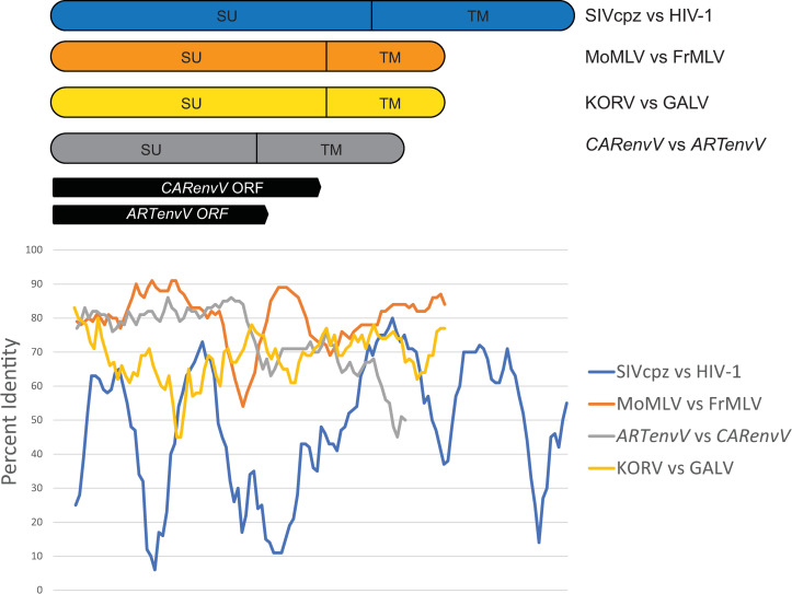
To illustrate the extraordinary similarity of ARTenvV and CARenvV sequences, we aligned the orthologs of the genes with the highest similarity (Ursus arctos and Monodon monoceros) and performed a sliding window analysis of percent nucleotide identity (Fig 7). Since the ORF of CARenvV is longer than ARTenvV, and neither of these genes includes the C-terminal end of the TM domain, we included additional nucleotides in this alignment beyond the stop codons of these ORFs using the nucleotide length of envV2 as a guide (Fig 7). For comparison, we performed the same pairwise analysis for env sequences of FrMLV and MoMLV which are derived from the laboratory mouse ecotropic MLV, for KoRV and GALV, which are closely related viruses likely acquired by koalas and gibbons from an intermediate rodent or bat species [24, 25], and for HIV-1 (NC_001802) and its closest relative SIVcpz (AF115393), related by the cross-species transmission of SIVcpz from chimpanzees to humans in the early 20th century [17–19].
Fig 7. CARenvV and ARTenvV show high nucleotide identity in SUenv.
Sliding window analysis of percent nucleotide identity along pairwise alignments of CARenvV and ARTenvV as well as the env genes of the indicated retroviruses. The location of the ORFs of CARenvV and ARTenvV relative to the sequence alignment, the relative sizes of the env genes as well as the location of the SU and the TM domains for each alignment are shown above the graph.
Results revealed that the high level of identity in the SU domains of ARTenvV and CARenvV (80–85%) is comparable to that seen for FrMLV and MoMLV (Fig 7). In contrast, the SU domains of KoRV and GALV show less than 70% identity, while HIV-1 and SIVcpz SUs show only about 60% nucleotide identity and < 70% identity for the entire length of env (Fig 7). This demonstrates that ARTenvV and CARenvV are much more closely related to each other than are the env genes of HIV-1 and SIVcpz or KoRV and GALV. These findings suggest that, by the time of their endogenization after cross-species transmission, the progenitors of ARTenvV and CARenvV had not undergone the same extent of mutational modifications that are observed in the SIV/HIV and GALV/KoRV pairings. Moreover, despite more than 60 million years of evolutionary divergence, ARTenvV and CARenvV ORFs are still up to 80% identical in nucleotide sequence suggesting that they were much closer in identity prior to their endogenization. Combined with the fact that orthologous copies of each ERV are absent outside of their respective orders, these findings indicate that the original ARTenvV and CARenvV ERVs likely descended from the same retrovirus that infected both the common ancestor of all the extant members of the order Artiodactyla and the common ancestor of the order Carnivora through an ancient cross-species transmission event.
Evidence of positive selection in CARenvV and ARTenvV
The adaptive evolutionary pathways of genes can be traced by comparing the ratio of the rate of nonsynonymous (dN) and synonymous (dS) changes of their orthologs in related species [69]. Similar to host genes, once endogenized and fixed in a lineage, ERV genes can be conserved under purifying/negative selection to retain a beneficial function leading to a dN/dS (ω) value below 1, or, they can evolve under diversifying/positive selection, resulting in adaptive changes and producing a ω value above 1 [70,71].
To identify the signatures of selection pressures in ARTenvV and CARenvV, we separately aligned the ORF orthologs from Artiodactyla and Carnivora. The vast majority of the nodes in the resulting maximum likelihood trees showed high bootstrap support for both ARTenvV and CARenvV with a few branches with low support in the infraorder Pecora for ARTenvV which may be due to its very high sequence similarity in this infraorder (S7 Fig). Additionally, orthologs of both ARTenvV and CARenvV form clear clusters in the major lineages of each order, including Pecora and Cetacea for ARTenvV (S7 Fig) and Caniformia and Feliformia for CARenvV (S8 Fig).
Use of the codeml program of PAML4 [72] revealed significant evidence of positive selection for both ARTenvV and CARenvV (p<0.01) (Table 1). Bayes empirical Bayes (BEB) analysis of posterior probabilities identified six sites for CARenvV and two for ARTenvV under positive selection with a posterior probability of >0.95 (Table 1) although no sites mapped to homologous sites in the two lineages. Three of these residues for CARenvV and one for ARTenvV were also found to be under positive selection by the MEME and FEL programs in the Datamonkey webserver [73]. At most sites, however,orthologs of both CARenvV and ARTenvV showed high sequence similarity, with pairwise ω values much below unity for the vast majority of the species (S5 and S6 Files). These results indicate that while both ARTenvV and CARenvV are highly conserved, they have each been subject to recurrent positive selection in their respective orders.
Table 1. PAML analysis of CARenvV and ARTenvV.
| Gene | Codon frequency | ω0a | M7-M8 | Tree lengthc | dN/dS (%) | Residuesd with dN/dS of >1 and pre of >0.95 | |
|---|---|---|---|---|---|---|---|
| 2δ | P value | ||||||
| CARenvV | f3 × 4 | 0.3 | 9.25 | 0.0098 | 2.78555 | 2.11 (3.45) | 56Q, 99Q, 131R*, 336M, 437Y*, 440N* |
| f3 × 4 | 1.7 | 9.25 | 0.0098 | 2.78555 | 2.11 (3.45) | 56Q, 99Q, 131R*, 336M, 437Y*, 440N* | |
| ARTenvV | f3 × 4 | 0.3 | 11.47 | 0.0032 | 2.82655 | 2.01 (3.61) | 240R, 305A* |
| f3 × 4 | 1.7 | 11.47 | 0.0032 | 2.82655 | 2.01 (3.61) | 240R, 305A* | |
a ωo indicates the initial seed value of ω used.
b 2δ, two times the difference of the natural log values of the maximum likelihood from pairwise comparisons of the different models.
c Tree length is defined as the sum of the nucleotide substitutions per codon at each branch.
d Residue numbers are based on dog (CARenvV) and cattle (ARTenvV) protein sequences.
e pr, posterior probability.
* Indicates residues identified under positive selection via the datamonkey webserver.
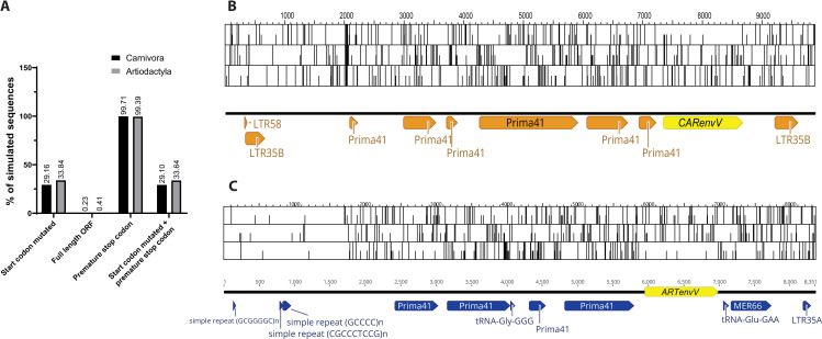
Preservation of a large ORF of an ancient retroviral env in two mammalian orders for more than 50 million years suggests that evolutionary pressures acted to maintain these ORFs. To determine the level of preservation expected for ORFs this size (1323 bp for CARenvV and 1029 bp for ARTenvV) under genetic drift and neutral evolution in these orders, we constructed ancestral sequences for both ARTenvV and CARenvV and simulated their neutral evolution using the mammalian neutral substitution rate [74]. These results revealed that less than 0.5% of the sequences maintained an intact ORF for both ARTenvV and CARenvV, and the vast majority of the sequences (>99%) contained a premature stop codon (Fig 8A). ORF maps of the ERVs that contain ARTenvV and CARenvV also reveal the unusual preservation of these env ORFs compared to the rest of their proviruses which contain numerous stop codons in all three frames (Fig 8B and 8C). It is important to note that since this analysis does not consider deletions or insertions, we expect the number of intact ORFs to be even lower than what was observed under neutral evolutionary pressure. These findings suggest that selection pressures during the evolution of Carnivora and Artiodactyla led to the preservation and co-option of the ARTenvV and CARenvV ORFs.
Fig 8. Preservation of CARenvV and ARTenvV ORF under neutral evolution.
A. Results of the Monte-Carlo simulations of CARenvV and ARTenvV evolution for 54 and 64 million years are shown, respectively, using the mammalian neutral substitution rate. The percentage of 100,000 simulated sequences that have the indicated properties are plotted. B. ORF map of the CARenvV ERV with all three forward reading frames is shown. CARenvV ORF is annotated in yellow and the Dfam designation of repeat elements is indicated in orange. C. ORF map of the ARTenvV ERV with all three reading frames is shown. ARTenvV ORF is annotated in yellow and the Dfam designation of repeat elements is indicated in blue. Potential start codons are indicated with a half-height line and stop codons are indicated with a full height line.
CARenvV and ARTenvV are expressed in a variety of tissues
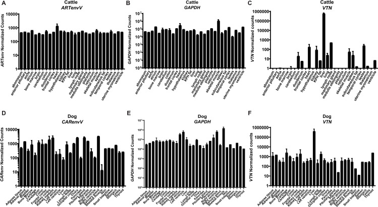
To determine the tissue-specific expression profile of CARenvV and ARTenvV, we performed in silico analyses of RNA sequencing data from the domestic dog (C. lupus familiaris) for CARenvV and from cattle (B. taurus) for ARTenvV. Both CARenvV and ARTenvV are expressed at similar levels in a wide variety of tissues (Fig 9). While CARenvV showed lower expression levels in dog skeletal muscle, liver, pancreas and right ventricle, this was not the case for ARTenvV in cattle (Fig 9).
Fig 9. CARenvV and ARTenvV are expressed in a wide variety of tissues.
In silico analyses of RNA sequencing data from 29 tissues from cattle (B. taurus) and 30 tissues from domestic dog (C. lupus familiaris) are shown for A. cattle ARTenvV, B. cattle GAPDH, C. cattle VTN, D. dog CARenvV, E. dog GAPDH and F. dog VTN. The mapping accuracy of the RNA sequencing reads, and the normalization of the read counts was confirmed using as controls a liver specific gene (VTN) (C. and F.) or a housekeeping gene (GAPDH) (B. and E.). Raw RNA sequencing data extracted from SRA projects PRJNA314981, PRJNA78827, PRJNA379574, PRJNA177791 and PRJNA314981 were mapped to the cattle or dog genome assembly and the counts that map to the coding region of each gene were extracted and normalized using DEseq2. KPH: Kidney, pelvic and heart.
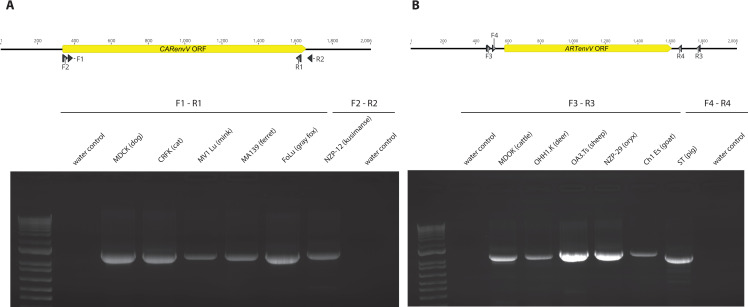
Next, we used RT–PCR and 5’ and 3’ RACE-PCR to determine the structure of the transcripts that encode CARenvV and ARTenvV. First, we probed total RNA extracted from six Carnivora species and six Artiodactyla species for the presence of CARenvV and ARTenvV, respectively, via RT—PCR. Transcripts were detected in all species tested (Fig 10) and confirmed by sequencing (S7 File). This analysis also showed that both ARTenvV and CARenvV transcripts are generated via splicing of two exons separated by approximately 4500 and 6500 bp, respectively, and that these genes contain a 3’ UTR ranging from 700 to 1200 bp (S9 and S10 Figs). These findings are also consistent with the computer-based annotation of these genes in RefSeq. For both ARTenvV and CARenvV, we found the 3’ end of the transcript to be the same among the species analyzed (S9 and S10 Figs), while the transcript start site was within a few nucleotides in different species for ARTenvV (S9 Fig). While 5’ RACE-PCR showed that the transcript start site of CARenvV is located within the 5’ LTR of the ERV, we observed significant variation in the location of the transcript start site within the first exon of CARenvV among different Carnivora species (S10 Fig). For ARTenvV, the transcript start site is located immediately downstream of a GC-rich region (S9 Fig).
Fig 10. CARenvV and ARTenvV expression in Artiodactyla and Carnivora cell lines.
A. and B. At the top is a schematic of the genomic region that contains A. CARenvV ORF or B. ARTenvV ORF with the locations of the primers used for RT-PCR indicated below the diagram. Below are the agarose gels showing the RT-PCR products of A. CARenvV and B. ARTenvV that were amplified from the total RNA of the indicated species with the primers shown in the upper panel. The gels shown are representative of at least two independent experiments.
CARenvV and ARTenvV protein expression
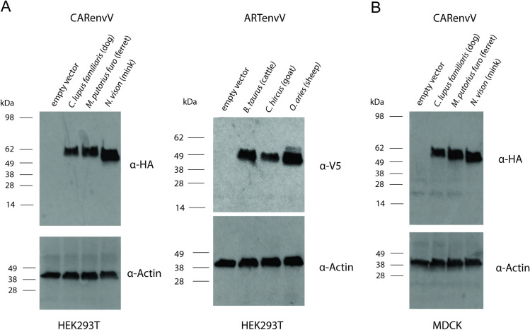
To determine whether CARenvV and ARTenvV genes have the capacity to produce envelope proteins, we cloned the ORF of CARenvV and ARTenvV from three species each into a CMV promoter-driven expression vector, adding C-terminal tags of HA to CARenvV and V5 to ARTenvV. Both CARenvV and ARTenvV proteins from transiently transfected HEK293T cells were detected in whole-cell lysates via immunoblotting (Fig 11A). Both ARTenvV and CARenvV contain multiple potential N-linked glycosylation sites, and their observed molecular weights are higher than the expected weights of 37.6 and 49.1, respectively, suggesting a glycosylation mediated shift. In addition, while the amino acid sequence of CARenvV (but not ARTenvV) contains a predicted furin cleavage site (Fig 6B), we did not observe any evidence of cleavage of CARenvV protein when expressed in HEK293T cells (Fig 11A). To determine whether CARenvV is subject to furin cleavage in cells from a Carnivora species, we expressed HA-tagged CARenvV from three species in MDCK cells using a retroviral delivery system. As shown in Fig 11B, we observed expression of CARenvV at the sizes expected for uncleaved proteins but observed no smaller cleavage products. These findings show that both CARenvV and ARTenvV ORFs can produce stable proteins under the control of a constitutive promoter when exogenously expressed.
Fig 11. Ectopic expression of CARenvV and ARTenvV.
A. Immunoblotting analysis of 293T cells transfected with the plasmids expressing empty vector or the indicated CARenvV and ARTenvV constructs. B. Immunoblotting analysis of MDCK cells transduced with the plasmids expressing empty vector or the indicated CARenvV constructs. The blots shown are representative of at least two independent experiments.
Discussion
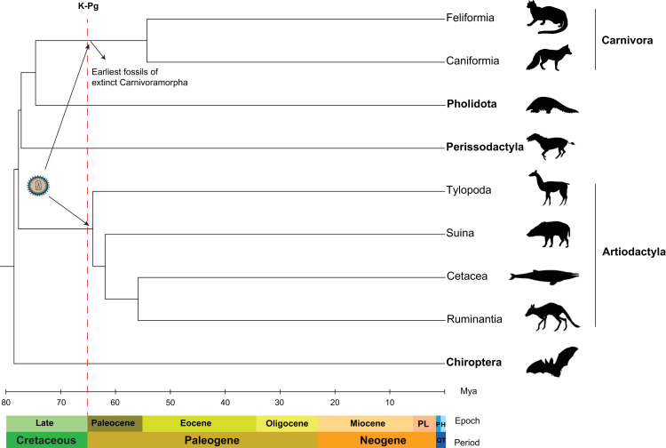
The Cretaceous–Paleogene (K–Pg) mass extinction event that is thought to have killed the non-avian dinosaurs approximately 66 million years ago represents a major turning point in the evolutionary history of Eutheria (placental mammals) [75]. While there are multiple models of the impact of the K-Pg event on the interordinal diversification of Eutheria [76], both the fossil record and molecular clock studies suggest that placental mammalian orders went through rapid diversification following the K-Pg event that continued throughout the Paleocene and Eocene epochs leading to the emergence of the modern taxa [77,78]. In this study, we presented multiple lines of evidence that a gamma-like retrovirus crossed the species barrier and was independently fixed in the genomes of the ancestral species of Artiodactyla and Carnivora during this significant period in the history of placental mammals (Fig 12). First, we found no evidence of ARTenvV and CARenvV orthologs in species outside of their respective orders including the Artiodactyla sister orders Perissodactyla and Pholidota. An alternative scenario based on a single endogenization event and vertical inheritance from the common ancestor of all these mammalian orders would have required three independent deletion events involving different ERVs in the common ancestors of three different mammalian orders. A much more likely scenario is that these ERVs were horizontally and independently acquired by the common ancestors of all extant Artiodactyla and Carnivora. This hypothesis is also bolstered by the fact that we found a highly similar ERV in separate orthologous locations in both Artiodactyla and Carnivora (S3 Fig) while other mammalian taxa lack this ERV subtype.
Fig 12. Cross-species transmission of an ancient gammaretrovirus around the K-Pg event.
The species cladogram shows the phylogenetic relationships for the indicated orders (in bold) that belong to the superorder Laurasiatheria. Suborders are identified for Carnivora and Artiodactyla. The tree and the divergence times were generated using TimeTree [82]. Geological timescales were derived from The International Chronostratigraphic Chart [119]. PL; pliocene, QT; quaternary, P; pleistocene, H; holocene.
Additionally, the preserved env ORFs of this ancient ERV (ARTenvV and CARenvV) found in the extant members of Artiodactyla and Carnivora display an unusually high sequence similarity (75–82%) in the ordinarily divergent SUenv domain that is much higher than the nucleotide identity observed between other pairs of closely related retroviruses that are formed as a result of a cross-species transmission event (e.g. HIV-1 and SIVcpz) [17–19]. It is not feasible for two ERVs that were not derived from the same virus to have such high nucleotide similarity in the otherwise highly variable SU domain of the env gene, indicating that ARTenvV and CARenvV were most likely formed as a result of an ancient cross-species transmission event involving the same gamma-like retrovirus.
Estimates from molecular clock studies using fossil calibrations indicate that the modern Carnivora diverged from their most recent common ancestor in the early Eocene epoch approximately 54 million years ago (Fig 12) [59,60]. However, paleontological studies group the extant Carnivora lineages with ancient extinct carnivorans into a stem-group called Carnivoramorpha based on phenotypic characteristics identified in the fossils [79]. This stem-group includes members that are thought to have lived during the Paleocene epoch (66 to 55 mya) based on the fossil record [80, 81]. In addition, molecular clock studies using both nuclear and mitochondrial genes put the divergence of the Artiodactyla from a common ancestor around the K-Pg event, in the early Paleocene or late Cretaceous between 67 and 61 mya [55,60,61,82]. Hence, it is conceivable that the exogenous retrovirus that produced ARTenvV and CARenvV transmitted directly between an ancestral Carnivoramorpha and Artiodactyla (Fig 12). Modern carnivorans are known to prey on even-toed ungulates, especially ruminants. The oldest known Artiodactyla fossil belongs to an extinct genus called Diacodexis [79,83], members of which resemble the modern mouse-deer (family Tragulidae), a small ruminant and a prey species of felines in Asia [84]. Although the remnants of Diacodexis do not appear in the fossil record until the Eocene epoch [83], it is feasible that ancestors of this genus that lived alongside the ancestors of Carnivoramorpha around the K-Pg event had a prey-predator relationship. Predation is a major mode of viral transmission between different mammalian species [85]. In fact, the major mode of transmission of SIVcpz to humans leading to the formation of HIV-1 is thought to be bushmeat hunting in western Africa which likely exposed hunters to the bodily fluids of SIVcpz-infected chimpanzees [86,87].
The possibility remains that cross-species transmission of this retrovirus happened after it was endogenized and fixed in one lineage. Some ERVs stay active even after being fixed in a population [28,88,89]. It is feasible that the exogenous gamma-like retrovirus that gave rise to ARTenvV became endogenized in the common ancestor of the Artiodactyla in the early Paleocene epoch and stayed replication-competent until the Eocene at which time the exogenous version of this ERV infected the germline of the common ancestor of the extant Carnivora. However, because we only found one extra copy of this ERV in both Artiodactyla and Carnivora, even if it stayed active for an extended period, it did not amplify or leave a recognizable fossil record in extant species. It is also possible that there were intermediate species involved in the transmission of this ancient virus. GALV and KORV are highly related gammaretroviruses hypothesized to have been acquired from an intermediate bat or rodent species [24,25,90]. Retroviral endogenization and fixation in a lineage is a rare event. Hence an intermediate species could have been infected with the progenitor gamma-like retrovirus of ARTenvV and CARenvV during the Paleocene epoch, and either this virus was not endogenized in this lineage or all of its members became extinct.
It is highly likely that evolutionary selection pressures kept the SU domain of ARTenvV and CARenvV intact, maintaining their unusual sequence similarity over more than 60 million years of evolution. Our sequence simulation analysis suggests that an ORF that is >1000 bp would acquire stop codons more than 99.5% of the time under neutral evolution in this 55–65 million year timeframe. However, the large ORFs in both ARTenvV and CARenvV are conserved with high sequence similarity in all lineages of their respective orders with only a few species acquiring early stop codons (Figs 2 and 4). Although we identified signatures of positive selection in both ARTenvV and CARenvV, dN/dS values were much below unity for the vast majority of the species. A few positively selected residues identified in CARenvV and ARTenvV were concentrated at the SU domain (3/6 for CARenvV and 2/2 for ARTenvV) even though none of them were located near each other. These residues may represent functional adaptations to the yet unidentified receptor of these env genes. env genes of circulating retroviruses tend to be under strong positive selection as they are in an arms race with the immune system of the host [91]. Our findings of limited positively selected residues for ARTenvV and CARenvV are consistent with these proteins being selected and conserved as host genes and are in concert with other co-opted env genes identified in mammals [34,37–42,48,92]. Future studies on the function of these env genes may reveal the functional importance of the positively selected residues we identified.
Our findings also offer additional clues to the origins of CARenvV and ARTenvV. Firstly, protein sequences of CARenvV and ARTenvV show similarity to human EnvV2 (Fig 6B), a co-opted ERV derived env gene found in simian primates [48,49] suggesting that these ERVs may share a common ancestor. In addition, repeat annotation of CARenvV and ARTenvV ERVs revealed that different portions of these sequences show similarity to two different primate ERV subfamilies, Prima41 and MER66 (Figs 1 and 3). This finding indicates that these ERVs may have experienced recombination. However, since both CARenvV and ARTenvV ERVs contain Prima41-like gagpol and Mer66-like env sequences, any potential recombination likely happened before endogenization and cross-species transmission.
While our results indicate that both ARTenvV and CARenvV are expressed in a variety of tissues, and the ORFs of these genes are capable of producing stable proteins when expressed from a constitutive promoter, the reason for the preservation of these retroviral genes for more than 60 million years is not clear. Most of the previously identified co-opted retroviral env genes contain TM domains and are fusogenic [31–38,40,42,48]. However, the transmembrane spanning portion of env is lost in both CARenvV and ARTenvV in all lineages. Hence, it is likely that both these genes acquired premature stop codons early in the evolution of each order before the divergence of the modern lineages and that the fusogenic function of these env genes either did not contribute to or conflicted with the basis for their co-option. Previous studies identified non-fusogenic/secreted co-opted env genes in mammalian genomes. For example, suppressyn, a truncated ERV derived Env protein found in simian primate genomes, is a soluble inhibitor of syncytin-1 induced cell fusion in the placenta [46]. While the widespread tissue expression profiles of CARenvV and ARTenvV make it unlikely that these genes are co-opted for a placenta specific function, we cannot rule out a function related to interference with circulating retroviruses that bind the same receptor, similar to some ERV envelope derived genes identified in mice and cats [93–95]. These questions may be addressed by the resolution of the tertiary structures of CARenvV, ARTenvV and EnvV2, and by the identification of the cell surface receptors for each protein. It is also important to note that while we detected the expression of CARenvV and ARTenvV ORFs by exogenously expressing the C-terminally tagged versions of these proteins, further studies using custom antibodies against these proteins in primary cells from relevant species could determine their endogenous expression levels and subcellular locations. Future studies may also determine the ancestral tropism/receptor of these envelope proteins by reconstructing the mutated TM region and testing fusion function in different cell types [96].
Our findings also revealed the presence of a retroduplicated, intronless copy of the ARTenvV gene in the members of the Hippotraginae, a subfamily in the order Artiodactyla. This copy of ARTenvV contains hallmarks of a LINE-1 mediated retrotransposition; an intronless copy of a gene at a different genomic location, a target site duplication at each side and a short series of As immediately downstream of the duplicated copy. LINE-1 mediated retrocopying of genes is a significant source of genomic diversity in mammals. Computational surveys done in the last two decades identified thousands of retroduplicated copies of genes including ERVs in the genomes of various mammals [97–100]. While the majority of these retrocopies are pseudogenes that have lost coding capacity, several retrogenes have been shown to express proteins, some of which have evolved new functions or become part of novel fusion genes by merging with other transcribed sequences [101–105]. Due to the extremely high sequence similarity (>97%) between the original and the retroduplicated copy in the O. dammah genome, we were not able to determine whether the retroduplicated copy is expressed in this species. However, all species belonging to the Hippotraginae with a genome in the database maintained an intact ORF of the retroduplicated ARTenvV. This may suggest that either this extra copy is still expressed in these species due to evolutionary pressures that maintain the ORF or simply that not enough time has passed since the retroduplication for the accumulation of enough mutations to neutralize this copy. Further analysis of RNAseq data from multiple tissues of O. dammah or other Hippotraginae species may determine if this intronless retroduplicated copy of ARTenvV is still expressed. We believe this is the first discovery of a retroduplicated copy of a co-opted retroviral env gene.
The oldest previously reported cross-species transmission events of retroviruses among mammals were proposed to have happened in the Oligocene epoch (24–34 million years ago) [8,28]. Our findings indicate a much older cross-species transmission of an ancient gamma-like retrovirus early in the age of mammals (Cenozoic Era) at least 64 million years ago, before the divergence of the extant lineages of Carnivora and Artiodactyla. These findings highlight the complex evolutionary relationship between retroviruses and the placental mammals as ARTenvV and CARenvV represent the first discovery of convergent co-option of the envelope gene from the same parental retrovirus in different mammalian orders.
Methods
Search for HERV-V related ERVs in mammalian genomes
The amino acid sequence of human envV2 (NP_001177984.1) was used as a probe for the tBLASTn search of the annotated mammalian genomes in the NCBI database (https://www.ncbi.nlm.nih.gov/genome/annotation_euk/all/) using the web based BLAST program available at the NCBI website (https://blast.ncbi.nlm.nih.gov). tBLASTn utilizes an amino acid sequence as a query and searches for all six frames of a given DNA sequence. The following parameters were used for tBLASTn: gap costs, 11 and 1 (existence and extension); matrix, BLOSUM62; conditional compositional score matrix adjustment; Expect threshold, 10−100. The DNA sequence of the hit from the initial tBLASTn search found in the genome of Ursus maritimus was extracted using the NCBI genome viewer (https://www.ncbi.nlm.nih.gov/genome/gdv/). This sequence was used in a subsequent BLAST search of the whole genome shotgun contigs of all Carnivora genomes housed in the NCBI genome database. The following parameters were used for BLASTn: gap costs, 5 and 2 (existence and extension); match/mismatch scores, +2/−3; repeat masking filter turned off; Expect threshold, 10−100.
To identify other mammalian ERVs that show similarity to CARenvV, the ORF of C. lupus familiaris CARenvV was used in a BLASTn search of annotated mammalian genomes excluding Carnivora. This search used the same parameters for BLASTn as above.
Reagents and plasmids
293T, MDBK (B. taurus), OHH1.K (O. hemionus), OA3.Ts (O. aries), Ch1 Es (C. hircus), ST (S. scrofa), NZP-29 (O. dammah), MDCK (C. lupus familiaris), CRFK (F. catus), Mv1 Lu (N. vison), FoLu (U. cineroargenteus) and NZP-12 (C. obscurus) cells were obtained from ATCC (Manassas, VA). Ferret (M. putorius furo) MA139 cells were obtained from Dr. Janet Hartley (NIAID, Bethesda, MD). Each cell line was grown in DMEM (Thermo Fisher, Waltham, MA) supplemented with 1% penicillin and 100 μg/mL streptomycin and 10% fetal bovine serum.
The PCR 2.1 TOPO and pcDNA-V5-TOPO plasmids were purchased from Thermo Fisher. The pLNCX2 and pVSV-G plasmids were obtained from Clontech (Mountain View, CA). The pCMVgagpol plasmid which encodes the gagpol gene of the Moloney murine leukemia virus was obtained from Cell Biolabs (San Diego, CA). The pcDNA 3.1 plasmid with a C-terminal HA tag was a kind gift from Dr. Klaus Strebel (NIAID, Bethesda, MD).
Anti-HA and anti-V5 mouse monoclonal antibodies were obtained from Thermo Fisher. Anti-Beta-actin rabbit polyclonal antibody was obtained from Sigma-Aldrich (St. Louis, MO). Goat anti-rabbit-HRP and rabbit anti-mouse-HRP antibodies were purchased from Southern Biotech (Birmingham, AL). G418 sulfate was obtained from Corning (New York, NY).
Database search for CARenvV and ARTenvV orthologs
CARenvV and ARTenvV ORF sequences from the C. lupus familiaris and B. taurus genome assemblies were used as probes in a BLAST search of 66 Carnivora and 125 Artiodactyla genome assemblies housed in the Genbank database. The following parameters were used for BLASTn: gap costs, 5 and 2 (existence and extension); match/mismatch scores, +2/−3; repeat masking filter turned off; Expect threshold, 10−75. 51 species of Carnivora that contained the BTN1A1 and BTN2A1 genes and 87 species of Artiodactyla that contained at least 25 kb long sequence surrounding the ARTenvV in a single scaffold/chromosome were used for further analysis (S1 Table).
DNA and RNA isolation
Total RNA was isolated from MDBK, OHH1.K, OA3.Ts, Ch1 Es, ST, NZP-29, MDCK, CRFK, MA139, Mv1 Lu, FoLu and NZP-12 cells using the PureLink RNA Mini Kit (Thermo Fisher), following the manufacturer’s instructions. Genomic DNA was isolated from OA3.Ts, MDBK and NZP-29 cells using PureLink Genomic DNA Mini Kit (Thermo Fisher), according to the manufacturer’s instructions.
5’ and 3’ RACE, RT-PCR, PCR and sequencing
RT—PCR for total RNA samples was performed using the Superscript III one step RT-PCR kit (Thermo Fisher) with the following program: 55°C for 30min; 95°C for 3min; 35 cycles of 95°C for 30s, 58°C for 30s, and 72°C for 120s; and 72°C for 5min. 5’ and 3’ RACE-PCR on total RNA isolated from cell lines was performed using SMARTer RACE 5’/3’ kit (Clontech) following manufacturer’s instructions. Primers used in this study are provided in S3 Table.
PCRs for amplifying the region surrounding the retrotransposed copy of ARTenvV ORF in genomic DNA samples were performed using Amplitaq Gold (Thermo Fisher) with the following program: 95°C for 3 min; 35 cycles of 95°C for 30 s, 60°C for 30 s, and 72°C for 90s; and 72°C for 5min using the relevant primers (S3 Table).
RT—PCR and PCR products were analyzed by 1% agarose gel electrophoresis and cloned into the PCR 2.1 TOPO plasmid (Thermo Fisher), following manufacturer’s instructions before sequencing. Gel images were captured using GENE FLASH (Syngene, Frederick, MD).
Plasmid construction for cDNA expression
For the construction of the plasmids that express C-terminally HA-tagged CARenvV and V5-tagged ARTenvV, total RNA from MDCK, Ch1 Es, OA3.Ts, Mv1 Lu, MDBK and MA139 cells were subjected to RT-PCR Superscript III one step RT-PCR kit (Thermo Fisher) with the following program: 55°C for 30 min; 95°C for 3 min; 35 cycles of 95°C for 30 s, 58°C for 30 s, and 72°C for 120 s; and 72°C for 5 min using the relevant primers indicated in S3 Table. PCR products were gel extracted using QIAquick gel extraction kit (QIAGEN, Germantown, MD). ARTenvV ORFs were cloned into pcDNA–V5 –TOPO plasmid with the TOPO cloning strategy using the manufacturer’s instructions and CARenvV ORFs were cloned into pcDNA3.1 plasmid with a C-terminal HA tag using EcoRI and EcoRV restriction enzymes.
Immunoblotting and detection of Env protein
293T cells were grown in a 12 well plate with 200,000 cells/well and transfected with 1000 ng of either of the following plasmids: pcDNA–ARTenvV(C. hircus)–V5, pcDNA–ARTenvV(O. aries)–V5, pcDNA–ARTenvV(B. taurus)–V5, pcDNA–CARenvV(C. lupus familiaris)–HA, pcDNA–CARenvV(N. vison)–HA, pcDNA–CARenvV(M. putorius furo)–HA and pcDNA–empty. Each transfection was done using Fugene HD (Promega). Three days after transfection, equal numbers of cells were lysed using RIPA buffer (Thermo Fisher) and lysates were sonicated with a Q700 sonicator (QSonica, Newtown, CT). For SDS-PAGE, equal volumes of lysates were loaded onto a 4–12% Bis Tris polyacrylamide gel, and immunoblotting was performed on a PVDF membrane after the transfer. Images of the immunoblots were produced using the ChemiDoc MP imaging system (Bio-Rad, Hercules, CA).
Retroviral transduction
For retrovirus mediated transduction of CARenvV into MDCK cells, CARenvV ORF sequences with an HA tag from the following species/plasmids were subcloned into the pLNCX2 retroviral plasmid using XhoI and HindIII restriction enzymes: pcDNA-CARenvV (C. lupus familiaris)–HA, pcDNA–CARenvV(N. vison)–HA, pcDNA–CARenvV (M. putorius furo)–HA.
For retroviral vector production, HEK293T cells were seeded at 70% confluency in 12 well plates and transfected with 500 ng of either of the following plasmids; pLNCX2–CARenvV (C. lupus familiaris)–HA, pLNCX2–CARenvV(N. vison)–HA, pLNCX2–CARenvV(M. putorius furo)–HA, pLNCX-empty and 400 ng pCMVgagpol and 100 ng pVSVG plasmids using Fugene HD. Three days post-transfection, the supernatant was collected and filtered through 0.45 μM filters (Millipore, Burlington, MA). 500 uL of the filtered supernatant was used to infect MDCK cells plated into a 6 well plate using 100000 cells/well. Three days post-transduction, 500 ug/mL G418 was added to the media and the transduced cells were grown under selection for three weeks before immunoblotting analysis was performed as described above.
Sequence alignment and phylogenetics
CARenvV and ARTenvV ORF sequences were aligned using MUSCLE as implemented in Geneious Prime using default settings. For pairwise analysis of aligned CARenvV-ARTenvV, FrMLV-MoMLV, KORV-GALV and HIV-1-SIVcpz env genes, SimPlot software was used with a 120 bp window size and step size of 20 bp [106]. Maximum-likelihood phylogenetic trees were generated using the RaxML program with the General Time Reversible + G + I model and 500 bootstraps for branch support [107].
For the analysis of the region of conserved synteny among Laurasiatheria for ARTenvV, CARenvV as well as additional copies of related ERVs, we examined the genomic region that encompasses BTN1A1 and BTN2A1 (CARenvV) as well as regions encompassing 150 kb downstream of SPTLC3 (the second Carnivora ERV copy), 50 kb downstream of CREM (ARTenvV) and 50 kb upstream of LDLRP1B (the second Artiodactyla ERV copy). These segments were extracted from the most recent Genbank assembly of each species (S1 Table). Multipipmaker was used for sequence alignment [108].
To determine the location and the sequence of the pol region for the RTase domain in the ERVs that contain CARenvV or ARTenvV, we extracted the genomic sequence 5000 bp upstream of the start codon of the CARenvV or ARTenvV ORF from four species (F. catus, C. lupus familiaris, B. taurus and B. acutorostrata). We queried these sequences via tBLASTn using the RTase domain of the Prima41 Pol protein sequence. The location of the RTase domain in the curated Prima41 Pol protein sequence (Dfam accession number: DF0001052.4) was identified by searching the Pfam [109] and Supfam [110] databases. The following parameters were used for the tBLASTn search: gap costs, 11 and 1 (existence and extension); matrix, BLOSUM62; conditional compositional score matrix adjustment; expect threshold, 10−20. Due to frameshifts accumulated over the evolution of these ERVs, the amino acid sequence from these searches was in different frames for some species. For the final alignment used in tree construction, amino acid sequences in different frames were juxtaposed to form the final sequence used for analysis. This strategy was also used to identify the RTase domain sequences of the ERV derived pol genes used for phylogenetic tree construction. RTase domain sequences of retrovirus sequences found in Genbank were identified either using the annotated sequence (where available) or by using the Pol protein sequence as a query for searches of the Pfam and Supfam databases. Amino acid alignments used for tree construction are provided in S2 (Env) and S3 (RTase) Files.
Secondary structure prediction for ARTenvV and CARenvV
Amino acid sequences of C. lupus familiaris CARenvV, B. taurus ARTenvV and human EnvV2 were used as queries for the Jpred secondary structure prediction server [111]. Jnetpred and Jnetconf graphs are extracted and used for determining secondary structure (Fig 6A). Hydropathicity scores for the same proteins were plotted using ProtScale (https://web.expasy.org/protscale/).
LTR dating estimation analysis
To estimate the date of integration for the ERV containing CARenvV we used the T = k/2r formula where T is the date of insertion, k is the divergence rate between the two LTRs and r is the neutral substitution rate of the species/lineage. For r, we utilized the previously calculated average mammalian neutral substitution rate of 2.22 x 10⁻⁹ [74]. Boundaries of LTRs were identified by aligning the 1000 bp surrounding the LTR35B sequences annotated by Dfam. This revealed a partial deletion in the 5’ LTR of the initially used C. lupus familiaris sequence. So, we used the F. catus CARenvV ERV LTRs for the final alignment and age calculation. We calculated the divergence (k) between 5’ and 3’ LTRs of this ERV for F. catus using the Tamura-Nei model [112] as implemented in MEGA 11 [113]. The alignment of the 5’ and 3’ LTRs is shown in S1 File.
Analysis of RNA sequencing data
For the in-silico analysis of the RNA sequencing data from B. taurus and C. lupus familiaris tissues, FastQ files were downloaded from the following projects at the NCBI Sequence Read Archive (SRA) database: C. lupus familiaris tissues; PRJNA396033, C. lupus familiaris placenta: PRJNA314981, C.lupus familiaris ovary and testes: PRJNA78827, B. taurus tissues: PRJNA379574, PRJNA177791, B. taurus placenta: PRJNA314981. Raw reads were trimmed using Trimmomatic v0.38 [3] and the quality of the trimmed reads was checked using FastQC (http://www.bioinformatics.babraham.ac.uk/projects/fastqc/). HISAT2 v2.1.0 [114] was used to align the trimmed reads to the C. lupus familiaris (GCF_014441545.1) or B. taurus genome assembly (GCF_002263795.1) using Refseq annotation files. Featurecounts was used together with these annotation files to obtain read counts of each gene [115]. DEseq2 [116] as implemented in the Galaxy platform [117] with default parameters was used to normalize the read count data obtained from featurecounts for each gene. In addition to CARenvV and ARTenvV, read count data was analyzed for a liver specific gene (VTN) and a housekeeping gene (GAPDH) for each species to control for mapping and normalization accuracy.
Maximum likelihood test for detecting positive selection
To test for codon evolution in CARenvV and ARTenvV ORF sequences, we used codeml of PAML 4.9 [72] in addition to two programs on the DataMonkey webserver: MEME, and FEL [73]. Separately aligned CARenvV and ARTenvV ORF sequences from 45 and 68 species respectively were manually inspected to exclude any indels that occurred in more than a few species. Likelihood ratio tests were performed to compare a pair of site-specific models: M8, a positive-selection model with beta distribution dN/dS values was compared to M7, another neutral model with beta distribution that does not allow positive selection. Chi-square analysis was done and the model that fit the data better was selected using a p value of 0.01. To detect the specific codons in CARenvV and ARTenvV that have evolved under positive selection, the F3x4 codon frequency model in codeml of PAML 4.9 was used, with two separate initial seed values of ω. Posterior probabilities of codons under positive selection were inferred using the BEB algorithm in the M8 model (Table 1). Alternative tests for positive-selection analyses were performed using the MEME and REL programs from DataMonkey webserver with recommended settings and p < 0.05.
Simulation of ARTenvV and CARenvV sequence evolution
A Monte Carlo simulation as implemented in Seq-Gen v1.3.4 [118] was utilized to determine the probability that the CARenvV and ARTenvV genes would retain an intact ORF under neutral evolution over 54 and 64 million years, respectively. Ancestral sequences for CARenvV and ARTenvV were inferred using PAML4 [72] and full length orthologs of CARenvV and ARTenvV. The mammalian neutral rate of evolution of 2.2 × 10⁻⁹ [74] was used to calculate the branch lengths of the trees used in Seq-Gen. Simulated sequences that contained a premature stop codon and/or a mutation in the start codon were counted for 100,000 iterations of the simulation. The transition/ transversion ratio was assumed to be 4.
Supporting information
(TXT)
(TXT)
(TXT)
(CSV)
Pairwise percentage nucleotide identity (lower triangle) and the pairwise dN/dS values (upper triangle) of CARenvV ORF of the indicated species of Carnivora are shown.
(XLSX)
Pairwise percentage nucleotide identity (lower triangle) and the pairwise dN/dS values (upper triangle) of ARTenvV ORF of the indicated species of Artiodactyla are shown.
(XLSX)
(TXT)
In blue are the repeat elements identified by the Dfam repeat database for ARTenvV ERV of B. taurus and C. dromaderius. In green are the computer annotated first exon of CCNY gene for each species. ARTenvV ORF of B. taurus is shown in yellow. A graph for pairwise percent nucleotide identity of each region is shown above the alignment. Dark green indicates 100% identity; green indicates >30% identity; red indicates <30% identity. This graph was generated using Geneious Prime 2020.0.5 (available at https://www.geneious.com).
(PDF)
In orange are ERV derived repeat elements identified by the Dfam repeat database for the additional copies of ARTenvV and CARenvV related ERVs of the indicated species. Location of the ERV segments that show >75% identity to ARTenvV or CARenvV ORFs are in blue.
(PDF)
Genomic segments containing the A. ARTenvV or B. CARenvV related additional ERVs and the linked A. LDLRP1B or B. SPTLC3 genes were extracted from the NCBI genome database for each of the indicated species and aligned using the MultiPipMaker alignment tool. Location of the A. ARTenvV or B. CARenvV related ERVs are shown in black boxes in the A. B. taurus or B. C. lupus familiaris reference assemblies. Regions with more than 75% identity are shown as red boxes. Regions with less than 75% and more than 50% identity are shown as green boxes. On the left is a cladogram representing the phylogenetic relationships between the species generated using TimeTree. Starting and ending coordinates of the depicted regions of A. B. taurus chromosome 2 (ARS-UCD1.2) and B. C. lupus familiaris chromosome 24 (ROS_Cfam_1.0) are shown in red.
(PDF)
(Upper panel) Genomic context of the second copy of ARTenvV is shown for the O. dammah genome (SCBI_Odam_1.1, scaffold NW_024070204.1). Refseq annotated genes are shown in green. The location of the second ARTenvV ORF copy is shown in black. (Lower panel) Genomic segments containing the second ARTenvV ORF and the DAB2 gene were extracted from the NCBI genome database for each of the indicated species and aligned using the MultiPipMaker alignment tool [108]. Alignment of the region that immediately surrounds ARTenvV ORF is shown. ARTenvV ORF is shown in a black box in the O. dammah reference assembly. Regions with more than 75% identity are shown as red boxes. Regions with less than 75% and more than 50% identity are shown as green boxes. The cladogram on the left represents the phylogenetic relationships between the species and was generated using TimeTree [82].
(PDF)
Alignment of the ARTenvV mRNA and the region that includes the second ARTenvV ORF is shown for O. dammah. Exons of ARTenvV mRNA are annotated in red, ARTenvV ORF is shown in green and target site duplications (TSD) are shown in yellow.
(PDF)
Upper panel shows the ARTenvV retroduplicated copy in the O. dammah genome with PCR primers indicated with arrows. The ARTenvV ORF is shown in yellow and the Refseq annotated gene in green. Lower panel shows the PCR products of the indicated species. The gel is representative of two independent experiments.
(PDF)
The ARTenvV ORF was aligned from the indicated species of Artiodactyla and a maximum likelihood phylogenetic tree was generated via RaxML with 500 replicates. Bootstrap values that are below 70 are shown at the indicated nodes. Two infraorder classifications are shown on the right. Scale bar represents 0.02 nucleotide substitutions per site.
(PDF)
CARenvV ORF was aligned from the indicated species of Carnivora and a maximum likelihood phylogenetic tree was generated via RaxML with 500 replicates. Bootstrap values that are below 70 are shown at the indicated nodes. Phylogenetic classifications for suborders of Carnivora are provided on the right. Scale bar represents 0.1 nucleotide substitutions per site.
(PDF)
Schematic of the genomic region that contains the annotated mRNA of B. taurus ARTenvV with the sequence of the indicated regions shown below. Alignment of the 5’ and 3’ end of the mRNA for the indicated species obtained via RACE-PCR is shown in the lower panel. Locations of the splice donor and acceptor sites, predicted poly A signal, and GT rich regions that follow the end of the transcripts are indicated.
(PDF)
Schematic of the genomic region that contains the annotated mRNA of F. catus CARenvV with the sequence of the indicated regions shown below. The alignment of the 5’ and 3’ end of the mRNA for the indicated species obtained via RACE-PCR is shown in the lower panel. Locations of the splice donor and acceptor sites, predicted poly A signal and T rich regions that follow the end of the transcripts are indicated.
(PDF)
(DOCX)
(DOCX)
(DOCX)
Acknowledgments
We thank Portion King and Shanika Kingston for DNA sequencing. We thank Klaus Strebel for the kind gift of pcDNA3.1 plasmid with a C-terminal HA tag.
Data Availability
All relevant data are within the manuscript and its Supporting Information files. Full length transcript sequences of CARenvV or ARTenvV from 12 species were deposited to Genbank under accession numbers OM892692 - OM892703.
Funding Statement
This work was funded by the Intramural Research Program (irp.nih.gov) of the National Institute of Allergy and Infectious Diseases (Grant No. AI000300-40) to C.A.K. The funders had no role in study design, data collection and analysis, decision to publish, or preparation of the manuscript.
References
- 1.Coffin JM, Hughes SH, Varmus H. Retroviruses. Plainview, N.Y.: Cold Spring Harbor Laboratory Press; 1997. xv, 843 p. p. [PubMed] [Google Scholar]
- 2.Dewannieux M, Heidmann T. Endogenous retroviruses: acquisition, amplification and taming of genome invaders. Curr Opin Virol. 2013;3(6):646–56. Epub 2013/09/06. doi: 10.1016/j.coviro.2013.08.005 . [DOI] [PubMed] [Google Scholar]
- 3.Johnson WE. Origins and evolutionary consequences of ancient endogenous retroviruses. Nat Rev Microbiol. 2019;17(6):355–70. Epub 2019/04/10. doi: 10.1038/s41579-019-0189-2 . [DOI] [PubMed] [Google Scholar]
- 4.Lander ES, Linton LM, Birren B, Nusbaum C, Zody MC, Baldwin J, et al. Initial sequencing and analysis of the human genome. Nature. 2001;409(6822):860–921. Epub 2001/03/10. doi: 10.1038/35057062 . [DOI] [PubMed] [Google Scholar]
- 5.Mouse Genome Sequencing C, Waterston RH, Lindblad-Toh K, Birney E, Rogers J, Abril JF, et al. Initial sequencing and comparative analysis of the mouse genome. Nature. 2002;420(6915):520–62. Epub 2002/12/06. doi: 10.1038/nature01262 . [DOI] [PubMed] [Google Scholar]
- 6.Lavialle C, Cornelis G, Dupressoir A, Esnault C, Heidmann O, Vernochet C, et al. Paleovirology of ’syncytins’, retroviral env genes exapted for a role in placentation. Philos Trans R Soc Lond B Biol Sci. 2013;368(1626):20120507. Epub 2013/08/14. doi: 10.1098/rstb.2012.0507 ; PubMed Central PMCID: PMC3758191. [DOI] [PMC free article] [PubMed] [Google Scholar]
- 7.Naville M, Volff JN. Endogenous Retroviruses in Fish Genomes: From Relics of Past Infections to Evolutionary Innovations? Front Microbiol. 2016;7:1197. Epub 2016/08/25. doi: 10.3389/fmicb.2016.01197 ; PubMed Central PMCID: PMC4977317. [DOI] [PMC free article] [PubMed] [Google Scholar]
- 8.Diehl WE, Patel N, Halm K, Johnson WE. Tracking interspecies transmission and long-term evolution of an ancient retrovirus using the genomes of modern mammals. Elife. 2016;5:e12704. Epub 2016/03/10. doi: 10.7554/eLife.12704 ; PubMed Central PMCID: PMC4798954. [DOI] [PMC free article] [PubMed] [Google Scholar]
- 9.Grandi N, Pisano MP, Demurtas M, Blomberg J, Magiorkinis G, Mayer J, et al. Identification and characterization of ERV-W-like sequences in Platyrrhini species provides new insights into the evolutionary history of ERV-W in primates. Mob DNA. 2020;11:6. Epub 2020/02/06. doi: 10.1186/s13100-020-0203-2 ; PubMed Central PMCID: PMC6995185. [DOI] [PMC free article] [PubMed] [Google Scholar]
- 10.Hayward A, Grabherr M, Jern P. Broad-scale phylogenomics provides insights into retrovirus-host evolution. Proc Natl Acad Sci U S A. 2013;110(50):20146–51. Epub 2013/11/28. doi: 10.1073/pnas.1315419110 ; PubMed Central PMCID: PMC3864273. [DOI] [PMC free article] [PubMed] [Google Scholar]
- 11.Parrish CR, Holmes EC, Morens DM, Park EC, Burke DS, Calisher CH, et al. Cross-species virus transmission and the emergence of new epidemic diseases. Microbiol Mol Biol Rev. 2008;72(3):457–70. Epub 2008/09/06. doi: 10.1128/MMBR.00004-08 ; PubMed Central PMCID: PMC2546865. [DOI] [PMC free article] [PubMed] [Google Scholar]
- 12.Smith GJ, Vijaykrishna D, Bahl J, Lycett SJ, Worobey M, Pybus OG, et al. Origins and evolutionary genomics of the 2009 swine-origin H1N1 influenza A epidemic. Nature. 2009;459(7250):1122–5. Epub 2009/06/12. doi: 10.1038/nature08182 . [DOI] [PubMed] [Google Scholar]
- 13.Leroy EM, Kumulungui B, Pourrut X, Rouquet P, Hassanin A, Yaba P, et al. Fruit bats as reservoirs of Ebola virus. Nature. 2005;438(7068):575–6. Epub 2005/12/02. doi: 10.1038/438575a . [DOI] [PubMed] [Google Scholar]
- 14.Li W, Shi Z, Yu M, Ren W, Smith C, Epstein JH, et al. Bats are natural reservoirs of SARS-like coronaviruses. Science. 2005;310(5748):676–9. Epub 2005/10/01. doi: 10.1126/science.1118391 . [DOI] [PubMed] [Google Scholar]
- 15.Yob JM, Field H, Rashdi AM, Morrissy C, van der Heide B, Rota P, et al. Nipah virus infection in bats (order Chiroptera) in peninsular Malaysia. Emerg Infect Dis. 2001;7(3):439–41. Epub 2001/06/01. doi: 10.3201/eid0703.010312 ; PubMed Central PMCID: PMC2631791. [DOI] [PMC free article] [PubMed] [Google Scholar]
- 16.Wallace RM, Gilbert A, Slate D, Chipman R, Singh A, Cassie W, et al. Right place, wrong species: a 20-year review of rabies virus cross species transmission among terrestrial mammals in the United States. PLoS One. 2014;9(10):e107539. Epub 2014/10/09. doi: 10.1371/journal.pone.0107539 ; PubMed Central PMCID: PMC4189788. [DOI] [PMC free article] [PubMed] [Google Scholar]
- 17.Gao F, Bailes E, Robertson DL, Chen Y, Rodenburg CM, Michael SF, et al. Origin of HIV-1 in the chimpanzee Pan troglodytes troglodytes. Nature. 1999;397(6718):436–41. Epub 1999/02/16. doi: 10.1038/17130 . [DOI] [PubMed] [Google Scholar]
- 18.Sharp PM, Hahn BH. Origins of HIV and the AIDS pandemic. Cold Spring Harb Perspect Med. 2011;1(1):a006841. Epub 2012/01/10. doi: 10.1101/cshperspect.a006841 ; PubMed Central PMCID: PMC3234451. [DOI] [PMC free article] [PubMed] [Google Scholar]
- 19.Huet T, Cheynier R, Meyerhans A, Roelants G, Wain-Hobson S. Genetic organization of a chimpanzee lentivirus related to HIV-1. Nature. 1990;345(6273):356–9. Epub 1990/05/24. doi: 10.1038/345356a0 . [DOI] [PubMed] [Google Scholar]
- 20.Lemey P, Pybus OG, Wang B, Saksena NK, Salemi M, Vandamme AM. Tracing the origin and history of the HIV-2 epidemic. Proc Natl Acad Sci U S A. 2003;100(11):6588–92. Epub 2003/05/14. doi: 10.1073/pnas.0936469100 ; PubMed Central PMCID: PMC164491. [DOI] [PMC free article] [PubMed] [Google Scholar]
- 21.Slattery JP, Franchini G, Gessain A. Genomic evolution, patterns of global dissemination, and interspecies transmission of human and simian T-cell leukemia/lymphotropic viruses. Genome Res. 1999;9(6):525–40. Epub 1999/07/13. . [PubMed] [Google Scholar]
- 22.Wolfe ND, Heneine W, Carr JK, Garcia AD, Shanmugam V, Tamoufe U, et al. Emergence of unique primate T-lymphotropic viruses among central African bushmeat hunters. Proc Natl Acad Sci U S A. 2005;102(22):7994–9. Epub 2005/05/25. doi: 10.1073/pnas.0501734102 ; PubMed Central PMCID: PMC1142377. [DOI] [PMC free article] [PubMed] [Google Scholar]
- 23.Locatelli S, Peeters M. Cross-species transmission of simian retroviruses: how and why they could lead to the emergence of new diseases in the human population. AIDS. 2012;26(6):659–73. Epub 2012/03/24. doi: 10.1097/QAD.0b013e328350fb68 . [DOI] [PubMed] [Google Scholar]
- 24.Denner J. Transspecies Transmission of Gammaretroviruses and the Origin of the Gibbon Ape Leukaemia Virus (GaLV) and the Koala Retrovirus (KoRV). Viruses. 2016;8(12). Epub 2016/12/22. doi: 10.3390/v8120336 ; PubMed Central PMCID: PMC5192397. [DOI] [PMC free article] [PubMed] [Google Scholar]
- 25.Hayward JA, Tachedjian M, Kohl C, Johnson A, Dearnley M, Jesaveluk B, et al. Infectious KoRV-related retroviruses circulating in Australian bats. Proc Natl Acad Sci U S A. 2020;117(17):9529–36. Epub 2020/04/15. doi: 10.1073/pnas.1915400117 ; PubMed Central PMCID: PMC7196810. [DOI] [PMC free article] [PubMed] [Google Scholar]
- 26.Niewiadomska AM, Gifford RJ. The extraordinary evolutionary history of the reticuloendotheliosis viruses. PLoS Biol. 2013;11(8):e1001642. Epub 2013/09/10. doi: 10.1371/journal.pbio.1001642 ; PubMed Central PMCID: PMC3754887. [DOI] [PMC free article] [PubMed] [Google Scholar]
- 27.van der Kuyl AC, Dekker JT, Goudsmit J. Discovery of a new endogenous type C retrovirus (FcEV) in cats: evidence for RD-114 being an FcEV(Gag-Pol)/baboon endogenous virus BaEV(Env) recombinant. J Virol. 1999;73(10):7994–8002. Epub 1999/09/11. doi: 10.1128/JVI.73.10.7994-8002.1999 ; PubMed Central PMCID: PMC112814. [DOI] [PMC free article] [PubMed] [Google Scholar]
- 28.Zhuo X, Feschotte C. Cross-Species Transmission and Differential Fate of an Endogenous Retrovirus in Three Mammal Lineages. PLoS Pathog. 2015;11(11):e1005279. Epub 2015/11/13. doi: 10.1371/journal.ppat.1005279 ; PubMed Central PMCID: PMC4643047. [DOI] [PMC free article] [PubMed] [Google Scholar]
- 29.Xu X, Zhao H, Gong Z, Han GZ. Endogenous retroviruses of non-avian/mammalian vertebrates illuminate diversity and deep history of retroviruses. PLoS Pathog. 2018;14(6):e1007072. Epub 2018/06/15. doi: 10.1371/journal.ppat.1007072 ; PubMed Central PMCID: PMC6001957. [DOI] [PMC free article] [PubMed] [Google Scholar]
- 30.Rivas-Carrillo SD, Pettersson ME, Rubin CJ, Jern P. Whole-genome comparison of endogenous retrovirus segregation across wild and domestic host species populations. Proc Natl Acad Sci U S A. 2018;115(43):11012–7. Epub 2018/10/10. doi: 10.1073/pnas.1815056115 ; PubMed Central PMCID: PMC6205466. [DOI] [PMC free article] [PubMed] [Google Scholar]
- 31.Mi S, Lee X, Li X, Veldman GM, Finnerty H, Racie L, et al. Syncytin is a captive retroviral envelope protein involved in human placental morphogenesis. Nature. 2000;403(6771):785–9. Epub 2000/02/29. doi: 10.1038/35001608 . [DOI] [PubMed] [Google Scholar]
- 32.Blaise S, de Parseval N, Benit L, Heidmann T. Genomewide screening for fusogenic human endogenous retrovirus envelopes identifies syncytin 2, a gene conserved on primate evolution. Proc Natl Acad Sci U S A. 2003;100(22):13013–8. Epub 2003/10/15. doi: 10.1073/pnas.2132646100 ; PubMed Central PMCID: PMC240736. [DOI] [PMC free article] [PubMed] [Google Scholar]
- 33.Cheynet V, Ruggieri A, Oriol G, Blond JL, Boson B, Vachot L, et al. Synthesis, assembly, and processing of the Env ERVWE1/syncytin human endogenous retroviral envelope. J Virol. 2005;79(9):5585–93. Epub 2005/04/14. doi: 10.1128/JVI.79.9.5585-5593.2005 ; PubMed Central PMCID: PMC1082723. [DOI] [PMC free article] [PubMed] [Google Scholar]
- 34.Dupressoir A, Marceau G, Vernochet C, Benit L, Kanellopoulos C, Sapin V, et al. Syncytin-A and syncytin-B, two fusogenic placenta-specific murine envelope genes of retroviral origin conserved in Muridae. Proc Natl Acad Sci U S A. 2005;102(3):725–30. Epub 2005/01/13. doi: 10.1073/pnas.0406509102 ; PubMed Central PMCID: PMC545540. [DOI] [PMC free article] [PubMed] [Google Scholar]
- 35.Heidmann O, Vernochet C, Dupressoir A, Heidmann T. Identification of an endogenous retroviral envelope gene with fusogenic activity and placenta-specific expression in the rabbit: a new "syncytin" in a third order of mammals. Retrovirology. 2009;6:107. Epub 2009/12/01. doi: 10.1186/1742-4690-6-107 ; PubMed Central PMCID: PMC2789053. [DOI] [PMC free article] [PubMed] [Google Scholar]
- 36.Dupressoir A, Vernochet C, Harper F, Guegan J, Dessen P, Pierron G, et al. A pair of co-opted retroviral envelope syncytin genes is required for formation of the two-layered murine placental syncytiotrophoblast. Proc Natl Acad Sci U S A. 2011;108(46):E1164–73. Epub 2011/10/29. doi: 10.1073/pnas.1112304108 ; PubMed Central PMCID: PMC3219115. [DOI] [PMC free article] [PubMed] [Google Scholar]
- 37.Cornelis G, Heidmann O, Bernard-Stoecklin S, Reynaud K, Veron G, Mulot B, et al. Ancestral capture of syncytin-Car1, a fusogenic endogenous retroviral envelope gene involved in placentation and conserved in Carnivora. Proc Natl Acad Sci U S A. 2012;109(7):E432–41. Epub 2012/02/07. doi: 10.1073/pnas.1115346109 ; PubMed Central PMCID: PMC3289388. [DOI] [PMC free article] [PubMed] [Google Scholar]
- 38.Cornelis G, Heidmann O, Degrelle SA, Vernochet C, Lavialle C, Letzelter C, et al. Captured retroviral envelope syncytin gene associated with the unique placental structure of higher ruminants. Proc Natl Acad Sci U S A. 2013;110(9):E828–37. Epub 2013/02/13. doi: 10.1073/pnas.1215787110 ; PubMed Central PMCID: PMC3587263. [DOI] [PMC free article] [PubMed] [Google Scholar]
- 39.Cornelis G, Vernochet C, Malicorne S, Souquere S, Tzika AC, Goodman SM, et al. Retroviral envelope syncytin capture in an ancestrally diverged mammalian clade for placentation in the primitive Afrotherian tenrecs. Proc Natl Acad Sci U S A. 2014;111(41):E4332–41. Epub 2014/10/01. doi: 10.1073/pnas.1412268111 ; PubMed Central PMCID: PMC4205661. [DOI] [PMC free article] [PubMed] [Google Scholar]
- 40.Redelsperger F, Cornelis G, Vernochet C, Tennant BC, Catzeflis F, Mulot B, et al. Capture of syncytin-Mar1, a fusogenic endogenous retroviral envelope gene involved in placentation in the Rodentia squirrel-related clade. J Virol. 2014;88(14):7915–28. Epub 2014/05/03. doi: 10.1128/JVI.00141-14 ; PubMed Central PMCID: PMC4097791. [DOI] [PMC free article] [PubMed] [Google Scholar]
- 41.Cornelis G, Vernochet C, Carradec Q, Souquere S, Mulot B, Catzeflis F, et al. Retroviral envelope gene captures and syncytin exaptation for placentation in marsupials. Proc Natl Acad Sci U S A. 2015;112(5):E487–96. Epub 2015/01/22. doi: 10.1073/pnas.1417000112 ; PubMed Central PMCID: PMC4321253. [DOI] [PMC free article] [PubMed] [Google Scholar]
- 42.Cornelis G, Funk M, Vernochet C, Leal F, Tarazona OA, Meurice G, et al. An endogenous retroviral envelope syncytin and its cognate receptor identified in the viviparous placental Mabuya lizard. Proc Natl Acad Sci U S A. 2017;114(51):E10991–E1000. Epub 2017/11/23. doi: 10.1073/pnas.1714590114 ; PubMed Central PMCID: PMC5754801. [DOI] [PMC free article] [PubMed] [Google Scholar]
- 43.Boso G, Buckler-White A, Kozak CA. Ancient Evolutionary Origin and Positive Selection of the Retroviral Restriction Factor Fv1 in Muroid Rodents. J Virol. 2018;92(18). Epub 2018/07/07. doi: 10.1128/JVI.00850-18 ; PubMed Central PMCID: PMC6146698. [DOI] [PMC free article] [PubMed] [Google Scholar]
- 44.Benit L, De Parseval N, Casella JF, Callebaut I, Cordonnier A, Heidmann T. Cloning of a new murine endogenous retrovirus, MuERV-L, with strong similarity to the human HERV-L element and with a gag coding sequence closely related to the Fv1 restriction gene. J Virol. 1997;71(7):5652–7. Epub 1997/07/01. doi: 10.1128/JVI.71.7.5652-5657.1997 ; PubMed Central PMCID: PMC191811. [DOI] [PMC free article] [PubMed] [Google Scholar]
- 45.Best S, Le Tissier P, Towers G, Stoye JP. Positional cloning of the mouse retrovirus restriction gene Fv1. Nature. 1996;382(6594):826–9. Epub 1996/08/29. doi: 10.1038/382826a0 . [DOI] [PubMed] [Google Scholar]
- 46.Sugimoto J, Sugimoto M, Bernstein H, Jinno Y, Schust D. A novel human endogenous retroviral protein inhibits cell-cell fusion. Sci Rep. 2013;3:1462. Epub 2013/03/16. doi: 10.1038/srep01462 ; PubMed Central PMCID: PMC3598002. [DOI] [PMC free article] [PubMed] [Google Scholar]
- 47.Boso G, Fleck K, Carley S, Liu Q, Buckler-White A, Kozak CA. The oldest co-opted gag gene of a human endogenous retrovirus shows placenta-specific expression and is upregulated in diffuse large B-cell lymphomas. Mol Biol Evol. 2021. Epub 2021/08/20. doi: 10.1093/molbev/msab245 . [DOI] [PMC free article] [PubMed] [Google Scholar]
- 48.Esnault C, Cornelis G, Heidmann O, Heidmann T. Differential evolutionary fate of an ancestral primate endogenous retrovirus envelope gene, the EnvV syncytin, captured for a function in placentation. PLoS Genet. 2013;9(3):e1003400. Epub 2013/04/05. doi: 10.1371/journal.pgen.1003400 ; PubMed Central PMCID: PMC3610889. [DOI] [PMC free article] [PubMed] [Google Scholar]
- 49.Kjeldbjerg AL, Villesen P, Aagaard L, Pedersen FS. Gene conversion and purifying selection of a placenta-specific ERV-V envelope gene during simian evolution. BMC Evol Biol. 2008;8:266. Epub 2008/10/02. doi: 10.1186/1471-2148-8-266 ; PubMed Central PMCID: PMC2567338. [DOI] [PMC free article] [PubMed] [Google Scholar]
- 50.Mi H, Ebert D, Muruganujan A, Mills C, Albou LP, Mushayamaha T, et al. PANTHER version 16: a revised family classification, tree-based classification tool, enhancer regions and extensive API. Nucleic Acids Res. 2021;49(D1):D394–D403. Epub 2020/12/09. doi: 10.1093/nar/gkaa1106 ; PubMed Central PMCID: PMC7778891. [DOI] [PMC free article] [PubMed] [Google Scholar]
- 51.Thomas PD, Campbell MJ, Kejariwal A, Mi H, Karlak B, Daverman R, et al. PANTHER: a library of protein families and subfamilies indexed by function. Genome Res. 2003;13(9):2129–41. Epub 2003/09/04. doi: 10.1101/gr.772403 ; PubMed Central PMCID: PMC403709. [DOI] [PMC free article] [PubMed] [Google Scholar]
- 52.Storer J, Hubley R, Rosen J, Wheeler TJ, Smit AF. The Dfam community resource of transposable element families, sequence models, and genome annotations. Mob DNA. 2021;12(1):2. Epub 2021/01/14. doi: 10.1186/s13100-020-00230-y ; PubMed Central PMCID: PMC7805219. [DOI] [PMC free article] [PubMed] [Google Scholar]
- 53.Dangel AW, Baker BJ, Mendoza AR, Yu CY. Complement component C4 gene intron 9 as a phylogenetic marker for primates: long terminal repeats of the endogenous retrovirus ERV-K(C4) are a molecular clock of evolution. Immunogenetics. 1995;42(1):41–52. Epub 1995/01/01. doi: 10.1007/BF00164986 . [DOI] [PubMed] [Google Scholar]
- 54.Johnson WE, Coffin JM. Constructing primate phylogenies from ancient retrovirus sequences. Proc Natl Acad Sci U S A. 1999;96(18):10254–60. Epub 1999/09/01. doi: 10.1073/pnas.96.18.10254 ; PubMed Central PMCID: PMC17875. [DOI] [PMC free article] [PubMed] [Google Scholar]
- 55.dos Reis M, Inoue J, Hasegawa M, Asher RJ, Donoghue PC, Yang Z. Phylogenomic datasets provide both precision and accuracy in estimating the timescale of placental mammal phylogeny. Proc Biol Sci. 2012;279(1742):3491–500. Epub 2012/05/26. doi: 10.1098/rspb.2012.0683 ; PubMed Central PMCID: PMC3396900. [DOI] [PMC free article] [PubMed] [Google Scholar]
- 56.Arnason U, Adegoke JA, Gullberg A, Harley EH, Janke A, Kullberg M. Mitogenomic relationships of placental mammals and molecular estimates of their divergences. Gene. 2008;421(1–2):37–51. Epub 2008/07/02. doi: 10.1016/j.gene.2008.05.024 . [DOI] [PubMed] [Google Scholar]
- 57.Bashir A, Ye C, Price AL, Bafna V. Orthologous repeats and mammalian phylogenetic inference. Genome Res. 2005;15(7):998–1006. Epub 2005/07/07. doi: 10.1101/gr.3493405 ; PubMed Central PMCID: PMC1172044. [DOI] [PMC free article] [PubMed] [Google Scholar]
- 58.Maeda N, Kim HS. Three independent insertions of retrovirus-like sequences in the haptoglobin gene cluster of primates. Genomics. 1990;8(4):671–83. Epub 1990/12/01. doi: 10.1016/0888-7543(90)90254-r . [DOI] [PubMed] [Google Scholar]
- 59.Hassanin A, Veron G, Ropiquet A, Jansen van Vuuren B, Lecu A, Goodman SM, et al. Evolutionary history of Carnivora (Mammalia, Laurasiatheria) inferred from mitochondrial genomes. PLoS One. 2021;16(2):e0240770. Epub 2021/02/17. doi: 10.1371/journal.pone.0240770 ; PubMed Central PMCID: PMC7886153. [DOI] [PMC free article] [PubMed] [Google Scholar]
- 60.Chen MY, Liang D, Zhang P. Phylogenomic Resolution of the Phylogeny of Laurasiatherian Mammals: Exploring Phylogenetic Signals within Coding and Noncoding Sequences. Genome Biol Evol. 2017;9(8):1998–2012. Epub 2017/08/24. doi: 10.1093/gbe/evx147 ; PubMed Central PMCID: PMC5737624. [DOI] [PMC free article] [PubMed] [Google Scholar]
- 61.Zurano JP, Magalhaes FM, Asato AE, Silva G, Bidau CJ, Mesquita DO, et al. Cetartiodactyla: Updating a time-calibrated molecular phylogeny. Mol Phylogenet Evol. 2019;133:256–62. Epub 2018/12/19. doi: 10.1016/j.ympev.2018.12.015 . [DOI] [PubMed] [Google Scholar]
- 62.Hancks DC, Kazazian HH Jr., Roles for retrotransposon insertions in human disease. Mob DNA. 2016;7:9. Epub 2016/05/10. doi: 10.1186/s13100-016-0065-9 ; PubMed Central PMCID: PMC4859970. [DOI] [PMC free article] [PubMed] [Google Scholar]
- 63.Wei W, Gilbert N, Ooi SL, Lawler JF, Ostertag EM, Kazazian HH, et al. Human L1 retrotransposition: cis preference versus trans complementation. Mol Cell Biol. 2001;21(4):1429–39. Epub 2001/02/07. doi: 10.1128/MCB.21.4.1429-1439.2001 ; PubMed Central PMCID: PMC99594. [DOI] [PMC free article] [PubMed] [Google Scholar]
- 64.Esnault C, Maestre J, Heidmann T. Human LINE retrotransposons generate processed pseudogenes. Nat Genet. 2000;24(4):363–7. Epub 2000/03/31. doi: 10.1038/74184 . [DOI] [PubMed] [Google Scholar]
- 65.Coffin J, Blomberg J, Fan H, Gifford R, Hatziioannou T, Lindemann D, et al. ICTV Virus Taxonomy Profile: Retroviridae 2021. J Gen Virol. 2021;102(12). Epub 2021/12/24. doi: 10.1099/jgv.0.001712 ; PubMed Central PMCID: PMC8744268. [DOI] [PMC free article] [PubMed] [Google Scholar]
- 66.Gifford R, Tristem M. The evolution, distribution and diversity of endogenous retroviruses. Virus Genes. 2003;26(3):291–315. Epub 2003/07/24. doi: 10.1023/a:1024455415443 . [DOI] [PubMed] [Google Scholar]
- 67.Gifford RJ, Blomberg J, Coffin JM, Fan H, Heidmann T, Mayer J, et al. Nomenclature for endogenous retrovirus (ERV) loci. Retrovirology. 2018;15(1):59. Epub 2018/08/30. doi: 10.1186/s12977-018-0442-1 ; PubMed Central PMCID: PMC6114882. [DOI] [PMC free article] [PubMed] [Google Scholar]
- 68.Henzy JE, Johnson WE. Pushing the endogenous envelope. Philos Trans R Soc Lond B Biol Sci. 2013;368(1626):20120506. Epub 2013/08/14. doi: 10.1098/rstb.2012.0506 ; PubMed Central PMCID: PMC3758190. [DOI] [PMC free article] [PubMed] [Google Scholar]
- 69.Yang Z, Bielawski JP. Statistical methods for detecting molecular adaptation. Trends Ecol Evol. 2000;15(12):496–503. Epub 2000/12/15. doi: 10.1016/s0169-5347(00)01994-7 ; PubMed Central PMCID: PMC7134603. [DOI] [PMC free article] [PubMed] [Google Scholar]
- 70.Meyerson NR, Sawyer SL. Two-stepping through time: mammals and viruses. Trends Microbiol. 2011;19(6):286–94. Epub 2011/05/03. doi: 10.1016/j.tim.2011.03.006 ; PubMed Central PMCID: PMC3567447. [DOI] [PMC free article] [PubMed] [Google Scholar]
- 71.Johnson WE. Rapid adversarial co-evolution of viruses and cellular restriction factors. Curr Top Microbiol Immunol. 2013;371:123–51. Epub 2013/05/21. doi: 10.1007/978-3-642-37765-5_5 . [DOI] [PubMed] [Google Scholar]
- 72.Yang Z. PAML 4: phylogenetic analysis by maximum likelihood. Mol Biol Evol. 2007;24(8):1586–91. Epub 2007/05/08. doi: 10.1093/molbev/msm088 . [DOI] [PubMed] [Google Scholar]
- 73.Weaver S, Shank SD, Spielman SJ, Li M, Muse SV, Kosakovsky Pond SL. Datamonkey 2.0: A Modern Web Application for Characterizing Selective and Other Evolutionary Processes. Mol Biol Evol. 2018;35(3):773–7. Epub 2018/01/05. doi: 10.1093/molbev/msx335 ; PubMed Central PMCID: PMC5850112. [DOI] [PMC free article] [PubMed] [Google Scholar]
- 74.Kumar S, Subramanian S. Mutation rates in mammalian genomes. Proc Natl Acad Sci U S A. 2002;99(2):803–8. Epub 2002/01/17. doi: 10.1073/pnas.022629899 ; PubMed Central PMCID: PMC117386. [DOI] [PMC free article] [PubMed] [Google Scholar]
- 75.Schulte P, Alegret L, Arenillas I, Arz JA, Barton PJ, Bown PR, et al. The Chicxulub asteroid impact and mass extinction at the Cretaceous-Paleogene boundary. Science. 2010;327(5970):1214–8. Epub 2010/03/06. doi: 10.1126/science.1177265 . [DOI] [PubMed] [Google Scholar]
- 76.Springer MS, Foley NM, Brady PL, Gatesy J, Murphy WJ. Evolutionary Models for the Diversification of Placental Mammals Across the KPg Boundary. Front Genet. 2019;10:1241. Epub 2019/12/19. doi: 10.3389/fgene.2019.01241 ; PubMed Central PMCID: PMC6896846. [DOI] [PMC free article] [PubMed] [Google Scholar]
- 77.Liu L, Zhang J, Rheindt FE, Lei F, Qu Y, Wang Y, et al. Genomic evidence reveals a radiation of placental mammals uninterrupted by the KPg boundary. Proc Natl Acad Sci U S A. 2017;114(35):E7282–E90. Epub 2017/08/16. doi: 10.1073/pnas.1616744114 ; PubMed Central PMCID: PMC5584403. [DOI] [PMC free article] [PubMed] [Google Scholar]
- 78.Wible JR, Rougier GW, Novacek MJ, Asher RJ. Cretaceous eutherians and Laurasian origin for placental mammals near the K/T boundary. Nature. 2007;447(7147):1003–6. Epub 2007/06/22. doi: 10.1038/nature05854 . [DOI] [PubMed] [Google Scholar]
- 79.Halliday TJ, Upchurch P, Goswami A. Resolving the relationships of Paleocene placental mammals. Biol Rev Camb Philos Soc. 2017;92(1):521–50. Epub 2017/01/12. doi: 10.1111/brv.12242 ; PubMed Central PMCID: PMC6849585. [DOI] [PMC free article] [PubMed] [Google Scholar]
- 80.Fox RC, Youzwyshyn GP. New Primitive Carnivorans (Mammalia) from the Paleocene of Western Canada, and Their Bearing on Relationships of the Order. J Vertebr Paleontol. 1994;14(3):382–404. doi: 10.1080/02724634.1994.10011566 PubMed PMID: WOS:A1994PT90800006. [DOI] [Google Scholar]
- 81.Sole F, Smith T. Dispersals of placental carnivorous mammals (Carnivoramorpha, Oxyaenodonta & Hyaenodontida) near the Paleocene-Eocene boundary: a climatic and almost worldwide story. Geol Belg. 2013;16(4):254–61. PubMed PMID: WOS:000327831700008. [Google Scholar]
- 82.Hedges SB, Marin J, Suleski M, Paymer M, Kumar S. Tree of life reveals clock-like speciation and diversification. Mol Biol Evol. 2015;32(4):835–45. Epub 2015/03/06. doi: 10.1093/molbev/msv037 ; PubMed Central PMCID: PMC4379413. [DOI] [PMC free article] [PubMed] [Google Scholar]
- 83.Orliac MJ, Benoit J, O’Leary MA. The inner ear of Diacodexis, the oldest artiodactyl mammal. J Anat. 2012;221(5):417–26. doi: 10.1111/j.1469-7580.2012.01562.x PubMed PMID: WOS:000309454500004. [DOI] [PMC free article] [PubMed] [Google Scholar]
- 84.Wilson DE, Reeder DM. Mammal species of the world: a taxonomic and geographic reference. 3rd ed. Baltimore: Johns Hopkins University Press; 2005. [Google Scholar]
- 85.Malmberg JL, White LA, VandeWoude S. Bioaccumulation of Pathogen Exposure in Top Predators. Trends Ecol Evol. 2021;36(5):411–20. Epub 2021/02/08. doi: 10.1016/j.tree.2021.01.008 . [DOI] [PubMed] [Google Scholar]
- 86.Kalish ML, Wolfe ND, Ndongmo CB, McNicholl J, Robbins KE, Aidoo M, et al. Central African hunters exposed to simian immunodeficiency virus. Emerg Infect Dis. 2005;11(12):1928–30. Epub 2006/02/21. doi: 10.3201/eid1112.050394 ; PubMed Central PMCID: PMC3367631. [DOI] [PMC free article] [PubMed] [Google Scholar]
- 87.Aghokeng AF, Ayouba A, Mpoudi-Ngole E, Loul S, Liegeois F, Delaporte E, et al. Extensive survey on the prevalence and genetic diversity of SIVs in primate bushmeat provides insights into risks for potential new cross-species transmissions. Infect Genet Evol. 2010;10(3):386–96. Epub 2009/04/28. doi: 10.1016/j.meegid.2009.04.014 ; PubMed Central PMCID: PMC2844463. [DOI] [PMC free article] [PubMed] [Google Scholar]
- 88.Ribet D, Harper F, Dupressoir A, Dewannieux M, Pierron G, Heidmann T. An infectious progenitor for the murine IAP retrotransposon: emergence of an intracellular genetic parasite from an ancient retrovirus. Genome Res. 2008;18(4):597–609. Epub 2008/02/08. doi: 10.1101/gr.073486.107 ; PubMed Central PMCID: PMC2279247. [DOI] [PMC free article] [PubMed] [Google Scholar]
- 89.Kozak CA. Origins of the endogenous and infectious laboratory mouse gammaretroviruses. Viruses. 2014;7(1):1–26. Epub 2014/12/31. doi: 10.3390/v7010001 ; PubMed Central PMCID: PMC4306825. [DOI] [PMC free article] [PubMed] [Google Scholar]
- 90.Greenwood AD, Ishida Y, O’Brien SP, Roca AL, Eiden MV. Transmission, Evolution, and Endogenization: Lessons Learned from Recent Retroviral Invasions. Microbiol Mol Biol Rev. 2018;82(1). Epub 2017/12/15. doi: 10.1128/MMBR.00044-17 ; PubMed Central PMCID: PMC5813887. [DOI] [PMC free article] [PubMed] [Google Scholar]
- 91.Yoshida I, Sugiura W, Shibata J, Ren F, Yang Z, Tanaka H. Change of positive selection pressure on HIV-1 envelope gene inferred by early and recent samples. PLoS One. 2011;6(4):e18630. Epub 2011/04/29. doi: 10.1371/journal.pone.0018630 ; PubMed Central PMCID: PMC3079721. [DOI] [PMC free article] [PubMed] [Google Scholar]
- 92.Heidmann O, Beguin A, Paternina J, Berthier R, Deloger M, Bawa O, et al. HEMO, an ancestral endogenous retroviral envelope protein shed in the blood of pregnant women and expressed in pluripotent stem cells and tumors. Proc Natl Acad Sci U S A. 2017;114(32):E6642–E51. Epub 2017/07/26. doi: 10.1073/pnas.1702204114 ; PubMed Central PMCID: PMC5559007. [DOI] [PMC free article] [PubMed] [Google Scholar]
- 93.Ikeda H, Laigret F, Martin MA, Repaske R. Characterization of a molecularly cloned retroviral sequence associated with Fv-4 resistance. J Virol. 1985;55(3):768–77. Epub 1985/09/01. doi: 10.1128/JVI.55.3.768-777.1985 ; PubMed Central PMCID: PMC255061. [DOI] [PMC free article] [PubMed] [Google Scholar]
- 94.Jung YT, Lyu MS, Buckler-White A, Kozak CA. Characterization of a polytropic murine leukemia virus proviral sequence associated with the virus resistance gene Rmcf of DBA/2 mice. J Virol. 2002;76(16):8218–24. Epub 2002/07/23. doi: 10.1128/jvi.76.16.8218-8224.2002 ; PubMed Central PMCID: PMC155147. [DOI] [PMC free article] [PubMed] [Google Scholar]
- 95.Ito J, Watanabe S, Hiratsuka T, Kuse K, Odahara Y, Ochi H, et al. Refrex-1, a soluble restriction factor against feline endogenous and exogenous retroviruses. J Virol. 2013;87(22):12029–40. Epub 2013/08/24. doi: 10.1128/JVI.01267-13 ; PubMed Central PMCID: PMC3807920. [DOI] [PMC free article] [PubMed] [Google Scholar]
- 96.Blanco-Melo D, Gifford RJ, Bieniasz PD. Co-option of an endogenous retrovirus envelope for host defense in hominid ancestors. Elife. 2017;6. Epub 2017/04/12. doi: 10.7554/eLife.22519 ; PubMed Central PMCID: PMC5388530. [DOI] [PMC free article] [PubMed] [Google Scholar]
- 97.Pan D, Zhang L. Burst of young retrogenes and independent retrogene formation in mammals. PLoS One. 2009;4(3):e5040. Epub 2009/03/28. doi: 10.1371/journal.pone.0005040 ; PubMed Central PMCID: PMC2657826. [DOI] [PMC free article] [PubMed] [Google Scholar]
- 98.Navarro FC, Galante PA. RCPedia: a database of retrocopied genes. Bioinformatics. 2013;29(9):1235–7. Epub 2013/03/05. doi: 10.1093/bioinformatics/btt104 ; PubMed Central PMCID: PMC3634192. [DOI] [PMC free article] [PubMed] [Google Scholar]
- 99.Navarro FC, Galante PA. A Genome-Wide Landscape of Retrocopies in Primate Genomes. Genome Biol Evol. 2015;7(8):2265–75. Epub 2015/08/01. doi: 10.1093/gbe/evv142 ; PubMed Central PMCID: PMC4558860. [DOI] [PMC free article] [PubMed] [Google Scholar]
- 100.Carelli FN, Hayakawa T, Go Y, Imai H, Warnefors M, Kaessmann H. The life history of retrocopies illuminates the evolution of new mammalian genes. Genome Res. 2016;26(3):301–14. Epub 2016/01/06. doi: 10.1101/gr.198473.115 ; PubMed Central PMCID: PMC4772013. [DOI] [PMC free article] [PubMed] [Google Scholar]
- 101.Wilson SJ, Webb BL, Ylinen LM, Verschoor E, Heeney JL, Towers GJ. Independent evolution of an antiviral TRIMCyp in rhesus macaques. Proc Natl Acad Sci U S A. 2008;105(9):3557–62. Epub 2008/02/22. doi: 10.1073/pnas.0709003105 ; PubMed Central PMCID: PMC2265179. [DOI] [PMC free article] [PubMed] [Google Scholar]
- 102.Malfavon-Borja R, Wu LI, Emerman M, Malik HS. Birth, decay, and reconstruction of an ancient TRIMCyp gene fusion in primate genomes. Proc Natl Acad Sci U S A. 2013;110(7):E583–92. Epub 2013/01/16. doi: 10.1073/pnas.1216542110 ; PubMed Central PMCID: PMC3574956. [DOI] [PMC free article] [PubMed] [Google Scholar]
- 103.Mu D, Yang H, Zhu JW, Liu FL, Tian RR, Zheng HY, et al. Independent birth of a novel TRIMCyp in Tupaia belangeri with a divergent function from its paralog TRIM5. Mol Biol Evol. 2014;31(11):2985–97. Epub 2014/08/20. doi: 10.1093/molbev/msu238 . [DOI] [PubMed] [Google Scholar]
- 104.Boso G, Shaffer E, Liu Q, Cavanna K, Buckler-White A, Kozak CA. Evolution of the rodent Trim5 cluster is marked by divergent paralogous expansions and independent acquisitions of TrimCyp fusions. Sci Rep. 2019;9(1):11263. Epub 2019/08/04. doi: 10.1038/s41598-019-47720-5 ; PubMed Central PMCID: PMC6677749. [DOI] [PMC free article] [PubMed] [Google Scholar]
- 105.Rheinemann L, Downhour DM, Bredbenner K, Mercenne G, Davenport KA, Schmitt PT, et al. RetroCHMP3 blocks budding of enveloped viruses without blocking cytokinesis. Cell. 2021;184(21):5419–31 e16. Epub 2021/10/02. doi: 10.1016/j.cell.2021.09.008 . [DOI] [PMC free article] [PubMed] [Google Scholar]
- 106.Lole KS, Bollinger RC, Paranjape RS, Gadkari D, Kulkarni SS, Novak NG, et al. Full-length human immunodeficiency virus type 1 genomes from subtype C-infected seroconverters in India, with evidence of intersubtype recombination. J Virol. 1999;73(1):152–60. Epub 1998/12/16. doi: 10.1128/JVI.73.1.152-160.1999 ; PubMed Central PMCID: PMC103818. [DOI] [PMC free article] [PubMed] [Google Scholar]
- 107.Stamatakis A. RAxML version 8: a tool for phylogenetic analysis and post-analysis of large phylogenies. Bioinformatics. 2014;30(9):1312–3. Epub 2014/01/24. doi: 10.1093/bioinformatics/btu033 ; PubMed Central PMCID: PMC3998144. [DOI] [PMC free article] [PubMed] [Google Scholar]
- 108.Elnitski L, Burhans R, Riemer C, Hardison R, Miller W. MultiPipMaker: a comparative alignment server for multiple DNA sequences. Curr Protoc Bioinformatics. 2010;Chapter 10:Unit10 4. Epub 2010/06/04. doi: 10.1002/0471250953.bi1004s30 . [DOI] [PubMed] [Google Scholar]
- 109.El-Gebali S, Mistry J, Bateman A, Eddy SR, Luciani A, Potter SC, et al. The Pfam protein families database in 2019. Nucleic Acids Res. 2019;47(D1):D427–D32. Epub 2018/10/26. doi: 10.1093/nar/gky995 ; PubMed Central PMCID: PMC6324024. [DOI] [PMC free article] [PubMed] [Google Scholar]
- 110.Pandurangan AP, Stahlhacke J, Oates ME, Smithers B, Gough J. The SUPERFAMILY 2.0 database: a significant proteome update and a new webserver. Nucleic Acids Res. 2019;47(D1):D490–D4. Epub 2018/11/18. doi: 10.1093/nar/gky1130 ; PubMed Central PMCID: PMC6324026. [DOI] [PMC free article] [PubMed] [Google Scholar]
- 111.Drozdetskiy A, Cole C, Procter J, Barton GJ. JPred4: a protein secondary structure prediction server. Nucleic Acids Res. 2015;43(W1):W389–94. Epub 2015/04/18. doi: 10.1093/nar/gkv332 ; PubMed Central PMCID: PMC4489285. [DOI] [PMC free article] [PubMed] [Google Scholar]
- 112.Tamura K, Nei M. Estimation of the number of nucleotide substitutions in the control region of mitochondrial DNA in humans and chimpanzees. Mol Biol Evol. 1993;10(3):512–26. Epub 1993/05/01. doi: 10.1093/oxfordjournals.molbev.a040023 . [DOI] [PubMed] [Google Scholar]
- 113.Tamura K, Stecher G, Kumar S. MEGA11: Molecular Evolutionary Genetics Analysis Version 11. Mol Biol Evol. 2021;38(7):3022–7. Epub 2021/04/24. doi: 10.1093/molbev/msab120 ; PubMed Central PMCID: PMC8233496. [DOI] [PMC free article] [PubMed] [Google Scholar]
- 114.Villesen P, Aagaard L, Wiuf C, Pedersen FS. Identification of endogenous retroviral reading frames in the human genome. Retrovirology. 2004;1:32. Epub 2004/10/13. doi: 10.1186/1742-4690-1-32 ; PubMed Central PMCID: PMC524368. [DOI] [PMC free article] [PubMed] [Google Scholar]
- 115.Liao Y, Smyth GK, Shi W. featureCounts: an efficient general purpose program for assigning sequence reads to genomic features. Bioinformatics. 2014;30(7):923–30. Epub 2013/11/15. doi: 10.1093/bioinformatics/btt656 . [DOI] [PubMed] [Google Scholar]
- 116.Love MI, Huber W, Anders S. Moderated estimation of fold change and dispersion for RNA-seq data with DESeq2. Genome Biol. 2014;15(12):550. Epub 2014/12/18. doi: 10.1186/s13059-014-0550-8 ; PubMed Central PMCID: PMC4302049. [DOI] [PMC free article] [PubMed] [Google Scholar]
- 117.Jalili V, Afgan E, Gu Q, Clements D, Blankenberg D, Goecks J, et al. The Galaxy platform for accessible, reproducible and collaborative biomedical analyses: 2020 update. Nucleic Acids Res. 2020;48(W1):W395–W402. Epub 2020/06/02. doi: 10.1093/nar/gkaa434 ; PubMed Central PMCID: PMC7319590. [DOI] [PMC free article] [PubMed] [Google Scholar]
- 118.Rambaut A, Grassly NC. Seq-Gen: an application for the Monte Carlo simulation of DNA sequence evolution along phylogenetic trees. Comput Appl Biosci. 1997;13(3):235–8. Epub 1997/06/01. doi: 10.1093/bioinformatics/13.3.235 . [DOI] [PubMed] [Google Scholar]
- 119.Cohen KM, Finney SC, Gibbard PL, Fan JX. The ICS International Chronostratigraphic Chart. Episodes. 2013;36(3):199–204. doi: 10.18814/epiiugs/2013/v36i3/002 PubMed PMID: WOS:000343953900002. [DOI] [Google Scholar]
Associated Data
This section collects any data citations, data availability statements, or supplementary materials included in this article.
Supplementary Materials
(TXT)
(TXT)
(TXT)
(CSV)
Pairwise percentage nucleotide identity (lower triangle) and the pairwise dN/dS values (upper triangle) of CARenvV ORF of the indicated species of Carnivora are shown.
(XLSX)
Pairwise percentage nucleotide identity (lower triangle) and the pairwise dN/dS values (upper triangle) of ARTenvV ORF of the indicated species of Artiodactyla are shown.
(XLSX)
(TXT)
In blue are the repeat elements identified by the Dfam repeat database for ARTenvV ERV of B. taurus and C. dromaderius. In green are the computer annotated first exon of CCNY gene for each species. ARTenvV ORF of B. taurus is shown in yellow. A graph for pairwise percent nucleotide identity of each region is shown above the alignment. Dark green indicates 100% identity; green indicates >30% identity; red indicates <30% identity. This graph was generated using Geneious Prime 2020.0.5 (available at https://www.geneious.com).
(PDF)
In orange are ERV derived repeat elements identified by the Dfam repeat database for the additional copies of ARTenvV and CARenvV related ERVs of the indicated species. Location of the ERV segments that show >75% identity to ARTenvV or CARenvV ORFs are in blue.
(PDF)
Genomic segments containing the A. ARTenvV or B. CARenvV related additional ERVs and the linked A. LDLRP1B or B. SPTLC3 genes were extracted from the NCBI genome database for each of the indicated species and aligned using the MultiPipMaker alignment tool. Location of the A. ARTenvV or B. CARenvV related ERVs are shown in black boxes in the A. B. taurus or B. C. lupus familiaris reference assemblies. Regions with more than 75% identity are shown as red boxes. Regions with less than 75% and more than 50% identity are shown as green boxes. On the left is a cladogram representing the phylogenetic relationships between the species generated using TimeTree. Starting and ending coordinates of the depicted regions of A. B. taurus chromosome 2 (ARS-UCD1.2) and B. C. lupus familiaris chromosome 24 (ROS_Cfam_1.0) are shown in red.
(PDF)
(Upper panel) Genomic context of the second copy of ARTenvV is shown for the O. dammah genome (SCBI_Odam_1.1, scaffold NW_024070204.1). Refseq annotated genes are shown in green. The location of the second ARTenvV ORF copy is shown in black. (Lower panel) Genomic segments containing the second ARTenvV ORF and the DAB2 gene were extracted from the NCBI genome database for each of the indicated species and aligned using the MultiPipMaker alignment tool [108]. Alignment of the region that immediately surrounds ARTenvV ORF is shown. ARTenvV ORF is shown in a black box in the O. dammah reference assembly. Regions with more than 75% identity are shown as red boxes. Regions with less than 75% and more than 50% identity are shown as green boxes. The cladogram on the left represents the phylogenetic relationships between the species and was generated using TimeTree [82].
(PDF)
Alignment of the ARTenvV mRNA and the region that includes the second ARTenvV ORF is shown for O. dammah. Exons of ARTenvV mRNA are annotated in red, ARTenvV ORF is shown in green and target site duplications (TSD) are shown in yellow.
(PDF)
Upper panel shows the ARTenvV retroduplicated copy in the O. dammah genome with PCR primers indicated with arrows. The ARTenvV ORF is shown in yellow and the Refseq annotated gene in green. Lower panel shows the PCR products of the indicated species. The gel is representative of two independent experiments.
(PDF)
The ARTenvV ORF was aligned from the indicated species of Artiodactyla and a maximum likelihood phylogenetic tree was generated via RaxML with 500 replicates. Bootstrap values that are below 70 are shown at the indicated nodes. Two infraorder classifications are shown on the right. Scale bar represents 0.02 nucleotide substitutions per site.
(PDF)
CARenvV ORF was aligned from the indicated species of Carnivora and a maximum likelihood phylogenetic tree was generated via RaxML with 500 replicates. Bootstrap values that are below 70 are shown at the indicated nodes. Phylogenetic classifications for suborders of Carnivora are provided on the right. Scale bar represents 0.1 nucleotide substitutions per site.
(PDF)
Schematic of the genomic region that contains the annotated mRNA of B. taurus ARTenvV with the sequence of the indicated regions shown below. Alignment of the 5’ and 3’ end of the mRNA for the indicated species obtained via RACE-PCR is shown in the lower panel. Locations of the splice donor and acceptor sites, predicted poly A signal, and GT rich regions that follow the end of the transcripts are indicated.
(PDF)
Schematic of the genomic region that contains the annotated mRNA of F. catus CARenvV with the sequence of the indicated regions shown below. The alignment of the 5’ and 3’ end of the mRNA for the indicated species obtained via RACE-PCR is shown in the lower panel. Locations of the splice donor and acceptor sites, predicted poly A signal and T rich regions that follow the end of the transcripts are indicated.
(PDF)
(DOCX)
(DOCX)
(DOCX)
Data Availability Statement
All relevant data are within the manuscript and its Supporting Information files. Full length transcript sequences of CARenvV or ARTenvV from 12 species were deposited to Genbank under accession numbers OM892692 - OM892703.


