Abstract
Intracranial pressure (ICP) monitoring and control is a cornerstone of neuroanesthesia and neurocritical care. However, because elevated ICP can be due to multiple pathophysiological processes, its interpretation is not straightforward. We propose a formal taxonomy of intracranial hypertension, which defines ICP elevations into three major pathophysiological subsets: increased cerebral blood volume, masses and edema, and hydrocephalus.
Increased cerebral blood volume increases ICP and arises secondary to arterial or venous hypervolemia. Arterial hypervolemia is produced by autoregulated or dysregulated vasodilation, both of which are importantly and disparately affected by systemic blood pressure. Dysregulated vasodilation tends to be worsened by arterial hypertension. In contrast, autoregulated vasodilation contributes to intracranial hypertension during decreases in cerebral perfusion pressure that occur within the normal range of cerebral autoregulation. Venous hypervolemia is produced by Starling resistor outflow obstruction, venous occlusion, and very high extracranial venous pressure. Starling resistor outflow obstruction tends to arise when cerebrospinal fluid pressure causes venous compression to thus increase tissue pressure and worsen tissue edema (and ICP elevation), producing a positive feedback ICP cycle.
Masses and edema are conditions that increase brain tissue volume and ICP, causing both vascular compression and decrease in cerebral perfusion pressure leading to oligemia. Brain edema is either vasogenic or cytotoxic, each with disparate causes and often linked to cerebral blood flow or blood volume abnormalities. Masses may arise from hematoma or neoplasia.
Hydrocephalus can also increase ICP, and is either communicating or non-communicating.
Further research is warranted to ascertain whether ICP therapy should be tailored to these physiologic subsets of intracranial hypertension.
Introduction
Neural function is the essence of human existence. Thus, preventing the loss of any neural element is a major goal of perioperative anesthesia and critical care management. Intracranial pressure (ICP) can be a surrogate of secondary brain injury, as high ICP has been correlated with high mortality and poor neurologic outcome.1 ICP monitoring and control have thus been the cornerstones of the neurocritical care management of brain-injured patients since the mid-20th century.2 However, interpretation of ICP is not straightforward as numerous disparate physiologic processes may concurrently contribute to the observed numerical ICP value. Arguably, etiologic contributors to elevated ICP and its physiologic consequences should be major factors in developing rational therapeutic strategies. Treatment strategies used in one cause of ICP elevation, for example hyperemia, may be ineffective or even harmful if applied in others, for example edema producing ischemia, or hydrocephalus. While there are numerous published protocols and guidelines with respect to ICP management, they generally do not emphasize the multiple and dissimilar pathophysiological processes of intracranial hypertension which might enable a more individualized approach to its management.
This review outlines evidence for distinct conceptual subsets of intracranial hypertension with supportive data from published literature and invasive and noninvasive monitoring from the University of Pennsylvania Neurointensive Care Unit (neuroICU). The neuromonitoring modalities employed in the latter are summarized in the supplemental digital content (SDC) (SDC 1: Methods and IRB approval for neuromonitoring used in University of Pennsylvania neuroICU patients). We synthesize information from the literature and our patient-based observations in support of a conclusion that ICP elevations can have multiple distinct yet interacting pathophysiological subsets. Moreover, we suggest an unexplored and novel research area focused on examining outcomes of ICP management, based on consideration of disparate structural and physiologic factors contributing to, and resulting from, elevated ICP.
Pathophysiology of Intracranial Hypertension
More than two centuries ago, the Monro-Kellie doctrine characterized the cranial vault as a fixed space comprised of three components: blood, cerebrospinal fluid (CSF), and brain tissue. It defined that the sum of volumes of brain, CSF, and intracranial blood is constant and, therefore, that an increase in volume in one compartment results in a compensatory decrease in the volume of another. Initially, such volume increase results in only a small or imperceptible increase in ICP, likely due to compensatory mechanisms that include spinal dislocation of CSF or extracranial dislocation of cerebral venous blood. However, beyond a critical threshold of increased volume in any intracranial compartment, further small volume increases can result in an exponential increase in ICP, indicated on the steeper part of the pressure-volume curve (SDC 2: Intracranial compliance curve). It is important to note that the proper physiologic terminology for describing a change in pressure for a given change in volume is elastance, although compliance is the generally used term to describe intracranial dynamics, i.e. poor compliance indicating increased change in pressure for a given change in volume. Poor compliance is further manifest in changes in the intracranial pressure waveform (SDC 3: ICP waveform changes with decrements in compliance).
Cerebral perfusion pressure (CPP) is the pressure gradient across the cerebrovascular bed that drives cerebral blood flow (CBF). It is generally calculated from the difference between mean arterial pressure (MAP) and ICP (or distal venous pressure if greater). Increases in ICP can decrease CPP, and therefore decrease CBF. If ICP increases to the level of critical closing pressure,3 CBF, ordinarily continuous throughout the cardiac cycle, becomes discontinuous and there is no flow during diastole. If ICP exceeds systolic blood pressure, intracranial circulatory arrest may occur4 (SDC 4: Cerebral oligemia with decreased CPP). Moreover, ICP gradients can result in brain tissue shift or herniation, and arterial occlusion.
Pathophysiological subsets of intracranial hypertension
Multiple disparate factors contribute to ICP elevations. This suggests that defining a taxonomy of ICP subsets may assist in framing the pathophysiology of ICP, and thus in focusing research efforts and devising appropriate therapy.
Abnormalities in ICP have a non-uniform genesis. In the context of abnormal intracranial compliance they are generally related to issues with cerebral blood volume, edema and masses, and CSF. The taxonomy of intracranial hypertension is summarized in Table 1. A more detailed description of this taxonomy (with an incomplete list of clinical examples) is provided below, and in subsequent sections of this review.
Table 1.
Taxonomy of Intracranial Hypertension
| Increased Blood Volume | Cerebral Arterial Hypervolemia | Autoregulated Active Vasodilation Dysregulated Passive Vasodilation |
| Cerebral Venous Hypervolemia | Starling resistor outflow obstruction Venous sinus obstruction Very high extracranial venous pressure |
|
| Masses and Edema | Brain tissue edema →oligemia |
Vasogenic edema Cytotoxic edema |
| Masses →oligemia |
Intracranial Neoplasia Hematoma |
|
| Increased CSF Volume | Hydrocephalus | Communicating Non-communicating |
- Increased blood volume
- cerebral arterial hypervolemia
- autoregulated active vasodilation
- neural activation
- rapid eye movement sleep
- seizure
- nociception
- dysautonomia
- fever
- agitation/delirium
- metabolic mediators
- hypercapnia
- arterial hypercapnia
- acetazolamide
- hypoglycemia
- hypoxemia
- anemia
- decreasing blood pressure-related vasodilation
- dysregulated passive arterial vasodilation
- endothelial and blood brain barrier injury
- liver failure
- posterior reversible encephalopathy syndrome
- malignant hypertension
- reperfusion hyperemia
- post arteriovenous malformation resection
- post carotid endarterectomy or carotid stent
- post endovascular thrombectomy
- post global cerebral ischemia
- cerebral venous hypervolemia
- Starling resistor outflow obstruction
- venous sinus obstruction
- extrinsic compression
- thrombosis
- pseudotumor cerebri
- very high extracranial venous pressure
- very high intrathoracic pressure
- superior vena cava syndrome
- right heart failure or obstruction
- digital jugular compression (Queckenstedt maneuver)
- Trendelenburg position
- Masses and edema
- brain tissue edema
- vasogenic edema
- trauma
- unremitting hyperemia
- tumors
- blood brain barrier and blood-CSF barrier injury
- inflammation
- cytotoxic edema
- post anoxic-ischemic disruption of ionic homeostasis
- hyperammonemia
- ornithine transcarbamylase deficiency
- severe liver failure
- reversal of osmolar gradients
- idiogenic osmoles
- inflammation
- masses
- intracranial neoplasia
- hematoma
- intraparenchymal hemorrhage
- subarachnoid hemorrhage
- subdural hemorrhage
- epidural hemorrhage
- Increased cerebrospinal fluid volume
- hydrocephalus
- communicating
- decreased CSF absorption
- increased CSF production
- non-communicating
- congenital abnormality
- intraventicular hemorrhage
- ventricular tumor or cyst
- posterior fossa compression (tumor, edema, or hematoma)
Idiopathic intracranial hypertension
While these pathophysiologic subsets provide a conceptual framework for approaching intracranial hypertension, it is important to be cognizant that the processes they describe seldom arise in isolation, but often in combination. For example, a hyperemic process may initially increase ICP but then contribute to subsequent edema or hemorrhage with further increases in ICP, but now with oligemia. A detailed description of these ICP subsets follows.
Increased blood volume
In the normal state, physiologic increases in cerebral blood volume do not increase ICP because of the compensatory capacitive mechanisms of the intracranial compartment (SDC 2: Intracranial compliance curve) (SDC 3: ICP waveform changes with decrements in compliance) (SDC 4: Cerebral oligemia with decreased CPP). However, with disturbed intracranial compliance, compensatory mechanisms are diminished and further small increases in the volume of any intracranial compartment then produces significant increases in ICP. Increased intravascular blood volume can be related to cerebral arterial and cerebral venous hypervolemia.
Cerebral arterial hypervolemia
Autoregulated active vasodilation
Autoregulation of CBF refers to the property of the brain to maintain relatively constant blood flow and oxygen supply in the face of changes in CPP, dynamically accommodating changes in both nutrient supply and cerebral metabolic requirements.5 In normotensive adults with intact autoregulation, CBF is maintained at a constant rate of about 50ml/min/100g when CPP varies within a classically described range of 50 to 150 mmHg. Notably, Drummond presents evidence and opinion suggesting that a lower limit of autoregulation of 70mmHg in healthy humans may be more appropriate.6
The key mechanism of myogenic autoregulation is change in cerebrovascular resistance by vasoconstriction and vasodilation in response to changes in CPP. With intact autoregulation, when a decrement in CPP approaches the lower limit of autoregulation vasodilation occurs to maintain CBF but, as CPP decreases below the lower limit of autoregulation, maximum cerebral vasodilation arises. This initially associates with a proportional CBF decrement as oxygen extraction increases to meet metabolic demand as a secondary compensation. However, further CBF decrease produces anaerobic cerebral ischemia and, ultimately, infarction. Conversely, elevated CPP within the autoregulatory range induces cerebral vasoconstriction (SDC 5: Autoregulation of cerebral blood flow).7, 8 When autoregulation is intact, an increase in CPP above its upper limit produces dysregulated elevated CBF; this is discussed in more detail below. The following are examples where physiologic autoregulatory vasodilation or hyperemia may contribute to intracranial hypertension.
Neural activation
CBF is normally coupled to brain metabolism to ensure adequate nutrient and oxygen supply to support neural activity. Conditions of abnormally increased metabolic demand can increase CBF, with an ICP elevation if compliance is abnormal. These include increased ICP in response to rapid eye movement sleep,9 seizure,10 noxious stimuli (e.g. suctioning, endotracheal intubation, neurological assessments),11 lung recruitment maneuvers,12 fever,13 and paroxysmal sympathetic hyperactivity.14 Examples are presented in the SDC of cerebral hyperemic responses to laryngoscopy and paroxysmal sympathetic activity (SDC 6: Hyperemic intracranial hypertension produced by laryngoscopy) (SDC 7: Hyperemic intracranial hypertension produced by sympathetic storm).
Metabolic mediators
Hypercapnia,15 hypoxemia,15 hypoglycemia,16 and anemia,17, 18 are all normally associated with hyperemic responses linked to maintenance of nutrient supply and removal of waste products from the brain. This is best illustrated in the well-known relationships between CBF and PaO2 and PaCO2 (SDC 8: PaO2 and PaCO2 alter CBF). These physiologic responses to metabolic mediators associated with hyperemia are normally well tolerated, but can increase ICP in certain circumstances. An example of retained CO2 leading to hyperemia in a neuroICU patient is shown in the supplementary digital content (SDC 9: Increase in CBF and PbO2 with increased CO2). In this example, induction of moderately increased PaCO2 through a decrease in minute ventilation increased CBF and brain tissue oxygen partial pressure; ICP was low before the CO2 increase and thus capacitive mechanisms averted a significant increase in ICP. Drugs that mimic these conditions, such as acute intravenous acetazolamide-induced tissue respiratory acidosis in a non-compliant brain, may also increase CBF19 with consequent hyperemia-related increases in ICP20 (SDC 10: Increased ICP after acetazolamide injection).
Anemia (hemoglobin < 9g/dl) produces compensatory vasodilation and hyperemia in preclinical models as well as in humans (SDC 11: Anemia induced cerebral hyperemia in rodents) (SDC 12: Anemia induced cerebral hyperemia in humans),17,18 and has also been associated with increased ICP.21
Decreasing blood pressure-related vasodilation
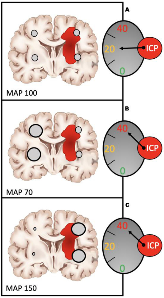
Lundberg in 1960 monitored ICP in hundreds of patients, identifying characteristic pressure waves.22 One of these waves, termed A waves by Lundberg and later referred to as plateau waves, (SDC 13: Lundberg intracranial pressure waves) occurs when ICP abruptly increases to nearly systemic blood pressure levels for about 10–30 minutes, occasionally accompanied by neurologic deterioration (SDC 14: Plateau waves caused by decreased blood pressure). Rosner and Becker8 suggest that two concurrent conditions of exponential physiology produce plateau waves: (1) ICP at the steep portion of the exponential ICP-intracranial volume relationship (SDC 2: Intracranial compliance curve), and: (2) preserved autoregulation in some parts of the brain such that cerebral blood volume increases exponentially with CPP decrements (SDC 15: Non-linear relationship of cerebral blood volume to blood pressure). This concurrent exponential physiology accounts for the abrupt and severe increase in ICP, which is characteristic of plateau waves. However, this “plateau wave physiology” likely also arises in less dramatic fashion as an element of hypervolemic ICP elevation. We have observed this phenomenon of an apparent inverse relationship between MAP and ICP during continuous multimodality neuromonitoring in NeuroICU patients (SDC 16: Plateau wave physiology in a neuroICU patient). Clearly, to develop plateau wave physiology there must be a significant portion of the brain with reactive vasculature, in the setting of an injured brain, with impaired compliance, as depicted in Figure 1 and the SDC (SDC 17: Role of heterogeneous autoregulation in plateau wave and dysregulation physiology).
Figure 1. Role of heterogeneous autoregulation in plateau wave and dysregulation physiology.
(A) A patient with heterogeneous injury is depicted with dysautoregulation on the injured side and intact autoregulation on the contralateral (uninjured) side. The injury and associated mass and edema effect increase ICP somewhat with associated decrement in intracranial compliance.
(B) In this physiologic context, a decrement in blood pressure produces vasodilation with increased cerebral blood volume on the uninjured side with a corresponding increase in ICP.
(C) In contrast, an increase in blood pressure causes vasoconstriction on the uninjured side, but dysregulated hyperemia and increased cerebral blood volume on the injured side. Notably, in this depiction, similar elevations in ICP are produced by distinctly different physiologic conditions.
ICP, intracranial pressure
Dysregulated passive arterial vasodilation
Primary CBF dysautoregulation with passive arterial vasodilation can arise secondary to a continuum of conditions associated with endothelial and blood brain barrier (BBB) injury. Such conditions include significant brain injury (ischemic or traumatic),23 acute liver failure,24 hyperperfusion states such as malignant hypertension/posterior reversible encephalopathy syndrome (PRES),25,26 arteriovenous malformation (AVM) resection with subsequent normal perfusion pressure breakthrough,27, 28 and post-carotid endarterectomy29 or endovascular thrombectomy with successful restoration of CBF.30 In addition, secondary dysregulation can arise in extremes of hypercapnia, hypoxemia, anemia, hypotension, and hypoglycemia, by which brain edema may be exacerbated. In all of these cases of extreme vasodilation, CBF is expected to vary directly with systemic blood pressure.
Endothelial and blood brain (and blood-CSF) barrier injury
Endothelial damage and disrupted BBB can result in dilated poorly reactive vascular beds and capillary leakage, respectively. The endothelial dysfunction dysregulates CBF, which then varies in proportion to CPP with consequent increased blood volume and ICP.31 (SDC 18: ICP increases with hypertension in the injured brain) (SDC 19: Loss of autoregulation with hyperemic intracranial hypertension). The injured BBB promotes hydrostatically-driven perivascular and interstitial vasogenic brain edema,32 ultimately resulting in oligemia.32
Liver failure
Aggarwal et al.24 documented the time course of CBF and hyperemic ICP during the progression of liver failure from mild to severe. The natural history encompasses impaired autoregulation leading to an early phase of significant hyperemia, followed by brain hypoxia and cerebral edema.33 The latter can progress to cause malignant intracranial hypertension, oligemia, and eventually brain death. The root cause is believed to be dysregulated hyperemia, attenuation of which may delay or prevent consequent edema.34
Posterior reversible encephalopathy syndrome
The pathogenesis of PRES has not been fully elucidated but is believed to involve cerebral hyperemia due to dysregulated vasodilation as a result of blood pressure exceeding autoregulatory limits, as well as BBB dysfunction due to endothelial damage; both result in vasogenic edema.26 PRES has been associated with several conditions, most notably uncontrolled hypertension, eclampsia, and immunosuppressive therapy.26, 35, 36 The natural progression of PRES occasionally leads to severe diffuse brain edema with malignant intracranial hypertension, intracerebral hemorrhage, and death.37, 38
Malignant hypertension
The impact on the brain of malignant hypertension has been known for some time and is very similar to, or the same as, PRES. It likely represents the result of hypertension beyond the upper limit of autoregulation, the consequence of which is high pressure hyperemia.25 This can lead to pinocytotic fluid transfer, injury to the capillary bed, vasogenic edema, and intracranial hypertension,39 and, eventually, malignant cerebral edema and oligemia.25, 40, 41
Reperfusion hyperemia
Several conditions of chronic or sub-acute ischemia or intracranial vascular hypotension with restoration of normal perfusion can produce a hyperemic state. This state can produce dysregulated vasodilation such that cerebral blood volume and ICP may vary directly with blood pressure. It can also create a potential for progression to BBB disruption, hemorrhage, intracranial hypertension and eventually, on a physiologic continuum, to oligemia and ischemia. This clearly indicates the potential adverse effects of hyperemia in baseline normal brain, which is perhaps also relevant to hyperemia associated with liver failure, PRES, and malignant hypertension.
Chronically hypotensive territories adjacent to AVMs sustain leftward shift of the normal cerebral autoregulation curve. Resumption of normal perfusion pressure after AVM resection is therefore believed to exceed the upper limit of autoregulation. This is commonly called normal perfusion pressure breakthrough and is analogous to the physiology of hypertensive hyperemia, leading to vasogenic edema and intraparenchymal hemorrhage.27, 28, 42
In patients with chronic carotid arterial stenosis, CBF is maintained by maximal distal arteriolar vasodilation. During carotid endarterectomy, immediately after unclamping the repaired artery, regional hyperemia can overwhelm autoregulatory mechanisms in areas of reduced vasoreactivity, which may lead to BBB disruption and protracted hyperemia with edema and hemorrhage.29 This pathophysiology is supported by reports of attenuation of reperfusion-associated complications through strict control of arterial blood pressure during carotid surgery.29
In the context of acute stroke with ischemic compromise to the BBB and endothelium, reperfusion hyperemia and microvascular injury can occur after interventions to restore perfusion. The consequence is an increased risk of hemorrhagic transformation and cerebral edema, contributing to ICP elevation. Notably, lower blood pressure in a population study is associated with less hyperemia and intraparenchymal hemorrhage, although increased risk of ischemia,30 whereas high blood pressure is associated with more intraparenchymal hemorrhage and worse functional outcomes.43 An example of an immediate hyperemic response during endovascular thrombectomy is shown in the SDC (SDC 20: Hyperemia with reperfusion after endovascular thrombectomy).
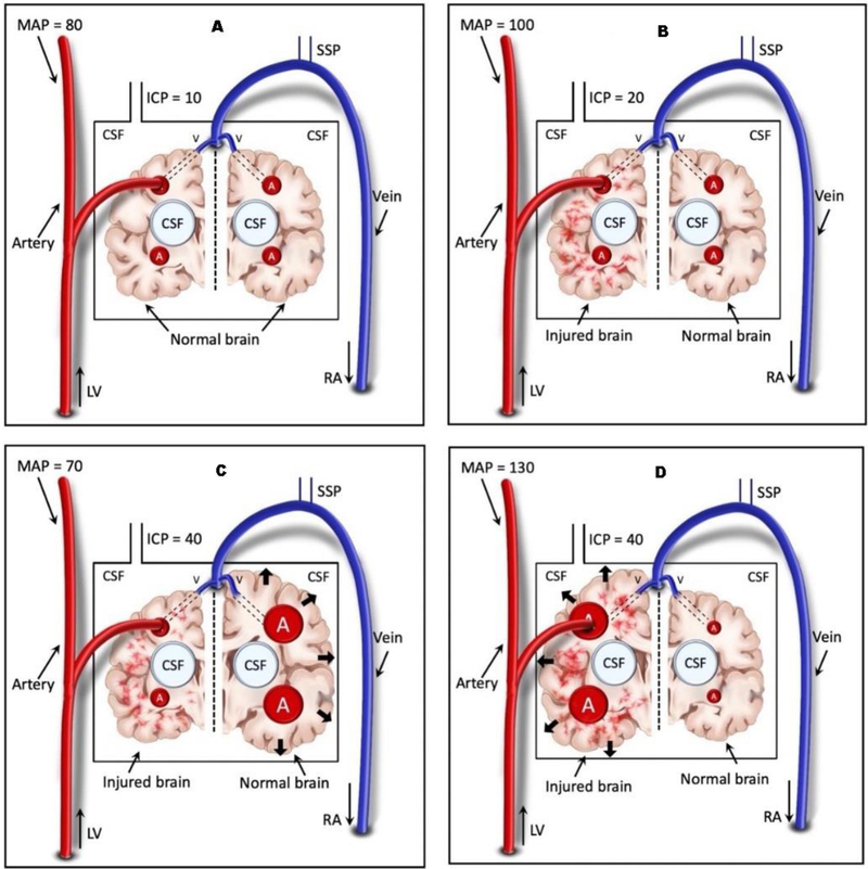
In summary, multiple reports clearly highlight that both regulated and dysregulated hyperemia can increase ICP. It thus appears that, depending on specific pathophysiology, both increasing and decreasing blood pressure can increase ICP in the appropriate clinical setting (Figure 2) (SDC 21: Hypervolemic intracranial hypertension with high and low blood pressure). In patients with both types of blood pressure-related hyperemia, this suggests the presence of an optimal CPP23 which might be considered as an element of hypervolemic ICP management strategy (SDC 22: Bimodal ICP response to blood pressure).
Figure 2. Hypervolemic intracranial hypertension with high and low blood pressure.
A. Normal uninjured brain with normal blood pressure and ICP
B. Injured brain (left) at a baseline ICP 20mmHg and MAP 100mmHg
C. Decreased blood pressure produces vasodilation of uninjured brain with ICP elevated to 40mmHg
D. Increased blood pressure produces vasodilation of injured brain with ICP elevated to 40 mmHg
Notably the same increase in ICP to 40mmHg associates with distinctly different physiologic conditions.
ICP, intracranial pressure; MAP, mean arterial blood pressure
Cerebral venous hypervolemia
Cerebral venous physiology is complex and incompletely understood. Nevertheless, an accepted notion is that increased cerebral venous or dural venous sinus pressures may increase brain tissue hydrostatic pressure and thereby contribute to vasogenic edema. Primary factors contributing to increased cerebral venous volume include functional Starling resistor type venous outflow obstruction, mechanical outflow obstruction, such as with venous thrombosis and very high extracranial venous pressure.
Starling resistor outflow obstruction
The Starling resistor concept, sometimes referred to as vascular waterfall phenomenon or hydraulic hypothesis, describes the situation where external tissue pressure is greater than internal vascular pressure. In such a situation hollow tubes such as blood vessels collapse and flow then becomes dependent on the difference between proximal upstream pressure and distal external extravascular pressure at the point of constriction.
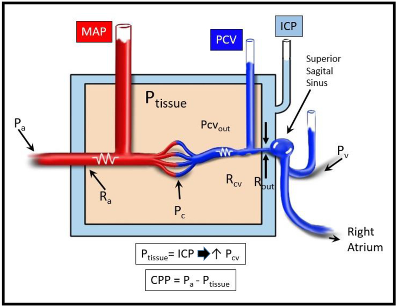
As suggested by Grande et al.44 and Huseby et al.45, increased ICP, from whatever etiology, can also produce a direct Starling resistor (vascular waterfall) type of constriction of venous outflow at the level of the cerebral veins proximal to the dural sinuses. This venous obstruction produces an increase in internal venous pressure, which maintains vein patency such that flow continues. However, it also increases tissue hydrostatic pressure, which then presents the potential to produce or exacerbate cerebral edema which further increases ICP with a positive feedback cycle thus produced (Figure 3) (SDC 23: Mechanism of vasogenic edema from elevated ICP producing a venous outflow obstruction).44, 46 This venous Starling resistor removes the effect on perfusion of distal downstream pressure (e.g. sagittal sinus), as long as outflow internal vascular pressure remains less than (or equal to) external tissue pressure (or ICP).47–50 Notably, sagittal sinus pressure is thought to be generally unaffected by ICP because of the unyielding walls of the sinus, whereas the thin-walled cerebral veins and lacunae are affected by ICP.49
Figure 3. Mechanism of vasogenic edema from elevated ICP producing a venous outflow obstruction.
Increased CSF pressure acts to produce a venous waterfall such that venous pressure and tissue pressure proximal to the venous obstruction increases as a consequence of elevated ICP. This in turn generates vasogenic edema (increased microvascular pressure, Pc) thereby increasing intracranial tissue volume to further increase ICP, resulting in a positive feedback cycle.44 In this physiologic situation, extracranial venous pressure must exceed ICP in order to affect intracranial venous dynamics.
Pa,arterial blood pressure; Pc, capillary pressure; Pcv, cerebral venous pressure; Pv, extracranial venous pressure; MAP, mean arterial pressure; ICP, intracranial pressure; Ptissue, brain tissue pressure; Ra, arterial resistance; Rcv, resistance of brain tissue veins; Rout, resistance at venous outflow at point of Startling resistor
This presents three conceptual situations relating venous outflow and ICP as follows (Pcv, cerebral venous pressure; Pss, sagittal sinus pressure; →, vascular waterfall):
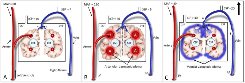
Normal ICP - MAP > Pcv > ICP > Pss. There is no tissue compression of cerebral veins and perfusion pressure driving CBF is determined by MAP - Pss (Figures 4A and 5A) (SDC 24: Oligemic intracranial hypertension) (SDC 25: Vasogenic edema from arterial and venous causes).
Elevated ICP - MAP > Pcv = ICP → Pss. This arises in the Lund model44 where increased ICP and external tissue pressure increase Pcv to a level equivalent to ICP. This permits continued flow but at higher Pcv, with consequent increased tissue hydrostatic pressure. In this case, perfusion pressure driving CBF is MAP - ICP (or Pcv). Pss, consequent to the vascular waterfall, is removed from the equation (Figures 3 and 5C) (SDC 23: Mechanism of vasogenic edema from elevated ICP producing a venous outflow obstruction) (SDC 25: Vasogenic edema from arterial and venous causes) .
Infarction/brain death: MAP = Pcv = ICP → Pss. There is no pressure gradient to support perfusion and Pss consequent to the vascular waterfall is removed from the equation (Figure 4B) (SDC 24: Oligemic intracranial hypertension).
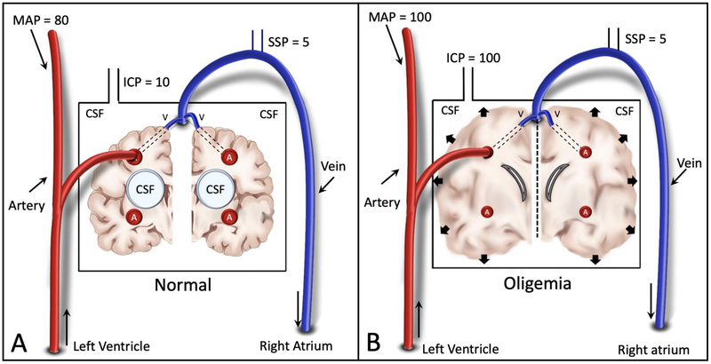
Figure 4. Oligemic Intracranial Hypertension.
A. Normal physiologic state with normal blood pressure and ICP with normal blood vessels.
B. ICP is increased to systemic blood pressure levels due to massive tissue edema, pinching off blood flow and compressing the ventricles
ICP, intracranial pressure
Figure 5. Vasogenic edema from arterial and venous causes.
A. Baseline state with normal blood pressure, venous pressure, and ICP.
B. Dysregulated cerebral hyperemia with hypertension, produces peri-arteriolar tissue vasogenic edema with elevated ICP
C. Increased venous pressure, at possible points noted by an asterisk, produces peri-venular tissue vasogenic edema with elevated ICP.
Note the depiction of similar levels of elevated ICP from distinctly different causes of vasogenic edema.
ICP, intracranial pressure
Venous Sinus Obstruction
One form of cerebral venous obstruction is believed to contribute to pseudotumor cerebri, or idiopathic intracranial hypertension (IHH).This is likely a mechanical venous constriction thought to contribute to the below-noted pathophysiology to increase ICP.51, 52 IHH likely has other contributing factors and is described in more detail subsequently.
The other primary factor in venous sinus obstruction is thrombosis. Cerebral venous sinus thrombosis is the collective term for thrombosis of the cerebral dural sinuses and cerebral veins,53, 54 and it can play a key role in the pathophysiology of intracranial hypertension. The physiology is complex and at least four interacting processes can be identified:
Given that CSF reabsorption occurs at arachnoid granulations in the cerebral venous sinuses, thrombosis of dural sinuses can impair CSF reabsorption resulting in increased CSF volume. This can cause communicating hydrocephalus and increased ICP.
Cerebral venous sinus thrombosis is believed to produce an increase in local venular and capillary pressures from venous outflow obstruction. This produces or exacerbates BBB disruption with consequent vasogenic edema and oligemia/ischemia.55, 56
Venular and capillary disruption can also result in intraparenchymal hemorrhage to increase ICP. If the hemorrhage includes the ventricular system, the process may be further complicated by non-communicating hydrocephalus.
Vasogenic edema with worsened intracranial hypertension can result in further venous outflow obstruction by a Starling resistor effect, further exacerbating the pathophysiologic process.
Very high extracranial venous pressure
Anything that increases venous pressure sufficient to fall within the range of ICP at the level of the cranium carries a theoretical risk of increased intracranial venous volume (Figure 3) (SDC 23: Mechanism of vasogenic edema from elevated ICP producing a venous outflow obstruction). Such situations may include high airway pressure, acute superior vena cava syndrome, right heart failure or obstruction, or Trendelenburg head-down position. The concept is dramatically demonstrated at the bedside where digital jugular compression (Queckenstedt maneuver) transiently increases ICP.57 A systematic overview of venous effects on ICP is provided by Wilson et. al58
Positive end expiratory pressure (PEEP) has been reported to increase ICP59 and this effect is often presumed to be related to venous outflow obstruction secondary to PEEP-related increased intrathoracic pressure. However, hydrostatically this can only arise when PEEP is very high and pulmonary compliance is sufficiently high to allow transmission of the PEEP to the intra-thoracic vasculature.60 Moreover, the increase in extracranial venous pressure must exceed ICP in order to impact intracranial venous outflow. Thus, an ICP of 20mmHg would mandate a PEEP-mediated increase in extracranial venous pressure of >20mmHg (~26 cmH20) in order to impact on ICP. Such high levels of PEEP are typically used only in the context of very low pulmonary compliance, in which case the airway pressure is not fully transmitted to the intra-thoracic vasculature.60 Further, the general situation of head-of-bed elevation would mandate even higher venous pressure in the thorax in order to affect intracranial venous blood volume. Thus, simple hydrostatic considerations suggest a minimal effect of PEEP when ICP is low, and a negligible effect when ICP is high, reflecting the notion that PEEP mediated increase in extracranial venous outflow pressure must exceed ICP in order to increase ICP.45 Nonetheless ICP has been noted to increase with application of PEEP in clinical situations.59 A synthesis of the literature leads to the conclusion that this is most likely a plateau wave type physiology, wherein PEEP produces a decrement in blood pressure which then produces compensatory cerebral arterial dilation in normally autoregulating cerebral vasculature (SDC 14: Plateau waves caused by decreased blood pressure) (SDC 15: Non-linear relationship of cerebral blood volume to blood pressure) (SDC 16: Plateau wave physiology in a neuroICU patient).
Masses and Edema
Increased intracranial tissue or extravascular blood volume are important causes of intracranial hypertension. In contrast to increased intravascular blood volume subsets of elevated ICP, masses and edema can directly produce oligemic intracranial hypertension and have immediate deleterious effects. The continuum of progressively decreased CPP from mass or edema producing oligemia observed with transcranial Doppler is illustrated in the SDC (SDC 4: Cerebral oligemia with decreased cerebral perfusion pressure). The three major causes of this situation are edema, and masses due to neoplasia or hemorrhage.
Brain Tissue Edema
Diffuse brain tissue edema is an important cause of intracranial hypertension and is commonly entwined with other causes of increased ICP. Brain tissue edema can produce severe oligemia as depicted in neuroICU patient neuromonitoring data, where a progressive rise in ICP post ischemia is accompanied by a decrease in CBF in progression to brain death (Figure 4) (SDC: 24, Oligemic intracranial hypertension) (SDC 26: Oligemic intracranial hypertension progressing to brain death). Brain edema is an abnormal accumulation of fluid within the brain parenchyma and is subdivided into two major categories - vasogenic and cytotoxic edema.61,62,63 Notably, both types of edema may arise concurrently.
Vasogenic edema
Vasogenic edema encompasses conditions associated with BBB breakdown, allowing movement of intravascular proteins, solutes and water through the damaged microvascular endothelial cells into the extracellular space. Etiologies of vasogenic edema are numerous and include traumatic brain injury, brain tumor, radiation necrosis, acute demyelination, dysregulated hyperemia, venous obstruction, and inflammatory/infectious processes (Figure 5) (SDC 25: Vasogenic edema from arterial and venous causes). In addition, vasogenic edema arises with increased blood pressure in the context of BBB and vascular injury. Moreover, as discussed above, intracranial hypertension from any cause with associated venous obstruction and increased tissue hydrostatic pressure promotes yet another type of vasogenic edema (Figure 5) (SDC 25: Vasogenic edema from arterial and venous causes). When brain edema overcomes the compensatory mechanisms, ICP, following an exponential relationship with volume of an intracranial compartment, can increase dramatically resulting in compromise of CBF with widespread ischemia or in herniation4, 64, 65 (Figure 4) (SDC 24: Oligemic intracranial hypertension) (SDC 26: Oligemic intracranial hypertension progressing to brain death).
Cytotoxic edema
Causes of cytotoxic edema include hypoxic-ischemic brain injury, traumatic brain injury, central nervous system infections, drug overdoses, renal and liver failure, spreading depolarization,66 ornithine transcarbamylase deficiency,67 and osmolar gradient syndromes as may be seen with hyperglycemia and hyponatremia. In such insults, processes arise to cause intracellular fluid translocation. In the case of hypoxia-ischemia, a primary insult leads to failure of ATP-dependent sodium-potassium and calcium pumps resulting in intracellular sodium (or other metabolite) accumulation, an osmotic gradient, fluid shift from extracellular to intracellular compartments, and eventual cellular swelling. Other syndromes may increase different intracellular osmoles to produce the same effect. Calcium can also accumulate within the cell, triggering an inflammatory cascade that recruits microglia and leads to free radical formation that ultimately results in the destruction of the BBB and concurrent vasogenic edema .61,62,63
Masses
Pathologic intracranial masses can increase ICP, eventually culminating in oligemia. Masses can be in the form of hematoma or neoplasm and, in both instances, the more rapid the growth of said mass, the more acute and profound the rise in ICP. Several types of hematomas can arise: intraparenchymal, subarachnoid, subdural, and epidural. Each hematoma and tumor type has unique characteristics, a detailed description of which is beyond the scope of this review.
Increased CSF Volume
Over a century ago, Dandy proposed the classic hypothesis of CSF hydrodynamics.68, 69 In brief, CSF is produced mainly by epithelial cells of the choroid plexus lining the cerebral ventricles using active transport, largely dependent on the enzyme carbonic anhydrase. CSF then circulates uni-directionally through the ventricular system, from the lateral ventricles to the foramen of Monro, into the third ventricle, aqueduct of Sylvius, the fourth ventricle, and finally exiting into the spinal subarachnoid space through the foraminae of Luschka and Magendie. The spinal subarachnoid space is in direct communication with the intracranial subarachnoid space from where CSF is passively absorbed down a pressure gradient through the arachnoid villi of dural venous sinuses into venous sinuses. In order for this process to occur, CSF pressure must exceed sagittal sinus venous pressure.70, 71 From the venous sinuses, CSF eventually enters into the systemic venous circulation. This classic description of CSF dynamics has recently been enhanced to incorporate the glymphatic system, a perivascular intraparenchymal pathway for CSF egress from the brain.72
Hydrocephalus
Hydrocephalus is a disruption of CSF formation, flow, or absorption characterized by excessive accumulation of CSF within the cerebral ventricular system and/or subarachnoid space. Regardless of cause, this can result in ventriculomegaly and intracranial hypertension. Hydrocephalus was historically classified by Dandy into two types - communicating and non-communicating (SDC 27: Hydrocephalus). Communicating hydrocephalus occurs due to excess CSF production or, more commonly, ineffective CSF absorption. For example, it can be a result of scarring or fibrosis in the subarachnoid space after infection, inflammation, or hemorrhage. Non-communicating hydrocephalus, on the other hand, occurs from obstruction of CSF flow within the ventricular system or its outlets, resulting in dilation of the ventricular system proximal to the point of obstruction.73 A role for the glymphatic system has been proposed for normal pressure hydrocephalus,72 but any role in other hydrocephalus types is not yet established.
Idiopathic intracranial hypertension
The exact pathophysiology of intracranial hypertension in IIH remains unknown. Hence it does not fit neatly into the taxonomic categories described above, although many aspects of IHH cut across them. Proposed mechanisms include excess CSF production, reduced CSF absorption, CSF outflow obstruction, increased brain tissue water content and abnormalities of vitamin A metabolism.74 It is also proposed that any mechanistic theory for IHH must account for its epidemiologic predilection for obese young females. Cerebral venous sinus stenosis is identified in a majority of IHH patients, raising the possibility of a causal contribution.51, 52 Given the limited understanding of its pathophysiology, management strategies for IIH focus on treatment of raised ICP with carbonic anhydrase inhibitors, alleviation of symptoms of raised ICP such as headaches and vision loss, and targeting potential contributing factors such as obesity. While there is no unifying hypothesis for IIH, future clinical trials will hopefully add to the existing knowledge of its pathophysiology.
Conclusion: Subset Oriented Treatment of Intracranial Hypertension
In this review we describe a physiologic taxonomy of intracranial hypertension and thereby illuminate the notion of several physiologic subsets contributing to raised ICP. Our review creates a tempting speculation that consideration of these various etiologies of intracranial hypertension may support focused and individualized diagnostic and therapeutic maneuvers which otherwise might not be considered. Some examples can be entertained. Diagnosis of intracranial hypertension with autoregulated hyperemia would support the use of hyperventilation, whereas dysregulated hyperemia would support aggressive blood pressure management. Oligemia as the cause of raised ICP would support osmotherapy or surgical decompression, but increased CSF volume warrants treatment with CSF diversion by ventriculostomy. We suggest that future research in ICP management should incorporate these concepts of ICP subsets to thereby decrease patient heterogeneity and lessen the likelihood of inconclusive results.
Supplementary Material
Supplemental Digital Content 10. Increased ICP after acetazolamide injection
Supplemental Digital Content 20. Hyperemia with reperfusion after endovascular thrombectomy
Supplemental Digital Content 21. Hypervolemic intracranial hypertension with high and low blood pressure
Supplemental Digital Content 22. Bimodal ICP response to blood pressure
Supplemental Digital Content 23. Mechanism of vasogenic edema from elevated ICP producing a venous outflow obstruction
Supplemental Digital Content 26. Oligemic intracranial hypertension progressing to brain death
Supplemental Digital Content 27. Hydrocephalus
Supplemental Digital Content 24. Oligemic intracranial hypertension
Supplemental Digital Content 25. Vasogenic edema from arterial and venous causes
Supplemental Digital Content 4. Figure. Cerebral oligemia with decreased CPP
Supplemental Digital Content 11. Anemia induced cerebral hyperemia in rodents
Supplemental Digital Content 12. Anemia induced cerebral hyperemia in humans
Supplemental Digital Content 3. Figure. ICP waveform changes with decrements in compliance
Supplemental Digital Content 5. Autoregulation of cerebral blood flow
Supplemental Digital Content 6. Hyperemic intracranial hypertension produced by laryngoscopy
Supplemental Digital Content 7. Hyperemic intracranial hypertension produced by sympathetic storm
Supplemental Digital Content 8. PaO2 and PaCO2 alter CBF
Supplemental Digital Content 9. Increase in CBF and PbO2 with increased CO2.
Supplemental Digital Content 13. Lundberg intracranial pressure waves
Supplemental Digital Content 14. Plateau waves caused by decreased blood pressure
Supplemental Digital Content 1. Text file: Methods and IRB approval for neuromonitoring used in University of Pennsylvania neuroICU patients
Supplemental Digital Content 15. Non-linear relationship of cerebral blood volume to blood pressure
Supplemental Digital Content 16. Plateau wave physiology in a NeuroICU patient
Supplemental Digital Content 17. Role of heterogeneous autoregulation in plateau wave and dysregulation physiology
Supplemental Digital Content 18. ICP increases with hypertension in the injured brain.
Supplemental Digital Content 19. Loss of autoregulation with hyperemic intracranial hypertension
Acknowledgements
Sources of support:
Department of Health and Human Services
National Institutes of Health
NATIONAL INSTITUTE OF NEUROLOGICAL DISORDERS AND STROKE
1R01NS082309-01A1
Footnotes
Conflicts of Interest
- Funded by 1R01NS082309-01A1
- Legal consultant; multiple legal and health care entities
- Royalty payments Oxford University Press and Elsevier
- Honoraria NIH study sections
- Editorial Board Neurocritical Care
- Editorial Board J Neurosurg Anesth
- Co-author on provisional patent number 17-8261 / 103241.000816 Trustees of the University of Pennsylvania
- Funded by 1R01NS082309-01A1
- CoAuthor on provisional patent number 17–8261 / 103241.000816 Trustees of the University of Pennsylvania
Other authors have no conflicts to declare
supplementary digital content
Supplemental Digital Content 1. Text file: Methods and IRB approval for neuromonitoring used in University of Pennsylvania neuroICU patients
Supplemental Digital Content 2. Figure: Intracranial compliance curve
Supplemental Digital Content 3. Figure. ICP waveform changes with decrements in compliance
Supplemental Digital Content 4. Figure. Cerebral oligemia with decreased CPP
Supplemental Digital Content 5. Autoregulation of cerebral blood flow
Supplemental Digital Content 6. Hyperemic intracranial hypertension produced by laryngoscopy
Supplemental Digital Content 7. Hyperemic intracranial hypertension produced by sympathetic storm
Supplemental Digital Content 8. PaO2 and PaCO2 alter CBF
Supplemental Digital Content 9. Increase in CBF and PbO2 with increased CO2.
Supplemental Digital Content 10. Increased ICP after acetazolamide injection
Supplemental Digital Content 11. Anemia induced cerebral hyperemia in rodents
Supplemental Digital Content 12. Anemia induced cerebral hyperemia in humans
Supplemental Digital Content 13. Lundberg intracranial pressure waves
Supplemental Digital Content 14. Plateau waves caused by decreased blood pressure
Supplemental Digital Content 15. Non-linear relationship of cerebral blood volume to blood pressure
Supplemental Digital Content 16. Plateau wave physiology in a NeuroICU patient
Supplemental Digital Content 17. Role of heterogeneous autoregulation in plateau wave and dysregulation physiology
Supplemental Digital Content 18. ICP increases with hypertension in the injured brain.
Supplemental Digital Content 19. Loss of autoregulation with hyperemic intracranial hypertension
Supplemental Digital Content 20. Hyperemia with reperfusion after endovascular thrombectomy
Supplemental Digital Content 21. Hypervolemic intracranial hypertension with high and low blood pressure
Supplemental Digital Content 22. Bimodal ICP response to blood pressure
Supplemental Digital Content 23. Mechanism of vasogenic edema from elevated ICP producing a venous outflow obstruction
Supplemental Digital Content 24. Oligemic intracranial hypertension
Supplemental Digital Content 25. Vasogenic edema from arterial and venous causes
Supplemental Digital Content 26. Oligemic intracranial hypertension progressing to brain death
Supplemental Digital Content 27. Hydrocephalus
Contributor Information
W Andrew Kofke, Department of Anesthesiology and Critical Care, University of Pennsylvania, Philadelphia, Pa.
Swarna Rajagopalan, Department of Neurology, West Virginia University, Morgantown, WV.
Diana Ayubcha, Department of Anesthesiology and Critical Care, University of Pennsylvania, Philadelphia, Pa.
Ramani Balu, Department of Neurology, University of Pennsylvania, Philadelphia, Pa.
Jovany Cruz-Navarro, Department of Anesthesiology, Baylor College of Medicine.
Panumart Manatpon, Department of Anesthesiology, University of Florida, Jacksonville, FL.
Elizabeth Mahanna-Gabrielli, Department of Anesthesiology and Critical Care, University of Pennsylvania, Philadelphia, Pa.
References
- 1.Miller JD, Becker DP, Ward JD, et al. Significance of intracranial hypertension in severe head injury. J Neurosurg 1977;47: 503–16. [DOI] [PubMed] [Google Scholar]
- 2.Schnitzer MS, Aschoff AA, Hacke W. Intracranial Pressure Monitoring In: Hacke W, Hanley DF, Einhäupl KM, et al. , eds. Neurocritical Care. Berlin, Heidelberg: Springer Berlin Heidelberg, 1994:90–97. [Google Scholar]
- 3.Dewey R, Pieper H, Hunt W. Experimental cerebral hemodynamics. Vasomotor tone, critical closing pressure, and vascular bed resistance. J Neurosurg 1974;41: 597. [DOI] [PubMed] [Google Scholar]
- 4.Hassler W, Steinmetz H, Gawlowski J. Transcranial Doppler ultrasonography in raised intracranial pressure and in intracranial circulatory arrest. J Neurosurg 1988;68: 745. [PubMed] [Google Scholar]
- 5.Aaslid R, Lindegaard KF, Sorteberg W, et al. Cerebral autoregulation dynamics in humans. Stroke 1989;20: 45–52. [DOI] [PubMed] [Google Scholar]
- 6.Drummond JC. The lower limit of autoregulation: time to revise our thinking? Anesthesiology 1997;86: 1431–3. [DOI] [PubMed] [Google Scholar]
- 7.Risberg J, Lundberg N, Ingvar DH. Regional cerebral blood volume during acute transient rises of the intracranial pressure (plateau waves). J Neurosurg 1969;31: 303–10. [DOI] [PubMed] [Google Scholar]
- 8.Rosner MJ, Becker DP. Origin and evolution of plateau waves. Experimental observations and a theoretical model. J Neurosurg 1984;60: 312–24. [DOI] [PubMed] [Google Scholar]
- 9.Pierre Kahn A, Gabersek V, Hirsch JF. Intracranial pressure and rapid eye movement sleep in hydrocephalus. Child’s Brain 1976;2: 156–166. [DOI] [PubMed] [Google Scholar]
- 10.Solheim O, Vik A, Gulati S, et al. Rapid and severe rise in static and pulsatile intracranial pressures during a generalized epileptic seizure. Seizure 2008;17: 740–743. [DOI] [PubMed] [Google Scholar]
- 11.Burney RG, Winn R. Increased cerebrospinal fluid pressure during laryngoscopy and intubation for induction of anesthesia. Anesthesia and Analgesia 1975;54: 687–690. [DOI] [PubMed] [Google Scholar]
- 12.Bein T, Kuhr LP, Bele S, et al. Lung recruitment maneuver in patients with cerebral injury: effects on intracranial pressure and cerebral metabolism. Intensive Care Med 2002;28: 554–8. [DOI] [PubMed] [Google Scholar]
- 13.Nyholm L, Howells T, Lewén A, et al. The influence of hyperthermia on intracranial pressure, cerebral oximetry and cerebral metabolism in traumatic brain injury. Upsala Journal of Medical Sciences 2017;122: 177–184. [DOI] [PMC free article] [PubMed] [Google Scholar]
- 14.Klug N, Hoffmann O, Zierski J, et al. Decerebrate rigidity and vegetative signs in the acute midbrain syndrome with special regard to motor activity and intracranial pressure. Acta Neurochirurgica 1984;72: 219–233. [DOI] [PubMed] [Google Scholar]
- 15.Sakabe T, SiesjÖ BK. The effect of indomethacin on the blood flow—metabolism couple in the brain under normal, hypercapnic and hypoxic conditions. Acta Physiologica Scandinavica 1979;107: 283–284. [DOI] [PubMed] [Google Scholar]
- 16.Arbeláez AM, Su Y, Thomas JB, et al. Comparison of Regional Cerebral Blood Flow Responses to Hypoglycemia Using Pulsed Arterial Spin Labeling and Positron Emission Tomography. PLoS ONE 2013;8. [DOI] [PMC free article] [PubMed] [Google Scholar]
- 17.Borgstrom L, Johannsson H, Siesjo B. The influence of acute normovolemic anemia on cerebral blood flow and oxygen consumption of anesthetized rats. Acta Physiol Scand 1975;93 (40: 505–14. [DOI] [PubMed] [Google Scholar]
- 18.Floyd T, McGarvey M, Ochroch E, et al. Perioperative changes in cerebral blood flow after cardiac surgery: influence of anemia and aging. Ann Thoracic Surg 2003;76 (6): 2037–42. [DOI] [PubMed] [Google Scholar]
- 19.Pindzola RR, Balzer JR, Nemoto EM, et al. Cerebrovascular reserve in patients with carotid occlusive disease assessed by stable xenon-enhanced ct cerebral blood flow and transcranial Doppler. Stroke 2001;32: 1811–7. [DOI] [PubMed] [Google Scholar]
- 20.Holl K, Heissler HE, Nemati N, et al. Effect of acetazolamide-induced endogenous volume pressure testing of the cerebrospinal system on intracranial pressure. Effect of Diamox® on intracranial pressure (ICP). Neurochirurgia 1990;33: 29–36. [DOI] [PubMed] [Google Scholar]
- 21.Mollan SP, Ball AK, Sinclair AJ, et al. Idiopathic intracranial hypertension associated with iron deficiency anaemia: A lesson for management. European Neurology 2009;62: 105–108. [DOI] [PubMed] [Google Scholar]
- 22.Lundberg N Continuous recording and control of ventricular fluid pressure in neurosurgical practice. Acta Psychiatr Scand Suppl 1960;36: 1–193. [PubMed] [Google Scholar]
- 23.Steiner LA, Czosnyka M, Piechnik SK, et al. Continuous monitoring of cerebrovascular pressure reactivity allows determination of optimal cerebral perfusion pressure in patients with traumatic brain injury. Crit Care Med 2002;30: 733–738. [DOI] [PubMed] [Google Scholar]
- 24.Aggarwal S, Obrist W, Yonas H, et al. Cerebral hemodynamic and metabolic profiles in fulminant hepatic failure: relationship to outcome. Liver Transpl 2005;11: 1353–60. [DOI] [PubMed] [Google Scholar]
- 25.Skinhoj E On the pathogenesis of hypertensive encephalopathy as revealed by cerebral blood flow studies in man. Progress in Brain Research 1977;47: 235–43. [DOI] [PubMed] [Google Scholar]
- 26.Bartynski WS. Posterior Reversible Encephalopathy Syndrome, Part 2: Controversies Surrounding Pathophysiology of Vasogenic Edema. 2008;29: 1043–1049. [DOI] [PMC free article] [PubMed] [Google Scholar]
- 27.Batjer HH, Devous MD Sr, Meyer YJ, et al. Cerebrovascular hemodynamics in arteriovenous malformation complicated by normal perfusion pressure breakthrough. Neurosurgery 1988;22: 503–509. [DOI] [PubMed] [Google Scholar]
- 28.Spetzler R, Wilson C, Weinstein P, et al. Normal perfusion pressure breakthrough theory. Clin Neurosurg 1978;25: 651–672. [DOI] [PubMed] [Google Scholar]
- 29.Bouri S, Thapar A, Shalhoub J, et al. Hypertension and the post-carotid endarterectomy cerebral hyperperfusion syndrome. European Journal of Vascular and Endovascular Surgery 2011;41: 229–237. [DOI] [PubMed] [Google Scholar]
- 30.Goyal N, Tsivgoulis G, Pandhi A, et al. Blood pressure levels post mechanical thrombectomy and outcomes in large vessel occlusion strokes. Neurology 2017;89: 540–547. [DOI] [PubMed] [Google Scholar]
- 31.Paulson OB, Strandgaard S, Edvinsson L. Cerebral autoregulation. Cerebrovasc Brain Metab Rev 1990;2: 161–92. [PubMed] [Google Scholar]
- 32.Kinoshita K Traumatic brain injury: pathophysiology for neurocritical care. Journal of Intensive Care 2016;4: 29. [DOI] [PMC free article] [PubMed] [Google Scholar]
- 33.Larsen FS, Knudsen GM, Hansen BA. Pathophysiological changes in cerebral circulation, oxidative metabolism and blood-brain barrier in patients with acute liver failure: Tailored cerebral oxygen utilization. Journal of Hepatology 1997;27: 231–238. [DOI] [PubMed] [Google Scholar]
- 34.Larsen FS, Wendon J. Brain edema in liver failure: basic physiologic principles and management. Liver transplantation. 2002;8: 983–989. [DOI] [PubMed] [Google Scholar]
- 35.Hinchey J, Chaves C, Appignani B, et al. A reversible posterior leukoencephalopathy syndrome. New England Journal of Medicine 1996;334: 494–500. [DOI] [PubMed] [Google Scholar]
- 36.Facchini A, Magnoni S, Civelli V, et al. Refractory intracranial hypertension in posterior reversible encephalopathy syndrome. Neurocrit Care 2013;19: 376–80. [DOI] [PubMed] [Google Scholar]
- 37.Facchini A, Magnoni S, Civelli V, et al. Refractory intracranial hypertension in posterior reversible encephalopathy syndrome. Neurocritical Care 2013;19: 376–80. [DOI] [PubMed] [Google Scholar]
- 38.Liman TG, Bohner G, Heuschmann PU, et al. The clinical and radiological spectrum of posterior reversible encephalopathy syndrome: the retrospective Berlin PRES study. Journal of Neurology 2012;259: 155–64. [DOI] [PubMed] [Google Scholar]
- 39.Bartynski WS. Posterior reversible encephalopathy syndrome, part 2: controversies surrounding pathophysiology of vasogenic edema. AJNR Am J Neuroradiol 2008;29: 1043–9. [DOI] [PMC free article] [PubMed] [Google Scholar]
- 40.Katsumata Y, Maehara T, Noda M, et al. Hypertensive encephalopathy: reversible CT and MR appearance. Radiation Medicine 1993;11: 160–3. [PubMed] [Google Scholar]
- 41.Skinhoj E, Strandgaard S. Pathogenesis of hypertensive encephalopathy. Lancet 1973;1: 461–2. [DOI] [PubMed] [Google Scholar]
- 42.Ogasawara K, Yoshida K, Otawara Y, et al. Cerebral blood flow imaging in arteriovenous malformation complicated by normal perfusion pressure breakthrough. Surgical Neurology 2001;56: 380–384. [DOI] [PubMed] [Google Scholar]
- 43.Mistry EA, Mistry AM, Nakawah MO, et al. Systolic Blood Pressure Within 24 Hours After Thrombectomy for Acute Ischemic Stroke Correlates With Outcome. Journal of the American Heart Association 2017;6. [DOI] [PMC free article] [PubMed] [Google Scholar]
- 44.Grande P, Asgeirsson B, Nordstrom C. Volume-targeted therapy of increased intracranial pressure: the Lund concept unifies surgical and non-surgical treatments. Acta Anaesth Scand 2002;46: 929–41. [DOI] [PubMed] [Google Scholar]
- 45.Huseby J, Luce J, Cary J, et al. Effects of positive end-expiratory pressure on intracranial pressure in dogs with intracranial hypertension. J Neurosurg 1981;55 (5): 704. [DOI] [PubMed] [Google Scholar]
- 46.Stokum JA, Gerzanich V, Simard JM. Molecular pathophysiology of cerebral edema. J Cereb Blood Flow Metab 2016;36: 513–38. [DOI] [PMC free article] [PubMed] [Google Scholar]
- 47.Permutt S RLR. Hemodynamics of collapsible vessels with toen: the vascular waterfall. J. Appl. Physiol 1963;18: 924–932. [DOI] [PubMed] [Google Scholar]
- 48.Ursino CAL M. A simple mathematical model of the interaction between intracranial pressure and cerebral hemodynamic. J. Appl. Physiol 1997;82: 1256–1259. [DOI] [PubMed] [Google Scholar]
- 49.Schaller B Physiology of cerebral venous blood flow: from experimental data in animals to normal function in humans. Brain Research Reviews 2004;46: 243–260. [DOI] [PubMed] [Google Scholar]
- 50.Luce JM HJ, Kirk W, Butler J. A Starling resistor regulates cerebral venous outflow in dogs. J Appl Physiol. 1982;53: 1496–503. [DOI] [PubMed] [Google Scholar]
- 51.Owler BK, Parker G, Halmagyi GM, et al. Pseudotumor cerebri syndrome: Venous sinus obstruction and its treatment with stent placement. Journal of Neurosurgery 2003;98: 1045–1055. [DOI] [PubMed] [Google Scholar]
- 52.Stienen A, Weinzierl M, Ludolph A, et al. Obstruction of cerebral venous sinus secondary to idiopathic intracranial hypertension. European Journal of Neurology 2008;15: 1416–1418. [DOI] [PubMed] [Google Scholar]
- 53.Ferro JM, Canhao P, Stam J, et al. Prognosis of cerebral vein and dural sinus thrombosis: results of the International Study on Cerebral Vein and Dural Sinus Thrombosis (ISCVT). Stroke 2004;35: 664–70. [DOI] [PubMed] [Google Scholar]
- 54.Saposnik G, Barinagarrementeria F, Brown RD Jr., et al. Diagnosis and management of cerebral venous thrombosis: a statement for healthcare professionals from the American Heart Association/American Stroke Association. Stroke 2011;42: 1158–92. [DOI] [PubMed] [Google Scholar]
- 55.Piazza G Cerebral venous thrombosis. Circulation 2012;125: 1704–9. [DOI] [PubMed] [Google Scholar]
- 56.Itrat A, Shoukat S, Kamal AK. Pathophysiology of cerebral venous thrombosis--an overview. J Pak Med Assoc 2006;56: 506–8. [PubMed] [Google Scholar]
- 57.Pearce JMS. Queckenstedt’s manoeuvre. Journal of Neurology, Neurosurgery and Psychiatry 2006;77: 728. [DOI] [PMC free article] [PubMed] [Google Scholar]
- 58.Wilson MH. Monro-Kellie 2.0: The dynamic vascular and venous pathophysiological components of intracranial pressure. Journal of Cerebral Blood Flow and Metabolism 2016;36: 1338–1350. [DOI] [PMC free article] [PubMed] [Google Scholar]
- 59.Shapiro H, Marshall L. Intracranial pressure responses to PEEP in head-injured patients. J Trauma 1978;18: 254. [DOI] [PubMed] [Google Scholar]
- 60.Suter PM, Fairley HB, Isenberg MD. Optimum End-Expiratory Airway Pressure in Patients with Acute Pulmonary Failure. New England Journal of Medicine 1975;292: 284–289. [DOI] [PubMed] [Google Scholar]
- 61.Unterberg AW, Stover J, Kress B, et al. Edema and brain trauma. Neuroscience 2004;129: 1021–1029. [DOI] [PubMed] [Google Scholar]
- 62.Klatzo I Pathophysiological aspects of brain edema. Acta Neuropathologica 1987;72: 236–239. [DOI] [PubMed] [Google Scholar]
- 63.Klatzo I Evolution of brain edema concepts. Acta Neruochir 1994;60: 3–6. [DOI] [PubMed] [Google Scholar]
- 64.Schnitzer MS, Alfred A. Aschoff, and Werner Hacke “Intracranial Pressure Monitoring”. Berlin Heidelberg: Springer; 1994. [Google Scholar]
- 65.Jha SK. Cerebral Edema and its Management. Med J Armed Forces India 2003;59: 326–31. [DOI] [PMC free article] [PubMed] [Google Scholar]
- 66.Dreier JP, Lemale CL, Kola V, et al. Spreading depolarization is not an epiphenomenon but the principal mechanism of the cytotoxic edema in various gray matter structures of the brain during stroke. Neuropharmacology 2018;134: 189–207. [DOI] [PubMed] [Google Scholar]
- 67.Wendell LC, Khan A, Raser J, et al. Successful management of refractory intracranial hypertension from acute hyperammonemic encephalopathy in a woman with ornithine transcarbamylase deficiency. Neurocritical Care 2010;13: 113–117. [DOI] [PubMed] [Google Scholar]
- 68.Dandy WE BK. An experimental and clinical study of internal hydrocephalus. J Am Med Assoc 1913;61: 2216–7. [Google Scholar]
- 69.Cushing H Studies on the cerebrospinal fluid. J Med Res 1914;31: 1–19. [PMC free article] [PubMed] [Google Scholar]
- 70.Forces LHW concerned in the absorption of the cerebrospinal fluid. Am J Physio 1935;114: 40–5. [Google Scholar]
- 71.Oreskovic D, Klarica M. The formation of cerebrospinal fluid: nearly a hundred years of interpretations and misinterpretations. Brain Res Rev 2010;64: 241–62. [DOI] [PubMed] [Google Scholar]
- 72.Rasmussen MK, Mestre H, Nedergaard M. The glymphatic pathway in neurological disorders. The Lancet Neurology 2018;17: 1016–1024. [DOI] [PMC free article] [PubMed] [Google Scholar]
- 73.Oreskovic D, Klarica M. Development of hydrocephalus and classical hypothesis of cerebrospinal fluid hydrodynamics: facts and illusions. Prog Neurobiol 2011;94: 238–58. [DOI] [PubMed] [Google Scholar]
- 74.Tabassi A, Salmasi AH, Jalali M. Serum and CSF vitamin A concentrations in idiopathic intracranial hypertension. Neurology 2005;64: 1893–1896. [DOI] [PubMed] [Google Scholar]
Associated Data
This section collects any data citations, data availability statements, or supplementary materials included in this article.
Supplementary Materials
Supplemental Digital Content 10. Increased ICP after acetazolamide injection
Supplemental Digital Content 20. Hyperemia with reperfusion after endovascular thrombectomy
Supplemental Digital Content 21. Hypervolemic intracranial hypertension with high and low blood pressure
Supplemental Digital Content 22. Bimodal ICP response to blood pressure
Supplemental Digital Content 23. Mechanism of vasogenic edema from elevated ICP producing a venous outflow obstruction
Supplemental Digital Content 26. Oligemic intracranial hypertension progressing to brain death
Supplemental Digital Content 27. Hydrocephalus
Supplemental Digital Content 24. Oligemic intracranial hypertension
Supplemental Digital Content 25. Vasogenic edema from arterial and venous causes
Supplemental Digital Content 4. Figure. Cerebral oligemia with decreased CPP
Supplemental Digital Content 11. Anemia induced cerebral hyperemia in rodents
Supplemental Digital Content 12. Anemia induced cerebral hyperemia in humans
Supplemental Digital Content 3. Figure. ICP waveform changes with decrements in compliance
Supplemental Digital Content 5. Autoregulation of cerebral blood flow
Supplemental Digital Content 6. Hyperemic intracranial hypertension produced by laryngoscopy
Supplemental Digital Content 7. Hyperemic intracranial hypertension produced by sympathetic storm
Supplemental Digital Content 8. PaO2 and PaCO2 alter CBF
Supplemental Digital Content 9. Increase in CBF and PbO2 with increased CO2.
Supplemental Digital Content 13. Lundberg intracranial pressure waves
Supplemental Digital Content 14. Plateau waves caused by decreased blood pressure
Supplemental Digital Content 1. Text file: Methods and IRB approval for neuromonitoring used in University of Pennsylvania neuroICU patients
Supplemental Digital Content 15. Non-linear relationship of cerebral blood volume to blood pressure
Supplemental Digital Content 16. Plateau wave physiology in a NeuroICU patient
Supplemental Digital Content 17. Role of heterogeneous autoregulation in plateau wave and dysregulation physiology
Supplemental Digital Content 18. ICP increases with hypertension in the injured brain.
Supplemental Digital Content 19. Loss of autoregulation with hyperemic intracranial hypertension


